 |
| About Bioline | All Journals | Testimonials | Membership | News |
|
||||||
|
||||||
Mem Inst Oswaldo Cruz, Rio de Janeiro, Vol. 101, No. 6, September ,2006, pp. 645-648 Antibacterial potentiality of Argemone mexicana solvent extracts against some pathogenic bacteria Indranil Bhattacharjee, Soroj Kumar Chatterjee, Soumendranath Chatterjee, Goutam Chandra+ Microbiology Research Unit, Parasitology Research
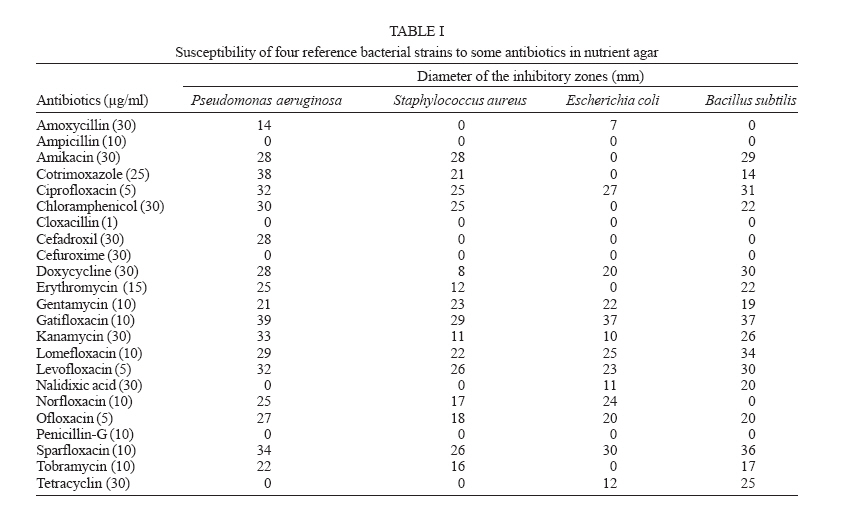
Laboratory, Department of Zoology, The University of Burdwan, Received 10 March 2006 Code Number: oc06109 The sensitivity of two Gram positive (Staphylococcus aureus and Bacillus subtilis) and two Gram negative (Escherichia coli and Pseudomonas aeruginosa) pathogenic multi-drug resistant bacteria was tested against the crude extracts (cold aqueous, hot aqueous, and methanol extracts) of leaves and seeds of Argemone mexicana L. (Papaveraceae) by agar well diffusion method. Though all the extracts were found effective, yet the methanol extract showed maximum inhibition against the test microorganisms followed by hot aqueous extract and cold aqueous extract. Key words: Argemone mexicana - medicinal plant - antibacterial activity
Medicinal plants being the effective source of both traditional and modern medicines, are genuinely useful for primary health care. Over the years, World Health Organization (WHO) advocated traditional medicines as safe remedies for ailments of both microbial and non-microbial origins (WHO 1978). Some antibiotics have become almost obsolete because of drug resistance (Ekpendu et al. 1994) and consequently new drugs must be sought for. Herbal treatment is one possible way to treat diseases caused by multidrug resistant bacteria (Olukoya et al.1993). Though pharmacological industries have produced a number of new antibiotics in the last three decades, yet resistance to these drugs by microorganisms has developed. In general, bacteria have the genetic ability to transmit and acquire resistance to drugs, which in turn may be utilized as therapeutic agents (Cohen 1992). The use of plant extracts and phytochemicals, with known antibacterial properties, may be of immense importance in therapeutic treatments. In the past few years, a number of studies have been conducted in different countries to prove such efficiency (Ikram & Inamul 1984, Almagboul et al. 1985, Sousa et al. 1991, Kubo et al. 1993, Shapoval et al. 1994, Artizzu et al. 1995, Izzo et al. 1995). According to the WHO (Santos et al.1995), medicinal plants would be the best source for obtaining a variety of drugs. About 80% population of the developed countries use traditional medicines, derived from medicinal plants. Therefore, such plants should be investigated thoroughly to determine their structural and functional properties, as well as the efficiency of various parts (Ellof 1998). Argemone mexicana L. (Papaveraceae), commonly known as prickly poppy, is used as a medicinal plant in several countries. In Mexico, the seeds are considered as an antidote to snake venom. In India, the smokes of the seeds are used to relieve toothache. The fresh yellow, milky seed extract contains protein-dissolving substances, effective in the treatment of warts, cold sores, cutaneous infections, skin diseases, itches, and also dropsy and jaundice (Chopra et al. 1986). The present study has been designed to determine the role of seeds and leaf extracts (cold aqueous, hot aqueous, and methanol extracts) of A. mexicana for potential antibacterial activity, if any, against two Gram positive bacteria (Staphylococcus aureus MTCC 2940 and Bacillus subtilis MTCC 441) and two Gram negative bacteria (Escherichia coli MTCC 739 and Pseudomonas aeru-ginosa MTCC 2453). MATERIALS AND METHODS The plant material used in this study consisted
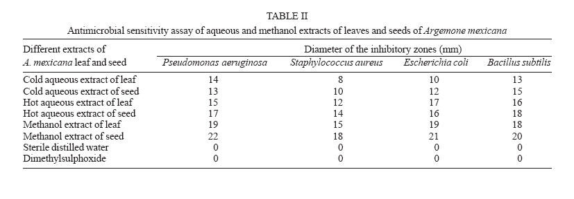
of leaves and seeds of A. mexicana, collected from a village
named Lakudi of Burdwan district (23°16'N, 87°54'E), Preparation and preservation of plant extract Cold aqueous extract - 50 g, each of the two samples, A. mexicana leaves and seeds, were weighed out and soaked separately into 200 ml cold water in a conical flask stoppered with rubber corks and left undisturbed for 24 h, then filtered off using sterile filter paper (Whattman no. 1) into a clean conical flask and subjected to water bath evaporation, where the aqueous solvent was evaporated at its boiling temperature of 100oC. The standard extracts obtained were then stored in a refrigerator at 4 oC for further use (Akueshi et al. 2002). Hot aqueous extract - 50 g, each of the two samples, A. mexicana leaves and seeds, were weighed out and soaked separately into 200 ml hot water which was then boiled for 30 min (Akueshi et al. 2002) and kept in a conical flask for 24 h undisturbed. The other steps were the same as followed in case of cold aqueous extract. Methanol extract - After drying, the plant materials were ground in a grinding machine (MX-110PN, Japan) in the laboratory. Exposure to sunlight was avoided to prevent the loss of active components; 200 ml of a methanol extraction fluid was mixed with 50 g, each of the powdered plant material. The mixtures were kept for 24 h in tightly sealed vessels at room temperature, protected from sunlight and mixed several times with a sterile glass rod. This mixture was filtered through Whattman no. 1 filter paper and the residue, adjusted to the required concentration (50 ml of methanol for the residue of 50 g of powdered plant material) with the extraction fluid for further extraction and it was repeated thrice and a clear colorless supernatant extraction liquid was finally obtained. The extracted liquid was subjected to rotary evaporation in order to remove the methanol. The semisolid extract produced was kept in a freezer at _ 80oC (REVCO model no. ULT 790-3-V32) overnight and then subjected to freeze drying for 24 h at _ 60oC in 200 ml vacuum. Then the extract was stored in an airtight container at 4oC in refrigerator for further use. All the dried extracts were exposed to UV rays (200-400 nm) for 24 h and checked frequently for sterility by streaking on nutrient agar plates (Chessbrough 2000). Antibacterial assay Disc diffusion method - Antibiogram was done by disc diffusion method (Bauer et al. 1966, NCCLS 1993) using commonly used antibiotics. The surfaces of the media were inoculated with bacteria from a broth culture. High potency bio-discs (Himedia) were placed on the agar. After 18 h of incubation at a specific temperature [(30 ± 1)oC for B. subtilis and 37oC for S. aureus, E. coli and P. aeruginosa], the plates were examined and the diameters of the inhibition zones were measured to the nearest millimeter. Agar-well diffusion method - The assay was conducted by agar well diffusion method (Perez et al. 1990). The bacterial strains grown on nutrient agar at 37oC for 18 h were suspended in a saline solution (0.85% NaCl) and adjusted to a turbidity of 0.5 Mac Farland standards (108 CFU/ml). The suspension was used to inoculate 90 mm diameter petri. Wells (6 mm diameter) were punched in the agar and filled with 50 µl of 2000 µg/ml extracts. The dissolution of the organic extracts (Methanol) was aided by 1% (v/v) DMSO and that of the aqueous extracts with water, which did not effect the growth of microorganisms, in accordance with our control experiments. Plates were incubated in air at 37oC for 24 h. Antibacterial activities were evaluated by measuring inhibition zone diameters. The experiments were conducted thrice. DMSO was taken as control for the methanol extracts. Sterile distilled water was taken as control for aqueous extracts, both hot and cold. Test microorganism - Four bacterial strains were used during the study. Gram positive bacteria include S. aureus MTCC 2940 and B. subtilis MTCC 441 and Gram negative bacteria include E. coli MTCC 739 and P. aeruginosa MTCC 2453. All the tested strains are reference strains and were obtained from The Microbiology Laboratory of Burdwan Medical College and Hospital. The bacteria were grown in nutrient broth (Himedia, M002) at 37oC and maintained on nutrient agar slants at 4oC. RESULTS Antibiogram of some common antibiotics against test microorganism - Antibiogram of the Gram positive and Gram negative bacteria revealed that all the bacterial strains were resistant to some widely used broad-spectrum antibiotics. However, all the bacteria were sensitive to the new generation antibiotics except B. subtilis because due to complex growth requirements, definitive NCCLS cut off values for antibiotics sucesptibility and resistance have not been established (TableI). All the values given are the mean of the three sets of observations and for the sake of convenience it has been rounded off. E. coli strain was resistant to and P. aeruginosa was sensitive to several antibiotics. Gatifloxacin was the most effective antibiotic against all the reference bacteria. Antimicrobial sensitivity assay of different extract - The antimicrobial screening of the plant extracts of the leaves and seeds of A. mexicana on P. aeruginosa, S. aureus, E. coli, and B. subtilis revealed that the seed extracts were more effective than those of the leaf extracts. The antibacterial activity of methanol extracts of A. mexicana (leaves and seeds) showed considerably more efficacy than the hot aqueous and cold aqueous extracts against all the reference bacterial strains. The methanol extracts of A. mexicana (leaves and seeds) showed maximum antibacterial activity against P. aeruginosa, followed by E. coli, B. sublitis, and S. aureus. On the contrary, aqueous extracts (cold and hot) of A. mexicana seeds showed maximum activity against B. subtilis, followed by P. aeruginosa, E. coli, and S. aureus. Again, cold aqueous extract of A. mexicana leaves showed highest efficacy against P. aeruginosa followed by B. subtilis, E. coli, and S. aureus where as in case of hot aqueous extract of A. mexicana leaves, maximum sensitivity was shown against E. coli, followed by B. subtilis, P. aeruginosa, and S. aureus (Table II). All the values given, are the mean of three sets of data and for the sake of convenience, it has been rounded off. DISCUSSION The methanol extracts of the leaves and seeds of the A. mexicana showed greater antibacterial activity than the corresponding water extracts. This finding is interesting, because in the traditional method of treating a bacterial infection, decoction of the plant parts or boiling the plant in water is employed. Whereas, according to present study, preparing an extract with an organic solvent was shown to provide a better antibacterial activity, in accordance with the results obtained by Nair et al. (2005). These observations may be attributed to two reasons: firstly, the nature of biological active components whose activity can be enhanced in the presence of methanol; secondly, the stronger extraction capacity of methanol could have produced greater number of active constituents responsible for antibacterial activity. The extracts of A. mexicana seeds under study showed greater antibacterial activity and the diameter of inhibition zone is higher than that of Ocimum gratissimum (Okigbo & Omodamiro 2005), Syzyium aromaticum seed, Cinnamomum cassia bark, Salvia officinalis leaf, Thymus vulgaris leaf, Rosmarinus officinalis leaf (Shanab et al. 2004), Sapindus emarginatus, Hibiscus rosa sinensis, Mirabilis jalapa, Rheo discolor, Nyctanthes arbortristis, Colocasia esculenta, Gracilaria corticata, Dictyota spps., Pulicaria wightiana (Nair et al. 2005). So A. mexicana plant can be used to discover bioactive natural products that will lead to the development of new pharmaceuticals. Such screening of various natural organic compounds and identification of active agents must be considered as a fruitful approach in the search of new herbal drugs. Moreover, seed extracts were more effective. But, in vivo study on this medicinal plant is necessary to determine toxicity of the active constituents, their side effects, serum-attainable levels, pharmacokinetic properties and diffusion in different body sites. The antimicrobial activities can be enhanced if the active components are purified and adequate dosage determined for proper administration. This may go a long way in preventing the administration of inappropriate concentrations, a common practice among many traditional medical practitioners. ACKNOWLEDGEMENTS To Department of Science and Technology, New Delhi, for providing instruments through FIST programme. REFERENCES
Copyright 2006 Instituto Oswaldo Cruz - Fiocruz The following images related to this document are available:Photo images[oc06109t2.jpg] [oc06109t1.jpg] |
| |||||||||
