 |
| About Bioline | All Journals | Testimonials | Membership | News |
|
||||||
|
||||||
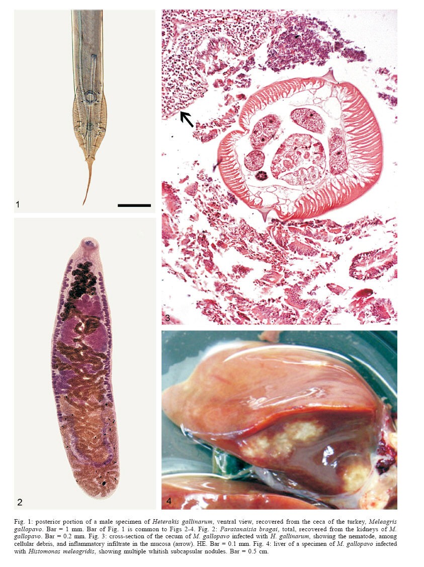
Mem Inst Oswaldo Cruz, Rio de Janeiro, Vol. 101, No. 6, September ,2006, pp. 677-681 Prevalence and pathology of the nematode Heterakis gallinarum, the trematode Paratanaisia bragai, and the protozoan Histomonas meleagridis in the turkey, Meleagris gallopavo Beatriz Brener, Rogério Tortelly*, Rodrigo Caldas Menezes**, Luís C Muniz-Pereira, Roberto Magalhães Pinto/+/++ +Corresponding author: rmpinto@ioc.fiocruz.br Received 9 May 2006 Code Number: oc06115 The prevalence of infection and associated pathology induced by two helminth and one protozoan species infecting Brazilian turkeys are reported. The intestinal nematode Heterakis gallinarum appeared with a prevalence of 70% in the infected birds, without gross lesions when not associated to the protozoan Histomonas meleagridis. Histological findings in the ceca were represented by the presence of H. gallinarum worms, intense chronic diffuse inflammatory processes with mononuclear and polymorphonuclear (heterophils) leucocyte infiltrations. The prevalence of the protozoan H. meleagridis associated to H. gallinarum was of 2.5% and microscopic examination revealed a severe inflammatory process in the liver and cecum with the presence of small clear areas with round eosinophilic parasites. Gross lesions were absent in turkeys infected with the renal digenetic trematode Paratanaisia bragai; the parasite was prevalent in 20% of the cases and cross-sections of the kidneys showed a remarkable distension of the collecting ducts with several worms in the lumen. The walls of the ducts presented a discrete heterophilic infiltrate among mononuclear cells. Key words: helminths - protozoan - turkeys - Meleagris gallopavo - pathology - Brazil In Brazil, reports of helminth infections occurring in turkeys are mostly restricted to general surveys of the parasites with no data on the associated pathology (Travassos 1965, Travassos et al. 1969, Vicente et al. 1995), in despite of the increasing economic importance of this bird for the ready-to-eat low fat food industry since the last decade. With respect to protozoans in this host there are no available reports of their occurrence in Brazilian turkeys to the date. Recently, Brener et al. (2006) studied the lesions caused by the gizzard nematode Cheilospirura hamulosa (Diesing, 1851) in turkeys from Brazilian backyard flocks, confirming its high pathogenicity, since this nematode species infects other galliform birds, mainly chickens and pheasants, provoking severe gross and microscopic lesions in these hosts. This paper deals with the prevalence and induced pathology of two helminth species, the intestinal nematode Heterakis gallinarum (Schrank, 1788), the renal digenetic trematode Paratanaisia bragai (Santos, 1934) Freitas, 1959, and the protozoan Histomonas meleagridis (Smith, 1895) in Brazilian turkeys. MATERIALS AND METHODS From May 2004 to October 2005, forty adult specimens, 19 males, 21 females of turkeys (Meleagris gallopavo Linnaeus, 1758), weight 950-8870 g, obtained from backyard flocks of different localities in the state of Minas Gerais, Brazil (19 animals), namely Candeias (20o46'01"S, 45o16'35"W), Caratinga (19o47'23"S, 42o08'21"W), Juiz de Fora (21o45'51"S, 43o21'01"W), Teixeiras (20o39'04"S, 42o51'24"W) and in the state of Rio de Janeiro, Brazil (21 animals), namely Cantagalo (21o58'52"S, 42o22'05"W), Maricá (22o55'10"S, 42o49'07"W), Niterói (22o53'00"S, 43o06'13"W) [Várzea das Moças district], Piraí (22o37'45"S, 43o53'53"W), Rio de Janeiro (22o54'10"S, 43o12'27"W) [Campo Grande district], Teresópolis (22o24'44"S, 42o57'56"W) were investigated for helminths and protozoans. After individual clinical evaluation, taking into account the general conditions, birds were killed by jugular section (hypobolemic shock) and submitted to necropsy in accordance to the technique of Zander et al. (1997). Organs (digestive and respiratory tracts, liver, spleen, kidneys, and eyes) were opened in Petri dishes containing 0.85% NaCl solution. One of the kidneys of each animal was kept uncut for histological purposes. Helminths were fixed either in hot (nematodes) or cold (compressed/uncompressed trematodes) AFA (ethanol 70oGL, 93 ml; formaldehyde, 5 ml; acetic acid, 2 ml). Portions of the parasitized organs were removed and immediately fixed in 10% formalin, to be further routinely processed for paraffin embedding. Five micrometers thick sections were stained with hematoxylin and eosin (HE). The recovered nematodes and trematodes were counted under a stereomicroscope. Some nematodes were clarified in acetic acid and phenol and mounted unstained in balsam; some trematodes were stained with alcoholic chloride carmine, dehydrated in an ethanol series (70-100o), cleared in phenol and mounted in balsam. Remaining samples of both helminths were preserved as wet material. Helminths were deposited in the helminthological collection of the Instituto Oswaldo Cruz (CHIOC). Classification of nematodes follows Vicente et al. (1995) and that of trematodes is in accordance with Travassos et al. (1969). Micrographs were obtained in a Zeiss Axyophot brightfield microscope. The development of this study has been authorized by the Committee of Ethics for the Use of Animals (CEUA/Fiocruz) no. P0095-01.
RESULTS Clinical signs were absent in turkeys parasitized
with the cecal nematode H. gallinarum alone (Fig.
1); the prevalence of infection was of 70% with a range of
intensity of 1-113 worms and a mean of 26 parasites; the microscopic
lesions were represented by intense cecal chronic diffuse inflammatory
processes with mononuclear and polymorphonuclear (heterophils)
leucocytes infiltrations, that extended discretely to the submucosa,
followed by edema. The mucosa presented multiple erosion foci together
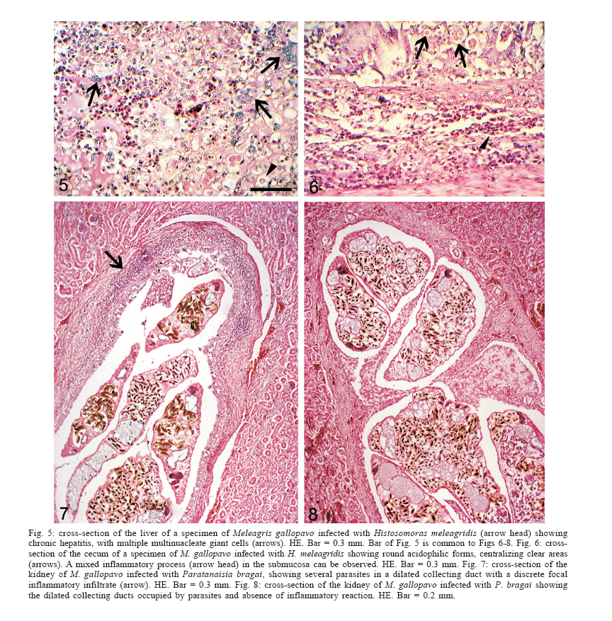
with parasites and cellular debris in transversal sections The prevalence of infection related to the association of H. gallinarum with the pleomorphic flagellate H. meleagridis was of 2.5% and the hepatic gross lesions consisted of solid nodules, of diameters ranging from about 0.5 to 1 cm (Fig. 4) that in cross-sections, appeared as whitish masses, whereas other histological findings were represented by severe and extensive granulomatous inflammatory process. The infiltrate presents a great amount of giant multinucleate cells, macrophages, epithelioid cells, lymphocytes and heterophils around small clear areas with round eosinophilic parasites identified to trophozoites of H. meleagridis; extensive parenchymal necrotic areas were also very outstanding (Fig. 5). In the cecum trophozoites of H. meleagridis were distributed among a severe inflammatory process extending from the mucosa to the muscular layer presenting a great amount of lymphocytes, macrophages and heterophils (Fig. 6). In the case of the renal trematode P. bragai (Fig. 2), the prevalence of infection was of 20% with a range of infection of 1-209 worms (from one of the kidneys only) and a mean of 38 parasites; clinical signs and gross lesions were not observed and the microscopic findings were related to a remarkable distension of the collecting ducts with several worms in the lumen (Figs. 7 and 8). The walls of the ducts presented a discrete heterophilic infiltrate among mononuclear cells (Fig. 7); in some cases, this reaction was absent (Fig. 8). Deposited specimens: H. gallinarum: CHIOC no. 35487 (wet material), 36809 a-b (whole mounts); P. bragai: CHIOC no. 35486 (wet material), 36808 a-e (whole mounts). DISCUSSION The nematode H. gallinarum has a wide geographical and host distribution and is often reported during avian helminth surveys. Although of common occurrence, few are the Brazilian studies related to the pathology induced by this parasite and associated lesions. The pioneer investigation in Brazil dealing with pathological aspects of H. gallinarum in birds of economic importance was that of Menezes et al. (2001), when guinea fowls (Numida meleagris Linnaeus, 1758) were considered. H. gallinarum was the most prevalent species in this host (100%), and the gross and microscopic lesions were not severe. Later, Menezes et al. (2003) reported data on the pathology of H. gallinarum and H. isolonche Linstow, 1906 in pheasants. Interestingly, concomitant infections, with these species caused more severe alterations than those observed when one of the species appeared alone. In association, the two nematode species of Heterakis determined severe cecal alterations characterized by necrosis of several areas with cholesterol clefts and giant cell granulomas in the intestinal submucosa and neoplastic nodules in the muscular and submucosa, and serosa, whereas in single infections, immature specimens of H. gallinarum were responsible for the occurrence of chronic difuse typhlitis, haemosiderosis, granulomas with necrotic center in the submucosa and leiomyomas in the submucosa, muscular and serosa of the caeca. Those previous data when compared to the present results indicate that turkeys are less severely affected by the parasitism even with H. gallinarum alone, than were the pheasants. Besides, experimental inoculations with strains of this nematode obtained from chickens and administered to turkeys were not successful, at least on what concerns the small size of worm burdens recovered and the low fecundity of females (Lund et al. 1970). This suggests that also Brazilian strains of H. gallinarum are either physiologically well adapted to the turkey inducing milder lesions or that the parasite attrition is higher in this host, promoting the destruction of a large number of larval H. gallinarum worms. To reinforce this hypothesis, it was observed that the animal with major pathological alterations, harbored 41 worms, of which most were larvae, indicating a recent infection; also, the lesions seem not to be related to the size of the burden, since in one of the animals with 113 adult parasites, the lesions were less severe. In the light of these findings it is to be supposed that migrating larvae are responsible for the severity of the lesions in the acute phase and that further, the lesions naturally recede. In despite of the inducement of rather mild lesions in turkeys infected with H. gallinarum alone, nematodes of this species are frequently associated to the presence of the protozoan Histomonas meleagridis, highly pathogenic to the avian hosts, and responsible for severe liver and cecal lesions; the protozoan is the cause of enterohepatitis or blackhead in turkeys. As widely known, eggs of H. gallinarum infected with throphozoites of H. meleagridis are the usual vectors for this protozoan (Springer et al. 1969, Lund & Chute 1973, Lund et al. 1975), in despite the fact that experimental attempts to infect turkeys with H. meleagridis in the absence of H. gal-linarum have succeed (Hu & McDougald 2003, Hu et al. 2004). The present pathological findings related to the association of H. gallinarum with H. meleagridis were compared to data after Wilkins and Lee (1974, 1976). Results obtained here refer to cellular and topographic changes of the liver and cecum in turkeys infected with H. meleagridis, thus confirming those previous investigations with respect to the pattern of the hepatic and cecal infections determined by H. meleagridis in this bird. These are the first data on the infection and pathology of H. meleagridis in Brazilian turkeys. In the case of the digenetic trematode Paratanaisia bragai, the first Brazilian pathological findings associated to this species were reported by Santos (1934) and Barretto and Filho (1942), being the latter, coincidentally obtained on the basis of specimens parasitizing turkeys from a suburban area of Rio de Janeiro. Data on these early studies were related to gross and microscopic lesions that consisted mainly of enlargement of the kidneys and dilatation of the renal collecting ducts with thick walls, multi-stratified epithelium and cellular infiltrate. Comparative data on the intensity of infection and pathology of P. bragai in the kidneys of different hosts and their distribution were presented by Pinto et al. (2004). More recently (Gomes et al. 2005) P. bragai was referred for the first time in the ring-necked pheasant, Phasianus colchicus L., 1758) from Brazil, together with pathological findings that consisted of absence of gross lesions and microscopic alterations very similar to those previously reported for other avian hosts (Pinto et al. 2004). Also in the ring-necked pheasants, the size of worm burdens was not related to the severity of the lesions. In this study, the parasitism of turkeys with the renal P. bragai reproduces the general pattern previously observed in other avian hosts as referred by Pinto et al. (2004) and by Gomes et al. (2005), and as far as data are concerned, P. bragai has never been referred in turkeys from other countries. ACKNOWLEDGEMENTS To Mr Rodrigo Méxas and Mr Bruno Eschenazi from the Laboratório de Produção e Tratamento de Imagens, Instituto Oswaldo Cruz-Fiocruz, for technical support with the figures. REFERENCES
Copyright 2006 Instituto Oswaldo Cruz - Fiocruz
The following images related to this document are available:Photo images[oc06115f1-4.jpg] [oc06115f5-8.jpg] |
| |||||||||
