 |
| About Bioline | All Journals | Testimonials | Membership | News |
|
||||||
|
||||||
Memórias do Instituto Oswaldo Cruz, Vol. 102, No.2, March 2007, pp. 159-164 Prevalence of oral hairy leukoplakia and epithelial infection by Epstein-Barr virus in pregnant women and diabetes mellitus patients cytopathologic and molecular study Adrianna Milagres, Eliane Pedra Dias/+, Débora dos Santos Tavares, Roberta Miranda Cavalcante, Vivian Antunes Dantas, Silvia Paula de Oliveira, José Paulo Gagliardi Leite* Programa de Pós-graduação
em Patologia, Hospital Universitário Antônio Pedro, Universidade
Federal Fluminense Rua Marquês de Paraná, 303 4º andar,
sala 1, 24033-900 Niterói, RJ, Brasil *Laboratório de Virologia,
Instituto Oswaldo Cruz-Fiocruz, Rio de Janeiro, RJ, Brasil Financial support: Capes, CNPq Received 3 October
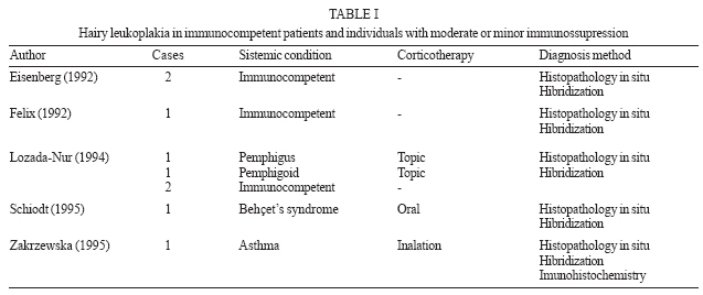
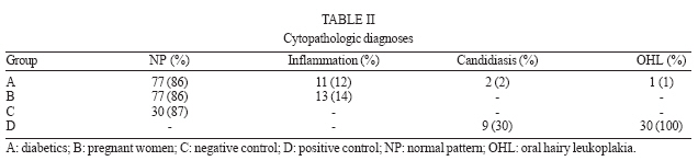
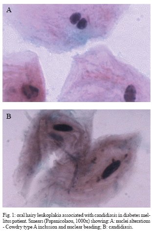
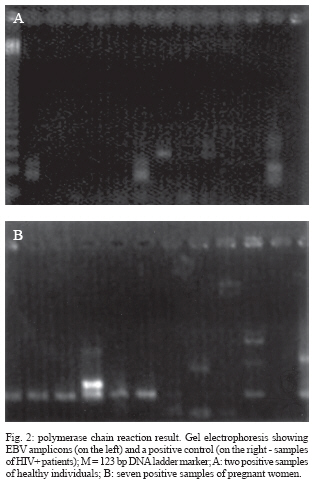
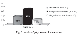
2006 Code Number: oc07028 Oral hairy leukoplakia (OHL) is generally reported in patients with severe immunosuppression, except for a few cases in individuals with moderate degree of immunodeficiency. It is a white lesion that appears mainly in the lateral border of the tongue, caused by Epstein-Barr virus (EBV). The nuclear changes caused by EBV (Cowdry A inclusion, ground glass and nuclear beading), observed in cytopathology, are specific and enough for the definitive diagnosis of OHL, independent of the identification of the virus. Here we investigated the prevalence of OHL and the presence of EBV-DNA in the lateral borders of the tongue from 90 pregnant women, 90 diabetes mellitus (DM) patients, 30 healthy individuals (negative group) and 30 HIV+ with OHL (positive group). Smears were analyzed by cytopathology and polymerase chain reaction (PCR). A case of subclinical OHL and candidiasis was identificated in a DM patient by cytopathologic analysis. PCR results demonstrated EBV-DNA in 65% of the pregnant women, in 35% of DM patients, and in 20% of the healthy individuals. We concluded that DM patients can develop OHL with a low prevalence. Furthermore, the prevalence of the EBV in lateral border of the tongue is larger in pregnant women than in healthy individuals. Key words: oral hairy leukoplakia - pregnant - diabetes mellitus - Epstein-Barr virus - cytopathology - polymerase chain reaction Oral hairy leukoplakia (OHL) is a white lesion caused by Epstein Barr virus (EBV) that occurs mainly in the lateral border of the tongue and it is often associated with severe immunodeficiency as in HIV infection, malignant tumor, and organ transplant recipients (Blomgren & Bäck 1996, Ammatuna et al. 2001, Casiglia & Woo 2002). However, according to the literature (Table I) there are cases of OHL in patients with moderate or minor immunosuppression (self-immune diseases) and also in immunocompetent individuals (Eisenberg et al. 1992, Felix et al. 1992, Lozada-Nur et al. 1994, Schiodt et al. 1995, Zakrzewska et al. 1995). Besides molecular methods to detect the presence of EBV in OHL suspect lesions, the diagnosis might be done through the identification of nuclear alterations that represent the cytopathic effect of EBV on keratinocytes: Cowdry A inclusion, ground glass and nuclear beading. Although these alterations are observed in both histopathological and cytopathological exams, the later one has been considered the best method to diagnose clinical and subclinical OHL (Fraga-Fernández & Vicandi-Plaza 1992, Migliorati et al. 1993, Dias et al. 2000). There is only one disease associated with EBV replicative infection in immunocompetent patients: infectious mononucleosis. Moreover, latent infection has been associated with several malignant and benign tumors in which EBV identification appears only when the disease is already installed (Gulley 2001, Sand et al. 2002). Reports on the seroprevalence of EBV infection are usually taken in healthy subjects, with the seropositivity in 70-90% (Ferres et al. 1995, Pancharoen et al. 2001, Ozkan et al. 2003). Nevertheless, the presence of EBV in oral epithelium has been investigated only by some authors: HIV-1+ subjects, prevalence of 11 to 67% (Mabruck et al. 1995, Scully et al. 1998, Triantos et al. 1998, Ammatuna et al. 2001), organ transplant recipients, 4-58% (Schimidt-westhausen et al. 1993, Ammatuna et al. 2001) and on healthy individuals, 7 to 90% (Mabruk et al. 1994, Scully et al. 1998, Triantos et al. 1998, Ammatuna et al. 2001, Sand et al. 2002). In diabetics, it has been observed a deficiency in polymorphonuclear leucocytes chemotaxy and abnormalities in phagocytic function of macrophages (Stiehm & Fulginiti 1980). In pregnant women, there is a suppression in cellular immune response and a decrease in peripheral lymphocytes count indicating a diminish of CD4 T cells and a concomitant increase of CD8 T cells (Nakamura et al. 1993, Priddy 1997). Considering that there is little information referring to OHL in non immunocompromised individuals and the several malignant tumors associated with EBV, this work aimed to investigate the prevalence of OHL and EBV epithelial infection in pregnant women and diabetic patients, considered here as individuals with minor immunodeficiency. MATERIALS AND METHODS This work was approved by ethics committee, and comprised 90 DM patients (group A) and 90 pregnant women (group B) attending, respectively, the Endocrinology and Gynecology and Obstetrics Departments at Hospital Universitário Antonio Pedro (HUAP) and Hospital Maternidade Fernando Magalhães. It has also been established a negative control group composed of 30 clinically healthy individuals and HIV seronegative (group C), and a positive control group of 30 patients (group D) HIV seropositive with OHL cytopatological diagnosis (data obtained from the files of the Pathological Anatomy Service at HUAP). Every patient was submitted to clinical anamnesis, oral examination and a smear of the lateral border of the tongue was collected using a sterile endocervical brush. The smears were then fixed in 96oGL alcohol and stained by Papanicolaou technique. The following aspects were evaluated by cytopathology: (a) cellularity (good or insufficient); (b) predominant cellular disposition (loose, in plaques, overlapped); (c) presence or absence of bacteria, polymorphonuclear (PMN), mononuclear (MN) cells and candidiasis; (d) presence or absence of Cowdry A inclusion, ground glass and nuclear beading. After collecting the cytological samples, the brushes had their tips sectioned and conditioned into Eppendorf tubes containing a TBS solution stored at 80oC. The brushes from 20 patients from group A, 20 from group B, 10 from group C, and 2 from group D were randomly selected to be submitted to polymerase chain reaction (PCR) analysis and the human β-globin gene was amplified in order to assess the adequacy of the specimen. EBV-DNA was extracted by means of DNAZOL® (Invitrogen) technique. NESTED-PCR was performed in all samples previously selected. Two primer sets that amplified a 297 bp or a 209 bp fragments from the EBNA-1 gene were used on these reactions (Cinque et al. 1993, Read et al. 1997). After initial denaturation for 5 min at 94oC followed by 2 min on ice, PCR mixture was added to 10 μl of the sample followed by the amplification program in the thermal cycler: 94oC for 2 min; 35 cycles (94oC for 30 s, 55oC for 30 s, 72oC for 30 s); one time (72oC for 10 min); 10oC forever; 5 μl of the PCR product was added to 45 μl of the NESTED mixture with the following cycle on the thermal cycler: 94oC for 45 s; 35 cycles (94oC for 20 s, 55oC for 30 s; 72oC for 30 s); one time (72oC for 10 min); 10oC forever. After this second amplification with inner primers, 10 μl of the amplified product was electrophoresed on a 1.5% agarose gel containing ethidium bromide. The results were photographed under UV illumination and considered positive when a band corresponding to the 209 bp DNA fragment was present. The PCR mixture used contained dNTP's (2,5 mM - Invitrogen®) 4 μl; 10X buffer (Invitrogen®) 5 μl; TAQ Platinum DNA polymerase 5UI (GIBCO®) 0.3 μl ; primers (20 µM) (pool) (Invitrogen®) 2 µl; MgCl2 (50 mM - Invitrogen®) 1.5 µl; H2O DNAse/RNAse free 27.2 µl and the NESTED one: dNTP's (2.5 mM - Invitrogen®) 4 µl; 10X buffer (Invitrogen®) 5 µl; MgCl2 (50mM - Invitrogen®) 1.5 µl; TAQ Platinum DNA polymerase 5UI (GIBCO®) 0.3 µl; primers (20 µM - pool - Invitrogen®) 2 µl; H2O DNAse/RNAse free 32.2 µl. (Cinque et al. 1993). RESULTS The clinical examination of all subjects from groups A, B, and C showed that the tongue and oral mucosa were normal. In the positive control group (group D), 11 patients (36.5%) presented a white lesion on borders of the tongue. Among these, 5 (16.5%) were bilateral, 3 (10%) on the right border and 3 (10%) on the left border. All samples presented enough material to cytopathological analysis, and the majority of them were considered of normal pattern (Table II). In groups A, B, and C only one case of OHL associated with candidiasis was identified (Fig.1) and two cases of candidiasis, both in the diabetic group. The PCR method (Fig.2) used identified epithelial infection on the lateral border of the tongue in 7 (35%) of 20 selected diabetics; 13 (65%) of 20 pregnant wo-men and in 2 (20%) of 10 selected healthy individuals (Fig.3). Comparing the percentage of EBV-DNA by PCR between pregnant women and diabetics, we found that there is no representative difference in the proportions of patients with EBV-DNA in the two groups (Fisher Exact Test, p > 0.05). On the other hand, the proportion of pregnant women to healthy individuals showed that the former is statistically bigger (65%) than the latter (20%) (Fisher Exact Test, p value = 0.025). Likewise, comparing diabetic and healthy subjects, no significant difference was detected (Fisher Exact Test, p > 0.05). All EBV-DNA negative samples were tested for β-globin and presented a positive result. DISCUSSION Although OHL and candidiasis are not considered lesions associated with an important morbidity or mortality, they are important hallmarks of immunodeficiency, specially in HIV infection (Cherry-Peppers et al. 2003, Greenspan et al. 2004, Chattopadhyay et al. 2005). The etiology of OHL has already been established, and EBV can be identified through electronic microscopy, in situ hybridization, immunohistochemistry, and PCR (Fraga-Fernández & Vicandi-Plaza 1992, Mabruk et al. 1995, Greenspan et al.1998, Gulley 2001). However, those techniques are very complex and expensive. The nuclear changes that represent EBV cytopathic effects can be visualized by histopathology and cytopathology (Fraga-Fernández et al. 1990, Fraga-Fernández & Vicandi-Plaza 1992, Migliorati et al. 1993, Dias et al. 1999, 2000). Considering that biopsy is an invasive procedure, we suggest that cytopathology should be the best choice to diagnose OHL. Among the advantages of the routine smears of the lateral border of the tongue for cytopathological exam are: (a) the diagnosis of inflammatory changes with the identification of the etiological agent (fungus and some bacteria) or cytopathic effects (HSV, CMV, EBV); (b) oncologic analysis of epithelial cells; (c) the possibility to use the material in different methods, including molecular and electronic microscopy studies. The development of OHL and candidiasis in patients with severe immunodeficiency, as in HIV seropositive, organ-transplanted subjects and in individuals presenting malignant neoplasias, is already well documented in the literature (Blomgren & Bäck 1996, Ammatuna et al. 2001, Casiglia & Woo 2002). Conversely, a relevant fact is that OHL findings in HIV seronegative patients, with low or moderate immunosuppression, or even in clinically healthy individuals are scarce (Eisenberg et al. 1992, Felix et al. 1992, Lozada-Nur et al. 1994, Schiodt et al. 1995, Zakrzewska et al. 1995). Considering the importance of candidiasis and OHL as markers and EBV association to several malignant neoplasias, it is pertinent to identify the frequency of EBV infection and candidiasis not only in healthy individuals, but also in those showing different degrees of immunosuppression. Our research grouped an important number of HIV-1 seronegative individuals (n = 210), being the first study to investigate the presence of OHL in diabetics and pregnant women. From this casuistic, 50 (24%) cases were submitted to EBV-DNA identification by PCR. OHL and candidiasis were identified only among diabetics and in a prevalence of 1 and 2%, respectively. The only case of OHL identified among all the samples studied, was related to a subclinic lesion in a 75-year-old diabetic man, who presented a hyperglycemic condition and chronic obstructive pulmonary disease since 1995, taking oral and systemic steroid (40 mg/day), characterizing a second condition of immunosuppression. These results are in agreement to the literature that considers OHL and candidiasis hallmarks of severe immunosuppression (Cherry-Peppers et al. 2003, Greenspan et al. 2004, Chattopadhyay et al. 2005). Studies reveal a high prevalence of oral candidiasis in diabetics. Amato and Pecora (1983) analyzing 50 DM type II patients, without any lesion on the oral cavity, identified Candida albicans hyphae in the saliva of 25 (50%) patients. Willis et al. (1999) demonstrated the presence of C. albicans in 77% of 414 DM patients using insulin, from which 40% did not present any clinical manifestation of candidiasis. Redinova and Zlobina (2002) performed an oral exam in 102 DM types I and II patients with, attesting the incidence of C. albicans in 78.4%, among these only 35% presented lesion on oral mucosa. Surprisingly, we identified only two cases (2.2%) without clinical manifestation of the lesion. One possible explanation for that can be related to the fact that we performed the examinations in a group of stable patients that received regular outpatient medical care and psycho-nutritional orientation. The prevalence of EBV on the tongue of healthy individuals, by PCR, varies from 10 to 90%. Mabruck et al. (1994), collected material from the lateral border of the tongue of 10 healthy patients, where they identified EBV-DNA in 9 (90%) of 10 cases. In the same way, Scully et al. (1998), identified in cytological smears of the tongue EBV-DNA in 4 (20%) out of 20 healthy subjects. Ammatuna et al. (2001), studying 30 healthy individuals who presented normal tongue clinical aspect, detected by PCR, EBV-DNA in 3 (10%) individuals. The major sample studied was published by Triantos et al. (1998) and included 40 individuals, where EBV was identified in the epithelial cells of the tongue in 4 (10%). Consequently, EBV identification in 20% of healthy individuals of our study is in accordance with the few studies already mentioned. This is the first study that investigates the presence of EBV-DNA on smears of the lateral border of the tongue of diabetics (35%) and pregnant women (65%). Our results showed an increase in the percentage of latent EBV infected patients, on the lateral border of the tongue, when compared to the percentage identified among healthy individuals. In pregnant women, it is reported only serological analyses, which in large series identify an average prevalence of EBV of 98% (Icart et al. 1981, Fleisher & Bolognese 1982, Le et al. 1983). Notwithstanding, in diabetics, serological studies are rare. (Hyoty et al. 1991, Jaeckel et al. 2002). The PCR analysis has called our attention because there were positive cases in only one of the borders of the tongue. All negative samples regarding the presence of EBV-DNA, were β-globin positive, that is, all of them presented enough material for analysis. This result may justify the unilateral identification of the OHL, clinical or subclinical, in several HIV+ patients. In addition, it suggests that EBV-DNA was not in the saliva; otherwise both samples should be positive. The results showed that EBV infection on smears of the lateral border of the tongue of individuals with minor immunodeficiency such as pregnant women and diabetics is considerably higher than in healthy subjects. Considering OHL a probable marker of immunodeficiency, PCR analysis to diagnose EBV latent infection and cytopathology to follow a possible passage to replicative phase could be useful in patients with different degrees of immunodeficiency. We believe that the investigation of the prevalence of EBV infection in the keratinocytes of the tongue, with proper identification of viral burden and respective phase of the infection, will determine the real contribution of OHL and EBV infection as markers of immunodeficiency and it would be an alternative tool in patients' clinical follow up. REFERENCES
Copyright 2007 Instituto Oswaldo Cruz - Fiocruz The following images related to this document are available:Photo images[oc07028f2.jpg] [oc07028t2.jpg] [oc07028f1.jpg] [oc07028t1.jpg] [oc07028f3.jpg] |
| |||||||||
