 |
| About Bioline | All Journals | Testimonials | Membership | News |
|
||||||
|
||||||
Memórias do Instituto Oswaldo Cruz, Vol. 102, No. 5, 2007, pp. 567-571 More productive in vitro culture of Cryptosporidium parvum for better study of the intra- and extracellular phases G Perez Cordón, C Marin, D Romero, C Rosales, M Sánchez Moreno, MJ Rosales+ Departamento
de Parasitologia, Facultad de Ciencias, Instituto de Biotecnología,
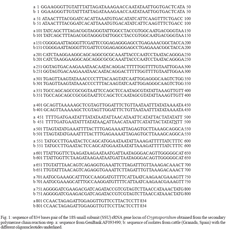
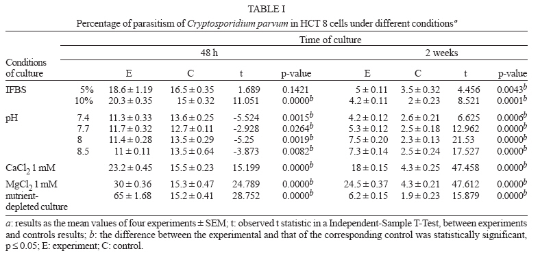
18071, Universidad de Granada, España Received 16 November 2006 Code Number: oc07090 The great difficulties in treating people and animals suffering from cryptosporidiosis have prompted the development of in vitro experimental models. Due to the models of in vitro culture, new extracellular stages of Cryptosporidium have been demonstrated. The development of these extracellular phases depends on the technique of in vitro culture and on the species and genotype of Cryptosporidium used. Here, we undertake the molecular characterization by polymerase chain reaction-restriction fragment lenght polymorphism of different Cryptosporidium isolates from calves, concluding that all are C. parvum of cattle genotype, although differing in the nucleotide at positions 472 and 498. Using these parasites, modified the in vitro culture technique for HCT-8 cells achieving greater multiplication of parasites. The HCT-8 cell cultures, for which the culture had not been renewed in seven days, were infected with C. parvum sporozoites in RPMI-1640 medium with 10% IFBS, CaCl2 and MgCl2 1 mM at pH 7.2. Percentages of cell parasitism were increased with respect to control cultures (71% at 48 h vs 14.5%), even after two weeks (47% vs 1.9%). Also, the percentage of extracellular stages augmented (25.3% vs 1.1% at 96 h). This new model of in vitro culture of C. parvum will enable easier study of the developmental phases of C. parvum in performing new chemotherapeutic assays. Key words: Cryptosporidium - in vitro - culture - extracellular stages Cryptosporidium infections have been reported in human and in most domestic animals. Most infections have been described in mammals and are attributed to bovine sources of C. parvum (Ramirez et al. 2004). Cattle have different species of Cryptosporidium: C. pestis (bovine genotype of C. parvum), C. bovis, C. andersoni, and Cryptosporidium "deer-like genotype" (Slapeta 2006). Although Slapeta (2006) proposed the re-classification of C. parvum as C. pestis, still exists controversy about this. The cattle genotype of C. parvum can infect other mammals, including human, and has been responsible of waterborne outbreaks as well as outbreaks among school children associated with farm visits (Monis & Thomson 2003, Becher et al. 2004). It is known that different Cryptosporidium isolates from various regions have different antigens, virulence, infectivity, and sensitivity to drugs as well as disinfectants (McDonald et al. 1991, Griffin et al. 1992, Fayer et al. 2000). Therefore, it is necessary to characterize the parasites molecularly in each region and know the parasites to be used in the in vitro culture. The first complete in vitro culture of C. parvum mentioning the appearance of extracellular stages was performed by Rosales et al. (1993) in cells Madin-Darby canine kidney MDCK cells. Afterwards, different cell lines were used as bovine fallopian tube epithelial cells (Yang et al. 1996), Caco-2, HT29, and HCT-8 (Maillot et al. 1997, Hijjawi et al. 2001, Hijjawi 2003), VELI cells (Lacharme et al. 2004), and Hijjawi et al. (2004) described in vitro development of C. parvum in host cell-free culture. Studies by Carreno et al. (1999), based on SSrRNA sequence, and Leander et al. (2003), based on SSU rDNA and b-tubulin sequences showed that the gregarines and Cryptosporidium formed a clade separate from the other major apicomplexa clade containing the coccidian. Hijjawi et al. (2002, 2004) reported the presence of extracellular gamont-like stages in the cycle of C. parvum and C. andersoni with HCT-8 cells or in RPMI-1640 maintenance medium devoid of host cells. Recent-ly, Rosales et al. (2005) has confirmed, by optical, Nomarski, and transmission electron microscopy images, the existence of extracellular trophozoite/gamont, stages in syzygy, zygotes, and spores with eight sporozoites during the in vitro culture of C. parvum on MDCK, HCT-8, and Vero cells as well as alveolar macrophages. It appears that, according to the methodology used in the in vitro culture and the Cryptosporidium species/genotype used, some extracellular phases or others are developed. These extracellular stages were few in number in all the reports, and thus it was considered necessary to review the culture technique for better study (Hijjawi et al. 2002, 2004, Rosales et al. 2005). In the present work, we undertake molecular characterization, using the polymerase chain reaction-restriction fragment (PCR-RFLP), of different Cryptospo-ridium isolates from calves, and we assay different factors in order to develop better technique of in vitro culture for HCT-8 cells, with the aim of studying the different phases (intra- and extracellular) of the biological cycle of Cryptosporidium with a more productive culture. MATERIALS AND METHODS Molecular characterization of Cryptosporidium species and genotypes - A total of 120 faecal samples were collected from calves of less than 21 days old from two dairy farms of Granada (Spain). All the samples were diarrheic. C. parvum oocysts were purified on a potassium bromide discontinuous gradient (Entrala et al. 2000). The parasites were stored in PBS at 4ºC until use. All the faecal samples with Cryptosporidium, n 17, were processed for molecular characterization of Cryptosporidium species and genotypes. DNA extraction and molecular characterization was done by PCR-RFLP according to Pérez-Cordón et al. (2005). Oocysts were digested using the technique of Robertson et al. (1993). Genomic DNA was extracted with phenol/choloroform/isoamyl alcohol as Ward et al. (2001). Cryptosporidium species and genotypes were determined by PCR of an SSUrRNA gene fragment and RFLP analysis as previously described (Xiao et al. 1999 a,b) but using the endonucleases SspI and VspI. The second PCR product of 819 to 837 pb was sequenced by Sistemas Genómicos, S.L. (Valencia, Spain) by single-strand sequencing of both ends of the insert performed with the amplification primers used during the PCR. Specific primers were designed from the sequences obtained during the previous sequencing phase (primer walking) and this step was repeated until one whole contig could be attained. Afterwards, the annealing and assembly of all the sequences obtained during the sequencing phase and contig editing was performed. Editing, deletion of all cloning vector sequences and settlement of all possible undetermined nucleotides was carried out. This sequence was confirmed by the program Needle (Neddleman-Wunsch global alignment), with the nucleotide sequences from the ribosomal RNA gene published in the database GenBank (AF093490). Modifications to the in vitro culture of C. parvum on cells - The in vitro culture of C. parvum on human ileocoecal adenocarcinoma (HCT 8) followed the methodology of Rosales et al. (1993) with different modifications: (1) the cell cultures were infected with sporozoites of C. parvum in RPMI-1640 medium with different concentrations of inactivated fetal bovine serum (IFBS), 2.5, 5, and 10%, and the cultures were maintained with this medium; (2) CaCl2 and MgCl2 1 mM were added to the culture medium; (3) cell cultures were infected with sporozoites in RPMI-1640 at different pH values (7.4, 7.7, 8.0, 8.5); (4) the HCT-8 cells in culture medium that had not been renewed in seven days (i.e. nutrient depleted) were infected. All the cell cultures infected were maintained for two weeks. Cultures were observed with Nomarski and light microscope (100x) every 24 h and interrupted at 48 h and at two weeks by fixation with methanol, whereupon they were stained with alcian-blue and Giemsa (Rosales et al. 1994) for observation with light microscopy to calculate the percentage of parasitized cells. About 1000 cells were counted. After the analysis of the results, the best conditions for the development of the parasite were selected and an overall experiment was conducted with the following technique: the HCT-8 cell cultures for which the culture had not been renewed in seven days were infected with C. parvum sporozoites in RPMI-1640 medium with 10% IFBS, CaCl2, and MgCl2 1 mM at pH 7.2. These cultures were maintained two weeks in the same culture medium and examined every 24 h under the Nomarski and light microscope. For the evaluation of parasite number and stage differentiation, some of the cultures were interrupted at 48, 72, 96, and two weeks. The glass disks with the infected cells were removed, fixed with methanol, and then stained with alcian-blue and Giemsa (Rosales et al. 1994) for observation with light microscopy. All the cells in each microwell were counted and the percentage of parasitized cells and of different C. parvum development stages were calculated. Supernatant of the infected culture was centrifuged at 1000 g for 5 min and extracellular stages were observed and counted with Nomarski microscopy. All the experiments were conducted four times and in all cases control HCT-8 cells were infected with C. parvum sporozoites following the technique of Rosales et al. (1993). Independent-sample T-test was applied in order to compare the percentage of parasitism of experimental and control conditions. RESULTS AND DISCUSSION The sequencing of the second product of the 834-pb PCR of all the samples was identical. All the faecal samples studied by PCR-RFLP contained the same species of Cryptosporidium. This sequence (Fig. 1b) presented 99.8% similarity with the C. parvum sequence of cattle genotype, published in GenBanK AF093490 (Fig. 1a). Differences in the nucleotides appeared only at positions 472 and 498 (Fig. 1b underlined). After the parasites were identified, a series of modifications were made in the in vitro culture technique of Rosales et al. (1993) with the aim of achieving more productive cultures for better study of the extracellular stages of C. parvum, which have been few in number in all previous the studies (Rosales et al. 1993, 2005, Hij-jawi et al. 2002, 2004), and for new chemotherapeutic assays. In 1993, we indicated the existence of extracellular forms in the in vitro culture of C. parvum on MDCK cells (Rosales et al. 1993). This finding was supported afterwards by Hijjawi et al. (2002, 2004). The in vitro culture technique of C. parvum of Rosales et al. (1993) enabled the observation of the different extracellular stages, such as trophozoites/gamont, stages in syzygy, zygotes, and spores with eight sporozoites, as demonstrated several years later (Rosales et al. 2005). These developmental phases appeared in the supernatant of the cultures with all the cells that had been assayed (MDCK, HCT 8, Vero, alveolar macrophages), although the number rose in the case of the HCT-8 cells, as the percentage of the rest of the C. parvum developmental phases also increased. On the other hand, Hijjawi et al. (2002, 2004) detected only gamont-like extracellular stages, using HCT-8 cells or in host cell-free cultures. The appearance of more extracellular phases following the methodology of Rosales et al. (1993) than with that of Hijjawi et al. (2002, 2004) may be due to the use of excystation techniques and different cultures or else to the different composition of RPMI-1640 medium used during the culture. In the cultures of Hijjawi et al. (2002, 2004) bovine bile, glucose, and ascorbic acid were added to the RPMI-1640 medium, and some of the these compounds could limit the development of some extracellular stages of C. parvum. In this work, calf genotype C. parvum was cultured on HCT-8 cells and different modifications of the RPMI-1640 culture medium were made during the infection of HCT-8 cells with C. parvum sporozoites and during the parasite-cell interaction (two weeks). Thus, changes in pH and different IFBS concentrations were assayed, CaCl2, MgCl2 were added to the culture medium, and cells were subjected to nutrient depletion. The results (Table I) reflect that the most effective approach was to infect cells cultured in nutrient-depleted RPMI-1640 medium. The nutrient depleted cells were infected more easily with the C. parvum sporozoites in the fresh culture medium, parasitism percentages being 65% and control 15.2%. Also, the parasitism percentages increased when CaCl2 was added to the medium (23.2 vs 15.5% in control) and somewhat more with MgCl2 (30 vs 15.3% control), good results also being achieved in the maintenance of the continuous culture for two weeks (24.5 vs 4.3%). As percentages of cell parasitism were increased, extracellular stages of C. parvum augmented too. This was not surprising, as the calcium and magnesium at a concentration of 1 mM in the form of CaCl2 and MgCl2 increased the mobility of Toxoplasma gondii sporozoites and C. parvum (Hamer et al. 1994). Changes in pH 7.4, 7.7, 8 or 8.5 demonstrated that the most effective was pH 7.2 of the controls as Rosales et al. (1993). Higher percentages were also observed in cultures where a greater quantity of IFBS was added (10%), giving values of 20.3 vs 15% in control, which contained 2.5% IFBS. Supplementation of coccidial cultures with IFBS or other serum proteins affected coccidian motility and development (Upton et al. 1994). Afterwards, the appropriate factors were selected and C. parvum was cultured in vivo by the following technique: the HCT-8 cells for which the culture medium had not been renewed for seven days were infected with C. parvum sporozoites in RPMI-1640 medium with 10% IFBS, pH 7.2 with CaCl2 and MgCl2 1 mM, and were maintained with this medium for two weeks. The results (Table II) showed very high parasitism percentages (71% in the experimental culture vs 14.5% in the control) which remained high after two weeks (47 vs 1.9%). All the like cycle stages identified by Rosales et al. (2005) developed, both intracellular as well as extracellular, and the percentages of all these are reflected in Table II. Extracellular stages appeared after 72 h of in vitro culture, as indicated in other studies (Rosales et al. 1993, 2005, Hijjawi et al. 2002) in a low number in the controls (2.1% at 72 h) but the percentage of extracellular stages increased in the experimental cultures, peaking at 96 h (25.3%). The origin of extracellular stages of C. parvum is not known, but it is possible that when we infected the culture with sporozoites, some penetrated the host cell and developed into circular trophozoites while other sporozoites developed extracellular motile trophozoite stages. Trophozoites of complementary types associated with each other to form syzygy and after this, formation of zygotes and sporogenesis occurred. This in vitro culture technique for C. parvum in HCT 8 permits greater parasite development, raising the percentages of parasitized cells and of all the developmental phases of the parasite. This will enable better study of the extracellular stages or allow the use of these culture for therapeutic cultures. REFERENCES
Copyright 2007 Instituto Oswaldo Cruz - Fiocruz The following images related to this document are available:Photo images[oc07090t1.jpg] [oc07090t2.jpg] [oc07090f1.jpg] |
| |||||||||
