 |
| About Bioline | All Journals | Testimonials | Membership | News |
|
||||||
|
||||||
Memórias do Instituto Oswaldo Cruz, Vol. 102, No. 7, November, 2007, pp. 883-885 Short Communication Thalidomide failed to inhibit angiogenesis and fibrosis in hepatic schistosomiasis of the mouse Camila Bião Lima, Karen Brasil Iglesias, Zilton A Andrade+ Laboratório
de Patologia Experimental, Centro de Pesquisas Gonçalo Moniz-Fiocruz,
Rua Valdemar Falcão 121, 40295-001 Salvador, BA, Brasil Financial support: Papes III - Fiocruz, Pronex Received
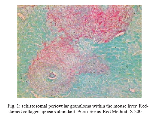
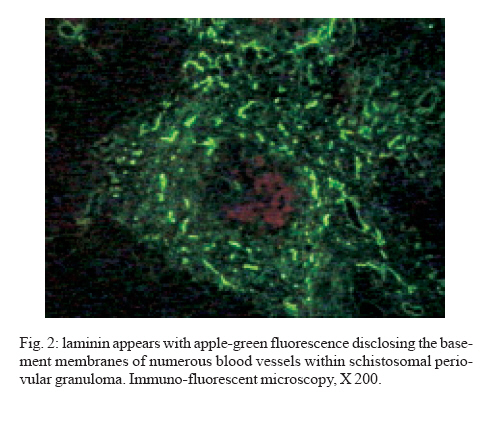
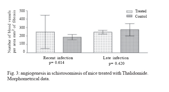
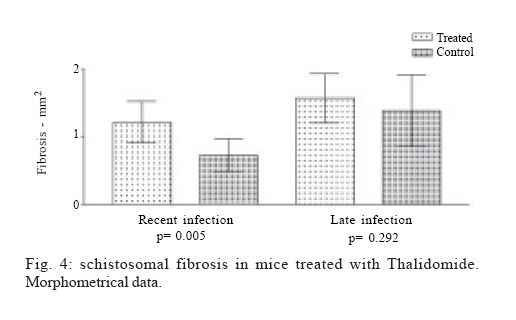
21 May 2007 Code Number: oc07159 The relationship between angiogenesis and fibrosis has been demonstrated in several pathological conditions, one of them being schistosomiasis. To observe whether suppression of angiogenesis would interfere with fibrosis, Thalidomide, an anti-angiogenesis drug, was administered during 30 consecutive days to mice with experimental schistosomiasis. Computerized morphometric measurements of fibrosis, and the counting of blood vessels from hepatic schistosomal lesions did not significantly differ when treated animals and their controls were compared at the end of the experiments. These rather unexpected results are presented under the understanding that they may be of interest during further studies on the anti-angiogenesis properties of thalidomide, and the relationship between angiogenesis and fibrosis. Key words: Thalidomide - schistosomiasis - angiogenesis - fibrosis The relationship between angiogenesis and fibrosis has recently been emphasized (Rosmorduc et al. 1999). It has also been suggested that anti-angiogenesis drugs may soon be a common and rational way to treat fibrosis due to chronic hepatic diseases, since current therapies are not yet satisfactory (Lai & Adams 2005). Although there are a great deal of indirect evidences to support such expectation (D´Amato et al. 1994, Muriel et al. 2003), a direct demonstration that depression of angiogenesis will interfere with fibrosis formation is still lacking. To test this possibility, the behavior of both fibrosis and angiogenesis were measured after treatment with thalidomide, a supposed anti-angiogenesis drug (D'Amato et al. 1994), in the murine model of schistosomiasis. Angiogenesis and fibrosis appear associated and prominent in hepatic periovular granulomas (Baptista & Andrade 2005), as well as in periportal ("pipestem") fibrosis (Silva et al. 2006). In addition, in vitro studies have revealed that soluble egg antigens (SEA) induce proliferation of endothelial cells (Freedman & Ottesen 1988), and also up-regulate vascular endothelial growth factor and angiogenesis (Loeffler et al. 2002). Sixty Swiss mice, 24 males and 36 females, weighing approximately 24 g, were maintained in separate metal cages, with free access to a commercial balanced diet. Infection was made with 50 freshly eliminated Schistosoma mansoni cercariae (Feira de Santana strain) per animal, by the transcutaneous route. The animals were divided into two groups, according to the time thalidomide was started: a Recent Group, with 20 animals receiving the drug at the 25th day from cercarial exposure, when the first changes of acute disease are about to take place (Andrade & Azevedo 1987); and a Late Group, with 40 animals, with treatment initiated at day 49 post-infection, when numerous periovular granulomas are already undergoing modulation (Silva et al. 2000). Thalidomide was used as the pure salt (100 mg/kg day), dissolved into a 2% saline solution of DMSO (Dimethyl sulphoxide, Sigma). Each animal received 0.3 ml of this solution (100mg/kg), at the dorsal subcutaneous tissue, daily, during 30 consecutive days. At completion of treatment the animals were anesthetized with xylazine and ketamine and sacrificed by exsanguinations. Their livers were moderately enlarged and presented whitish dots at the external or cut surfaces, revealing that all had been infected, which was confirmed with smash preparations microscopically showing the presence of schistosome eggs. Portions of the liver tissue were fixed in neutral 10% formalin and embedded in paraffin. Microtome sections were stained with hematoxylin and eosin, and with Sirius-red method for collagen. Other fresh portions were embedded in Tissue-Tek, frozen in liquid nitrogen, and cut in a cryostat at minus 20oC, for immunofluorescence studies with rabbit anti-laminin antibodies, and fluoresceinated anti-rabbit-immunoglobulin. Fibrosis was evaluated in Sirius-red stained sections (Fig. 1). The five most representative red-stained areas on the slides were demarcated with a cursor and measured by means of a computerized system (Axion Vision 3.1, Carl Zeiss Vision GmbH), totaling an area of 0.7 mm2 per each animal (Fig. 1). The same method was applied for counting blood vessels, which basement membrane appeared marked by apple green fluorescence for laminin (Fig. 2). The number of vessels within the demarcated areas, which contained periovular granulomas, was counted (Fig. 2). For statistical analysis the non-parametric two-tail T test was used with a computerized system (Graph Pad Prism, version 3.00). Confidence interval was 95%, and the p< 0.05 was considered statistically significant. Results are depicted on Fig. 3 and 4, indicating measurements made on fibrosis and angiogenesis, in mice treated with thalidomide and in their respective non-treated controls, both during recent and late infections. For reason not yet understood, there was a statistically significant increase of fibrosis in thalidomide-treated animals with recent schistosome infection, but except for that, no differences were noted in the amount of fibrosis or in the number of blood vessels when thalidomide-treated animals and their respective controls were compared, during recent or old schistosome infection. The relationship of angiogenesis and fibrosis is not only evident in the granulation tissue during wound repair, but has recently been demonstrated to play a role in the pathogenesis of fibrosis during some pathological conditions (Rosmorduc et al. 1999, Souza et al. 2006). The trial now attempted failed to demonstrate the anti-angiogenesis properties of thalidomide, and no action upon the amount of fibrosis could be demonstrated. The thalidomide used was the chemically pure salt, a courtesy from Dr Euzenir Sarno. Perhaps it would function in other systems. Further studies on this line would be worthwhile. It is hoped that this communication will stimulate others to investigate this subject in other systems or with more effective anti-angiogenesis drugs. REFERENCES
Copyright 2007 Instituto Oswaldo Cruz - Fiocruz The following images related to this document are available:Photo images[oc07159f3.jpg] [oc07159f4.jpg] [oc07159f2.jpg] [oc07159f1.jpg] |
| |||||||||
