 |
| About Bioline | All Journals | Testimonials | Membership | News |
|
||||||
|
||||||
Memórias do Instituto Oswaldo Cruz, Vol. 103, No. 1, February, 2008, pp. 31-38 Screening of Amazonian plants from the Adolpho Ducke forest reserve, Manaus, state of Amazonas, Brazil, for antimicrobial activity Ana Lúcia Basílio Carneiro, Maria Francisca Simas Teixeira1, Viviana Maria Araújo de Oliveira2, Ormezinda Celeste Cristo Fernandes3, Gláucia Socorro de Barros Cauper4, Adrian Martin Pohlit4/+ Departamento
de Morfologia 1Laboratório de Micologia, Instituto
de Ciências Biológicas, Universidade Federal do Amazonas,
Manaus, AM, Brasil 2Fundação de Hematologia
e Hemoterapia do Amazonas-HEMOAM, Manaus, AM, Brasil 3Centro
de Pesquisa Leônidas e Maria Deane-Fiocruz, Manaus, AM, Brasil 4Laboratório de Princípios Ativos da Amazônia-LAPAAM,
Coordenação de Pesquisa em Produtos Naturais-CPPN,
Instituto Nacional de Pesquisa da Amazônia-INPA, Av. André
Araújo 2936, 69011-970 Manaus, AM, Brasil Finnancial support: PPG-7/CNPq (557106/2005-2), PPBio/MCT/INPA Received
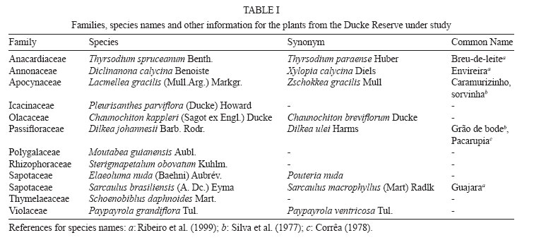
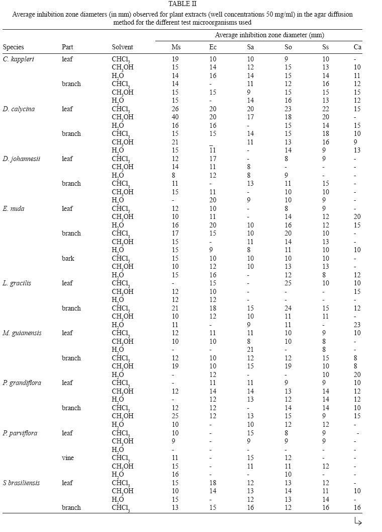
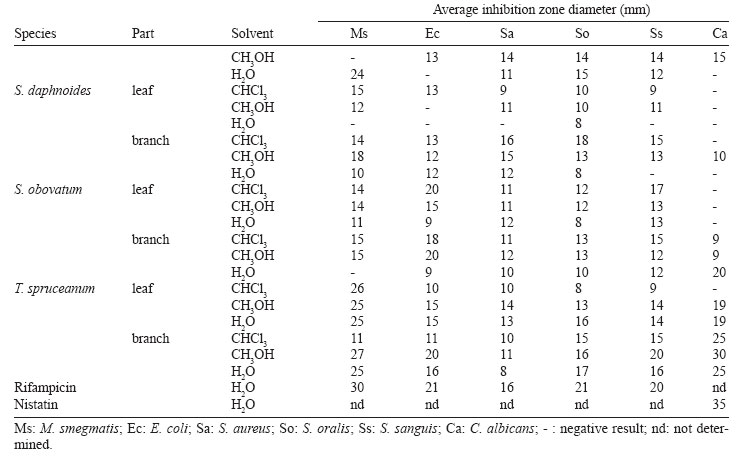
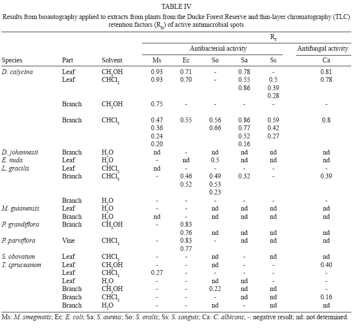
15 May 2007 Code Number: oc08005 Tropical forests are species-rich reserves for the discovery and development of antimicrobial drugs. The aim of this work is to investigate the in vitro antimicrobial potential of Amazon plants found within the National Institute on Amazon Research's Adolpho Ducke forest reserve, located in Manaus, state of Amazonas, Brazil. 75 methanol, chloroform and water extracts representing 12 plant species were tested for antimicrobial activity towards strains of Mycobacterium smegmatis, Escherichia coli, Streptococcus sanguis, Streptococcus oralis, Staphylococcus aureus and Candida albicans using the gel-diffusion method. Active extracts were further evaluated to establish minimum inhibitory concentrations (MIC) and antimicrobial profiles using bioautography on normal-phase thin-layer chromatography plates. Diclinanona calycina presented extracts with good antimicrobial activity and S. oralis and M. smegmatis were the most sensitive bacteria. D. calycina and Lacmellea gracilis presented extracts with the lowest MIC (48.8 µg/ml). D. calycina methanol and chloroform leaf extracts presented the best overall antimicrobial activity. All test organisms were sensitive to D. calycina branch chloroform extract in the bioautography assay. This is the first evaluation of the biological activity of these plant species and significant in vitro antimicrobial activity was detected in extracts and components from two species, D. calycina and L. gracilis. Key words: plant extract - antibacterial - antifungal - Diclinanona calycina - Lacmellea gracilis - bioautography The Amazon region is the largest tropical forest in the world. It occupies almost half the South American continent and is a center of biological diversity. Aside from its importance in the global ecological equilibrium and as a location of increasing ecological tourism, this region is a very rich source of species for agriculture, plant domestication and medicinal applications. Approximately 125,000 plant species are in tropical forests which continue to be a great reservatory for the discovery of new bioactive molecules and phytotherapeutic agents. However, the pharmacological potential of only about 1% of all tropical plant species has been evaluated. Also, Brazil has catalogued only about 0.4% of its flora. It is estimated that ca. 60% of all commercially available or clinical-phase antitumor and antimicrobial drugs are of natural origin (Montanari & Bolzani 2001, Gurib-Fakim 2006, Turolla & Nascimento 2006). Despite the existence of publications on the traditional (Fenner et al. 2006, Giorgetti et al. 2007) and pharmaceutical (Moreira et al. 2006) uses of plant species found in Brazil there are still relatively few scientific studies on the pharmacological and toxicological properties of plants from Brazil. There are also very few examples of the discovery of bioactive substances from Brazilian biodiversity which have served as prototypes for development of phytotherapeutic agents and pharmaceuticals in Brazil. Furthermore, many active substances present in native plant extracts from Brazil have still not been identified so this is an area which needs to be explored (Moreira et al. 2006). As examples of Brazilian plant-derived commercial products one should mention Ierobina®, which is used in the treatment of indigestion (Botion et al. 2005), and the anti-inflammatory phytotherapeutic agent Acheflan® (Giorgetti et al. 2007). The relatively scarce interaction between mainly public universities and companies in Brazil as well as Brazilian patent law have been drawbacks for transformation of research results into developed products and patents (Moreira et al. 2006, Giorgetti et al. 2007). Native Amazon forest plants have been poorly studied and there are no good estimates of the number of species which have been studied or which have application to human health. The use of flora from this region has been modest in relation to its strategic value in the development of local products. Our group has been actively screening medicinal and other plants found in the Brazilian Amazon region for larvicidal and insecticidal (Pohlit et al. 2004) as well as cytotoxic (Quignard et al. 2003, Quignard et al. 2004) properties, among other biological activities. Many plant species have demonstrated antibacterial (Koo et al. 2000, Melo et al. 2006) and antifungal (Motsei et al. 2003, Gayoso et al. 2004) properties. However, little research has focused on the evaluation of plant species for activity against microorganisms from the oral cavity (Pereira et al. 2005, Bandeira et al. 2006). This is especially true for plants from the Amazon region. Therefore, there is a general lack of scientific investigation which can provide more realistic knowledge of the potential of Amazon biodiversity. As a means of contributing to knowledge of the pharmacological potential of the Amazon flora, the present work evaluated 12 Amazonian plant species - having no known previous pharmacological study- for in vitro antimicrobial activity in bacteria and a fungus species associated with the human oral cavity diseases, as well as other diseases. MATERIALS AND METHODS Plant selection - Species identified by the Flora Project (Ribeiro et al. 1999) in the National Institute on Amazon Research's Adolpho Ducke forest reserve in Manaus, state of Amazonas, Brazil were initially contemplated for study. These plant specimens had been previously catalogued so that their botanic identities (based on voucher samples at the Instituto Nacional de Pesquisa da Amazônia-INPA Herbarium) and exact locations in the Ducke reserve were readily available. After a literature search, several hundred plant species for which no ethnobotanic, phytochemical, pharmacological or medicinal information were available were identified. From this larger group of plants, the 12 plant species were chosen for the purposes of this study (Table I) based on: (1) the ease of access and availability of sufficient plant materials in the Ducke reserve; and (2): their broad representative value of the local, unexplored flora (the species under study belong to 11 distinct botanic families). Plant collection - Plants were collected between January and June 2004 in the Ducke reserve. Plant specimens were located in the field based on the Ducke reserve trail map (Flora Project 2006) and geographical coordinates were obtained from the Flora Project database. Plant materials (leaves, branches, vine or bark) were initially dried in an air-conditioned, dehumidified room, then further dried in an oven at ca. 40ºC for a total of ca. seven days, and then finally ground. Preparation of extracts - Plant methanol and chloroform extracts were prepared by continuous extraction of ground plant material in a soxhlet apparatus for 18 h (3 x 6 h) followed by rotary evaporation and freeze-drying. Water extracts were prepared by infusion followed by filtration and total evaporation of the filtrate. Extracts were stored in a freezer at -20ºC. Microorganisms used - Standardized strains from the American type culture collection (ATCC) and the Department of Antibiotics, Federal University of Pernambuco (DAUPE) were used in bioassays. The Gram-positive bacteria were Mycobacterium smegmatis (ATCC 607), Streptococcus oralis (ATCC 10557), Streptococcus sanguis (ATCC 15300) and Staphylococcus aureus (DAUPE). The Gram-negative bacterium was Escherichia coli (DAUPE 224). Antifungal activity was evaluated using a clinical strain of Candida albicans from the collection of the Department of Parasitology, Federal University of Amazonas. Organisms were maintained at 4ºC on brain heart agar (bacteria) and sabouraud (SAB) (C. albicans). For the antibacterial tests, organisms were grown overnight in brain heart infusion followed by incubation at 37ºC. Before the test, C. albicans was cultured on SAB at 37ºC for 48 h. Antimicrobial susceptibility testing - Evaluation of the antimicrobial activity of plant extracts was carried out in Petri dishes using the agar diffusion method (Alves et al. 2000, Chah et al. 2006, Melo et al. 2006) by perforating the culture medium and charging each cavity with extract dissolved in dimethylsulphoxide (DMSO), ethanol or sterilized distilled water at a concentration of 50 mg/ml. For antibacterial tests, 25 ml of Mueller Hinton (MH) culture medium was used. For antifungal tests, SAB was used (C. albicans). Microbial suspensions were prepared and the density was adjusted in the tube to 1 on the McFarland scale. After solidification, medium was inoculated with microbial suspension (100 µl) with the aid of a swab. After 10 min, the agar was perforated so as to yield five circular, equidistant cavities (diameter 6 mm each). An aliquot of extract (50 µl of a 50 mg/ml solution) and positive (rifampicin and nistatin) and negative (solvent blank) controls were transferred to cavities. After the incubation period (24 and 48 h) at 37ºC, plates were examined and extract activity was evaluated by measurement of inhibition zone diameters (in mm). Tests were performed in triplicate and average halo diameters were determined. Rifampicin (1 mg/ml) was used as positive control for bacteria and nistatin (1 mg/ml) was used for tests involving C. albicans. The negative controls were DMSO, ethanol and sterilized, distilled water in accordance with the solvents used to dissolve each extract. A negative control containing culture medium was also used. Determination of minimum inhibitory concentrations (MIC) - Extracts considered very active in the above susceptibility tests (inhibition halo > 20 mm in the agar diffusion test) were next evaluated to determine MIC. This was carried out by dilution in solid culture medium by adapting techniques proposed by Melo et al. (2006). For the test, 35 ml of agar were prepared. MH was used for bacteria and SAB for C. albicans, using homogenization with 1.5 ml of the microorganism suspension having a density equal to 1 on the McFarland scale. Thirty 4 mm diameter orifices were prepared and inoculated, in triplicate, with 15 µl of each of a series of 10 dilutions of each crude extract stock solution (50 mg/ml). MICs correspond to the lowest concentration which inhibited the growth of the microorganism. Thin-layer chomatography (TLC)- Aluminum-backed commercial TLC silica gel GF254 plates (MERCK) were used. The extracts were applied to plates and eluted with chloroform/acetone (90:10). Eluted TLC plates were observed under ambient lighting and illuminated with an ultraviolet lamp at 254 and 366 nm. Bioautography - Active extracts were evaluated through the bioautography technique adapted from Alves et al. (2001) and Holetz et al. (2002). Briefly, microorganism culture (1 on McFarland's scale, 500 µl) and developing agent (2,3,5-triphenyltetrazolium chloride, 1 ml) in aqueous solution (this reagent aids in the observation of inhibition zones) were added to the culture medium (25 ml) at 50ºC. After homogenization, this mixture was poured over eluted chromatograms and these were placed in Petri dishes and incubated at 37ºC. Plates were observed after incubation for 24 and 48 h. Inhibition was evidenced by clear zones on the plates where no ostensible microorganism growth had occurred. RESULTS In the screening for antimicrobial activity, after a period of 24 h, the largest average inhibition halos resulted from the action of the methanol and chloroform extracts of Diclinanona calycina. S. oralis e M. smegmatis were the most sensitive organisms in this test. Of the 75 extracts evaluated for inhibitory effects on M. smegmatis, only ten presented negative results (no inhibition halos), while 11 allowed for the formation of inhibition halos with diameters greater than 20 mm. Extracts of Pinus parviflora presented slight or no activity towards the microorganisms used in testing. These and other results are presented in Table II (a, b). Antimicrobial substances used as positive controls presented inhibition halos as expected, differing from negative controls (DMSO, water, ethanol blanks), which in general did not present inhibition halos. This preliminary screening of all extracts permitted triage of promising antimicrobial extracts (presenting inhibition halo diameter > 20 mm). 34 halos with diameters > 20 mm, corresponding to 23 different extracts, were observed. Active extracts were further tested in dilution for the determination of minimum inhibition concentrations (MIC) by diffusion in solid culture medium. D. calycina e Lacmella gracilis presented the lowest MIC values (48.8 µg/ml). D. calycina leaf methanol and chloroform extracts presented the best inhibitory effects on E. coli, S. oralis, S. aureus and S. sanguis. L. gracilis branch chloroform extract presented the lowest MIC on M. smegmatis (48.8 µg/ml). Under the experimental conditions used, none of the eight tested extracts inhibited C. albicans growth (Table III). Results of bioautography screening revealed several (mainly chloroform) extracts which exhibited inhibition zones corresponding to substances of differing polarities/retention factors (Rfs) (Table IV). D. calycina extracts presented the best bioautography results. All microorganisms tested in this study were sensitive to D. calycina branch chloroform extract and most of these microorganisms (M. smegmatis, S. oralis, S. aureus and S. sanguis) showed sensitivity to more than one chemical component of this extract. In this way, screening and MIC determinations by diffusion in agar and bioautography permitted identification of active extracts containing antibacterial components. In this way, it was observed that in screening of extracts for antimicrobial activity it can be very important to use a variety of test methods which can, as in the present case, reveal information about the most active extracts and also reveal information on the number of bioactive components. DISCUSSION Microorganisms selected for this study are important human oral cavity pathogens or are of interest because they represent diseases which are of interest to public health. S. sanguis, for example, is one of the predominant colonizers of teeth. It has been isolated from human dental plaque, tongue, saliva, root canals, periapical and periodontal infections and it causes endocarditis. Cavities produced by S. sanguis occur principally in tooth fissures (Hamada & Slade 1980, Coykendall 1989, Piovano 1999, Wade et al. 2005). E. coli is recommended as control group in antimicrobial susceptibility tests for enterobacteria. Also, E. coli and S. aureus are included among the most common agents of hospital infections and are detected in cases of meningitis and are potential respiratory pathogens (Gendron et al. 2000, Melo-Souza, 2000, Esmerino et al. 2005). M. smegmatis is an important test model for initial, primary screening for antimycobacterial activity, which is important in the search for drugs with potential anti-tuberculosis effects. Mycobacterium tuberculosis (tuberculosis causing agent) is usually used at a later stage for further studies. M. smegmatis (ATCC 607) has been employed in bioassays and is cited many times, erroneously, in many publications as M. tuberculosis 607. It is chosen as a model for tuberculosis since it does not present the pathogenic properties ascribed to M. tuberculosis and it exhibits rapid growth, in contrast to M. tuberculosis, with which slow growth is normally associated (Reyrat & Kahn 2001, Newton et al. 2002, Okunade et al. 2004, Pauli et al. 2005). Several screening studies have been performed to evaluate in vitro antimycobacterial activity of plant extracts. Tosun et al. (2004), using M. tuberculosis H37Ra as test microorganism, and an ethnobotanic approach for plant selection, evaluated extracts from 44 plant species belonging to 17 families and found that 23 extracts inhibited growth at concentrations from 50 to 200 µg/ml. Billo et al. (2005) evaluated 22 plants (55 extracts) used in traditional medicine for treatment of symptoms potentially related to tuberculosis, representing 16 families, using M. bovis BCG as test organism, and found that five species exhibited activity towards this test organism. Many plant species present inhibition zones of differing diameters, however, size difference of the inhibition zone depends primarily upon these factors: (a) diffusion capacity of substances (present in the extracts) in the agar medium, (b) antimicrobial activity of diffused substances, (c) origin of microorganisms, (d) pH of substrates in plates, (e) density of inoculation, and (f) growth and metabolic activity of microorganisms in the medium. This suggests that inhibitory activity is not necessarily proportional to the inhibition zone diameter, especially when comparing different extracts. Inhibition zone diameter can further be associated with polarities of substances which make up the tested extracts and also with cell wall composition of test organisms since Gram-positive bacteria present cell walls with lower lipid levels than do Gram-negative bacteria (Pauli et al. 2005, Bandeira et al. 2006). Mycobacterium cell walls have elevated levels of high molecular weight lipids. This feature of cell wall structure seems to function as a barrier to the direct absorption of polar compounds (Pauli et al. 2005) and may be responsible for the more promising results obtained in the present study for chloroform extracts which have the lowest polarity among the extracts tested. The present study made use of simple, rapid and inexpensive techniques for in vitro evaluation of antimicrobial activity. A similar method was used in recent studies to evaluate the activity of plant extracts and fractions in strains of S. aureus, E. coli and Pseudomonas aeruginosa (Chah et al. 2006), Streptococcus mitis, Streptococcus mutans and S. sanguis (Melo et al. 2006), as well as, C. albicans and other fungi which cause opportunistic infections (Lima et al. 2005). Alves et al. (2000), using the same method employed in the present study, namely diffusion in agar (wells were 7 mm in diameter), considered extracts with inhibition zones with diameters between 9 and 12 mm to be partially active, between 13 and 18 mm to be active, and greater than 18 mm to be very active. In the present study, extracts having inhibition zones with diameters greater than 20 mm were considered to be very active. Bioautography revealed promising extracts from D. calycina. These results not only confirmed the activity of the leaf chloroform extract of this species detected in the other assays, but also revealed the antimicrobial potential of substances present in the branchwood chloroform extract, which were active in all microorganisms tested. Bioautography also revealed six extracts having components with inhibitory effects towards C. albicans. This activity towards C. albicans observed in bioautography contrasts strikingly with the results of MIC evaluation using this same species which revealed no significant activity for any of the extracts. A considerable difference was shown to exist in the results obtained using diffusion in agar and bioautography. For example, L. gracillis branch chloroform extract, which presented the lowest MIC (48.8 µg/ml) using M. smegmatis in the diffusion method, was inactive in bioautography. On the other hand, D. calycina branch chloroform extract, which presented inhibition zone diameter > 20 mm and good inhibitory activity (MIC < 97.7 µg/ml) only towards M. smegmatis, under bioautography conditions, presented active (growth inhibiting) substances (multiple Rfs) for all microorganisms evaluated. Based on these results, D. calycina (Annonaceae) e L. gracilis (Apocynaceae) are promising antimicrobial plant species which need further study to reveal the identity of the antimicrobial substances present in their extracts. It should be stressed that the antimicrobial activity presented for these plant species is new information, having no basis in popular use or direct relation to previously published laboratory studies. These results contribute to the knowledge of bioactive species in the Amazon flora which, through further study, can reveal new lead compounds for drug development. REFERENCES
Copyright 2008 Instituto Oswaldo Cruz - Fiocruz The following images related to this document are available:Photo images[oc08005t4.jpg] [oc08005t3.jpg] [oc08005t1.jpg] [oc08005t2b.jpg] [oc08005t2a.jpg] |
| |||||||||
