 |
| About Bioline | All Journals | Testimonials | Membership | News |
|
||||||
|
||||||
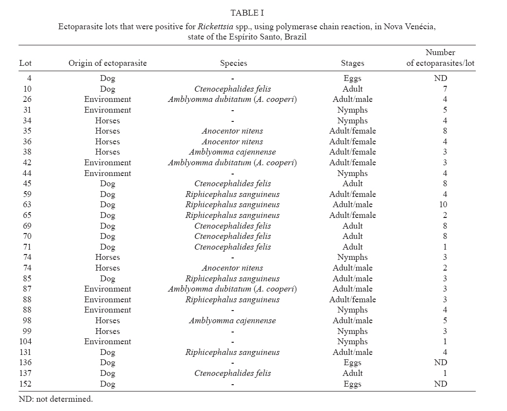
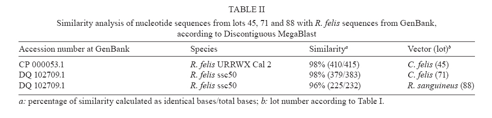
Memórias do Instituto Oswaldo Cruz, Vol. 103, No. 2, March, 2008 , pp. 191-194 Molecular identification of Rickettsia felis in ticks and fleas from an endemic area for Brazilian Spotted Fever KA Oliveira, LS Oliveira, CCA Dias1, A Silva Jr1, MR Almeida1, G Almada2, DH Bouyer3, MAM Galvão4, CL Mafra/+ Laboratório
de Bioquímica e Biologia Molecular de Agentes Infecciosos
e Parasitários, Departamento de Bioquímica e Biologia
Molecular 1Laboratório de Virologia Molecular
Animal, Bioagro, Universidade Federal de Viçosa, 36570-000
Viçosa, MG, Brasil 2Vigilância Epidemiológica,
Secretaria Estadual de Saúde, ES, Brasil 3Center
for Biodefense and Emerging Infectious Diseases, University of Texas
Medical Branch, Galveston, USA 4Laboratório de
Epidemiologia Molecular, Escola de Nutrição, Universidade
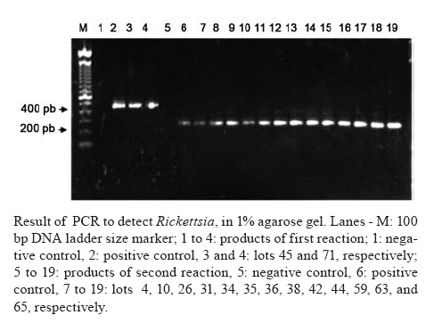
Federal de Ouro Preto, MG, Brasil Financial support: Center of Biodefense and Emerging Infectious Diseases, University of Texas Medical Branch at Galveston, USA, CAPES, CNPq Received 5 October 2007 Code Number: oc08032 Rickettsioses are arthropod-borne diseases caused by parasites from the Order Rickettsiales. The most prevalent rickettsial disease in Brazil is Brazilian Spotted Fever (BSF). This work intends the molecular detection of those agents in ectoparasites from an endemic area of BSF in the state of Espírito Santo. A total of 502 ectoparasites, among them Amblyomma cajennense, Amblyomma dubitatum (A. cooperi), Riphicephalus sanguineus, Anocentor nitens and Ctenocephalides felis, was collected from domestic animals and the environment and separated in 152 lots according to the origin. Rickettsia sp. was detected in pools of all collected species by amplification of 17kDa protein-encoding gene fragments. The products of PCR amplification of three samples were sequenced, and Rickettsia felis was identified in R. sanguineus and C. felis. These results confirm the presence of Rickettsia felis in areas previously known as endemic for BSF, disease caused by Rickettsia rickettsii. Moreover, they show the needing of further studies for deeper knowledge of R. felis-spotted fever epidemiology and differentiation of these diseases in Brazil. Key words: Rickettsia felis - Rickettsia rickettsii - Ctenocephalides felis - Brazilian Spotted Fever - R. felis-spotted fever - tick Brazilian Spotted Fever (BSF) is the most prevalent rickettsial disease in Brazil (Lemos et al. 2001). The illness is caused by Rickettsia rickettsii, a bacterial organism belonging to the family Rickettsiaceae and to the Order Rickettsiales, whose members are intracellular mandatory microorganisms, many of them causing infections worldwide-distributed in humans and other vertebrate and invertebrate hosts. These organisms are classically transmitted to humans via arthropod vector bites. In Brazil, the tick Amblyomma cajennense is the main vector of Rickettsia rickettsii (Labruna et al. 2004) and reservoir of BSF (Aragão & Fonseca 1961, Figueiredo et al. 1999). Rickettsia felis-spotted fever is caused by R. felis, whose main vector is the cat flea, Ctenocephalides felis. Once in this host, the pathogen is maintained by transovarial transmission with no lethal effect, which represents a factor of great importance for the epidemiology of this emerging rickettsiosis (Azad et al. 1992, Rolain et al. 2003). Studies have demonstrated the presence of R. felis in Brazil by serology performed in human cases (Raoult et al. 2001) and C. felis infection confirmed by polymerase chain reaction (PCR) (Horta et al. 2006, Oliveira et al. 2002). The objective of this work was the molecular detection of rickettsiae circulation in ectoparasites collected from domestic animals and from domestic environments, following a BSF outbreak, in an endemic area of BSF. This study sought to contribute to a wider understanding and knowledge of the rickettsial diseases related to these agents in Brazil. MATERIALS AND METHODS Study area - The study began in November 2003, in the municipal district of Nova Venécia (18º43'38''S, 40º24'02''W), Northwestern of state of Espírito Santo, Brazil, which is considered an endemic area for BSF. Nova Venécia has an estimated population of 44,380 inhabitants (http://www.ibge.gov.br). From August 1st to October 31st 2003, 16 suspected cases of rickettsiosis and two confirmed cases by indirect immunofluorescence assay (IFA) for R. rickettsii (data supplied for Vigilance of Health Secretary, Espírito Santo, Brazil) were identified, as well as the occurrence of one death, in this municipal district. The majority of cases were concentrated in the rural community named Patrimônio do XV, with about 1,200 inhabitants. Collection and preparation of material for analysis - A total of 502 ectoparasites (ticks and fleas) were collected from domestic animals (24 dogs and 5 horses) and from the natural environment, in the nymph and adult stages, as well as eggs laid in a controlled chamber. The ectoparasites were hand-picked from the animals, using tweezers when necessary. CO2 traps were used to capture free-living ticks in sites detected as being the probable origin of infection, according to the epidemiologic inquiry, considering possible environments for contact between human and ticks and evidence for the presence of capybaras. The ectoparasites were separated in 152 lots according to the origin, and adult stages identified through a stereoscopic microscope using Aragão and Fonseca's (1961) taxonomic keys for ticks and Linardi and Guimarães' keys (2000) for fleas. At the laboratory, the ectoparasites were kept in a BOD incubator, at 25ºC for 2-5 days, for reactivation and multiplication of the rickettsial agents according to Hayes and Burgdorfer (1982), and stored at -20ºC until nucleic acid extraction. DNA extraction and PCR - After the surface sterilization by immersion in absolute ethanol for 10 min, the ectoparasites were washed in PBS, crushed in 200 µl of the same buffer, and the DNA extraction was performed using the method described by Billings et al. (1998). For the amplification of 434 bp-portion of the gene encoding Rickettsia genus-specific 17-kDa protein, the primers 17kD1 (5'- GCTCTTGCAACTTCTATGTT-3') and 17kD2 (5'-CATTGTTCGTCAGGTTGGCG-3') described by Webb et al. (1990) were used, under the following conditions: an initial stage at 94ºC for 3 min, 30 cycles at 95ºC for 1 min, 50ºC for 1 min, and 72ºC for 2 min, followed by an step at 72ºC for 5 min. The reactions were performed in 25 µl containing PCR 1X (InvitrogenTM) buffer, 0.4 mM of each primer, 1 mM of MgCl2, 0.05 mM of each dNTP, 1U of recombinant Taq DNA Polymerase (InvitrogenTM), and 2 µl of DNA from each sample or 0.5 µl of purified Rickettsia prowazekii DNA for positive control. The amplifications were initially carried out in 30 pools, each containing about 5 lots of ectoparasites. After detecting the positive pools, they were separated and each lot was subjected to a new PCR to identify the infected lots. Nested-PCR was used for the negative samples in the first amplification, using 1 µl of the first reaction as template and the pair of genus-specific primers 17kN1 (5'- CATTACTTGGTTCTCAATTCGGT-3') and 17kN2 (5'-GTTTTATTAGTGGTTACGTAA-3') described by Schriefer et al. (1994), to amplify an inner 232-pb region of the gene encoding the 17 kDa protein. The reagent concentrations and amplification conditions were the same as described on the first amplification reaction. The reaction products of PCR were visualized in 1% agarose gel. Sequencing of PCR products - The PCR amplification products were cloned into the vector pCR® 2.1-TOPO® (InvitrogenTM TOPO® TA Cloning® Kit), following the manufacturer's protocol. Plasmid DNA extraction was performed using the High Pure Plasmid Isolation Kit (Roche Diagnostic), following the manufacturer's instructions. The generated nucleotide sequences were edited with CHROMAS (http://www.mb.mahidol.ac.th/pub/chromas/chromas.htm) and compared with the corresponding homologous sequences available through GenBank, using Discontiguous Mega Blast (http://www.ncbi.nlm.nih.gov). RESULTS Ticks and fleas were identified as Amblyomma dubitatum (A. cooperi), A. cajennense, Riphicephalus sanguineus, Anocentor nitens and C. felis. In this study, 28 lots of ectoparasites contained Rickettsia DNA amplified by nested-PCR, from these lots two were positive in the first reaction and 26 were positive in the second reaction, using genus-specific primers (Table I). Figure shows the generated band patterns. The sequencing of PCR amplification products of ectoparasite lots 45, 71 and 88 (Table I) was successful. The generated nucleotide sequences had greater similarity with R. felis sequences deposited in GenBank, and the sequence obtained from R. sanguineus lot (lot 88) showed 96% similarity to R. felis, whereas the sequences obtained from lots 45 and 71, both C. felis, showed 98% similarity to R. felis (Table II). DISCUSSION These results present a contribution to the initial understanding of rickettsial ecology in the studied area. The amplification of gene fragments encoding the 17kDa protein showed that Rickettsia spp. circulates among all the ectoparasite species collected in the studied area, confirming the role of different ectoparasites in the maintenance of these organisms. The detection of R. felis in R. sanguineus and C. felis is corroborated by other studies carried out in Brazil (Galvão et al. 2003, 2006). Knowing that by IFA with specific antigens, the sera of patients with rickettsiosis during the outbreak showed a positive title for R. rickettsii, the presence of R. felis in ectoparasites from the studied area indicates the possibility of occurrence of R. felis-spotted fever, concomitant or not with BSF, in the studied area. Some studies (Phillip et al. 1978, Oliveira et al. 2002, Horta et al. 2007) have indicated that at least two species of pathogenic rickettsia can be circulating in the endemic foci of BSF. Horta et al. (2007) found evidences of at least four Rickettsia species (R. rickettsii, R. parkeri, R. felis and R. bellii) in the studied areas (4 endemic and 1 non-endemic for BSF), however serological evidences of rickettsial infection in humans and/or animals were found for only two species: R. rickettsii and R. parkeri. These results suggest that some BSF cases occurred in endemic areas may have been caused by other rickettsiae rather than R. rickettsii, and being R. rickettsii the sole antigen regularly used in BSF serological diagnosis, some human cases of BSF due to other rickettsiae of this group have been wrongly identified as BSF in Brazil (Horta et al. 2007). It is also known that the occurrence of cross-reaction between R. rickettsii and R. felis serology is common, as reported by Raoult et al. (2001). This fact could also explain the detection of R. felis in the studied area in this present work, where cases of rickettsiosis were diagnosed as BSF due to R. rickettsii, not excluding, however, the potential presence of this and/or other Rickettsia species in the analysed ectoparasites. It is important to point out that the patients' clinical diagnosis was not completely compatible with the BSF symptoms, therefore confirming the possibility of facing, in this area, a pathology of emerging character in Brazil - R. felis-spotted fever. These results emphasize the need for more specific techniques to the diagnosis of rickettsiosis that will allow a better characterization of these diseases in Brazil, as well as the development of strategies more compatible with the real situation of the country. ACKNOWLEDGEMENTS To the employees from Vigilance of Health Secretary, Health Ministry, Marcelo Yoshito Wada, Marcelo Santalucia and Antônio Lima Neto; Augusto Marchon Zago and Maxwel Marchito de Freitas from Health Secretary of the state of Espírito Santo; Marllus Cavalcante from Municipal Health Secretary of Nova Venécia; Marcelo Renan de Deus Santos and Gilberto Marcos Junior from the Universitary Center of Vila Velha and Celso Eduardo de Souza of SUCEN/SP, for their assistance in obtaining the ectoparasites. To Dr. David H Walker from UTMB for partial financial support given for this project and for previous review. REFERENCES
Copyright 2008 - Instituto Oswaldo Cruz - Fiocruz The following images related to this document are available:Photo images[oc08032f1.jpg] [oc08032t2.jpg] [oc08032t1.jpg] |
| |||||||||
