 |
| About Bioline | All Journals | Testimonials | Membership | News |
|
||||||
|
||||||
Memórias do Instituto Oswaldo Cruz, Vol. 103, No. 5, August 2008, pp. 455-462 Regional pattern of the molecular types of Cryptococcus neoformans and Cryptococcus gattii in Brazil Luciana TrillesI, II, *; Márcia dos Santos LazéraI; Bodo WankeI; Raquel Vasconcelos OliveiraIII; Gláucia Gonçalves BarbosaI; Marília Martins NishikawaIV; Bernardina Penarrieta MoralesI; Wieland MeyerII ILaboratório
de Micologia Financial support: CAPES, NH&MRC (ID 990738; ID 352303) Received 30 January
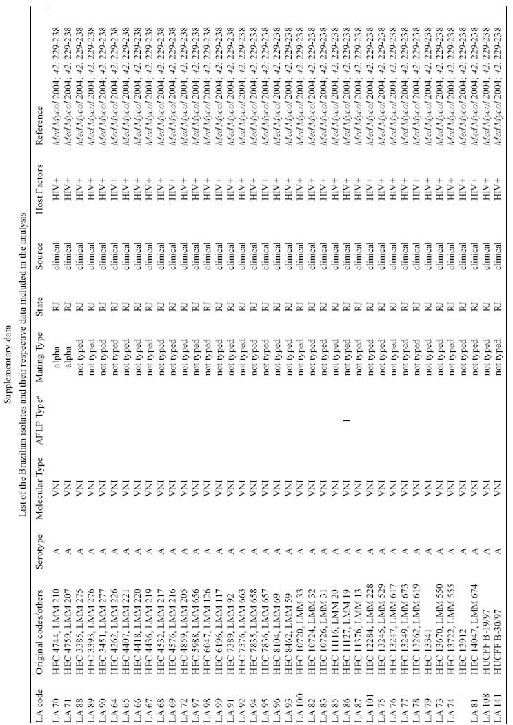
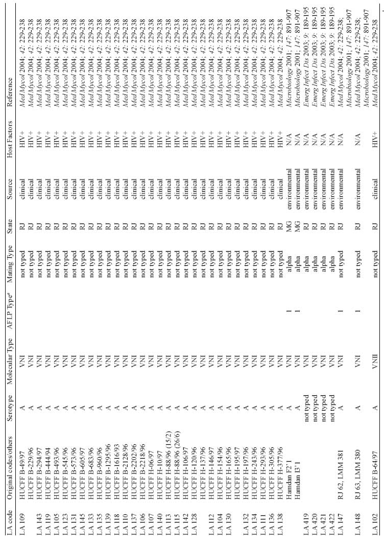
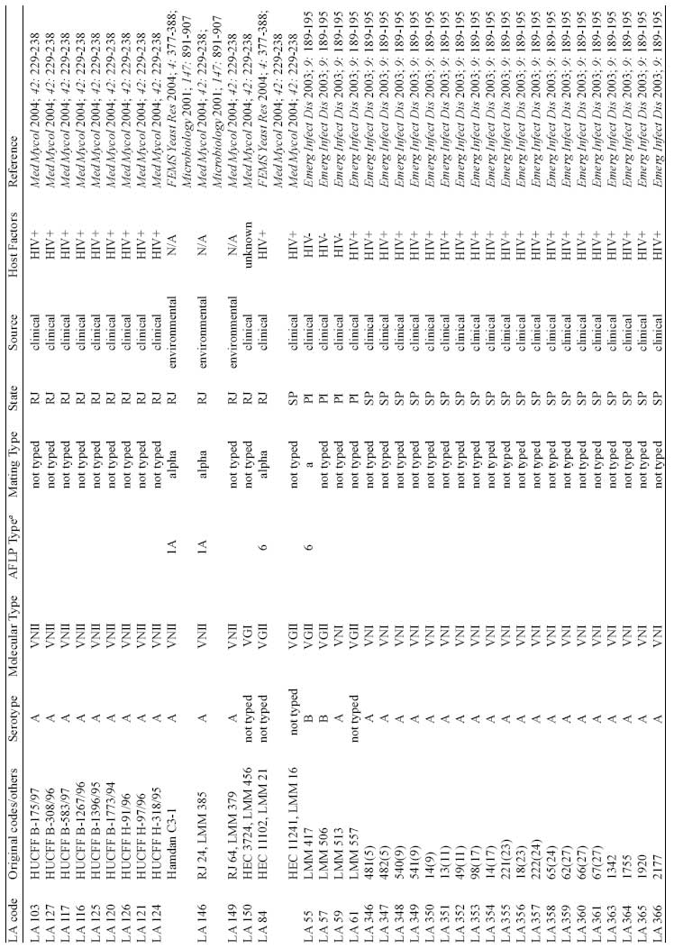
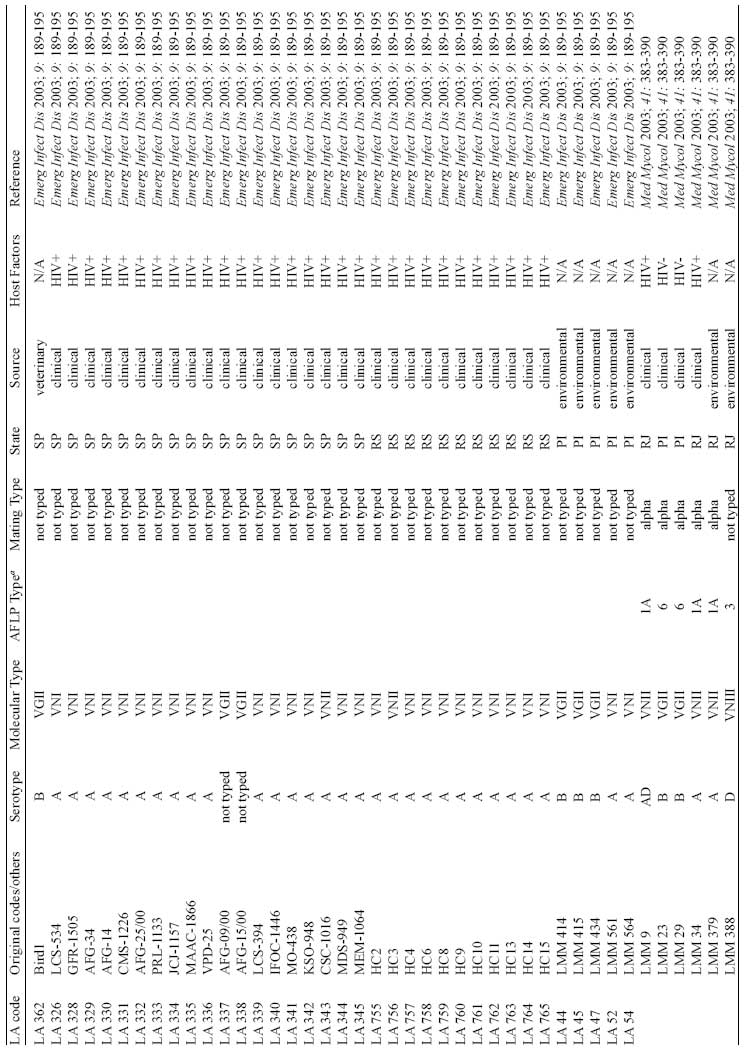
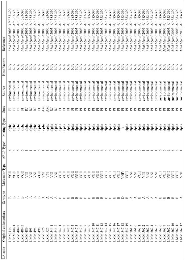
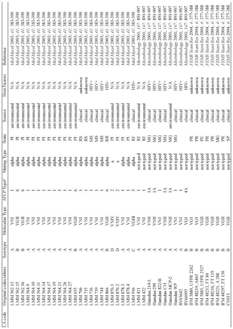
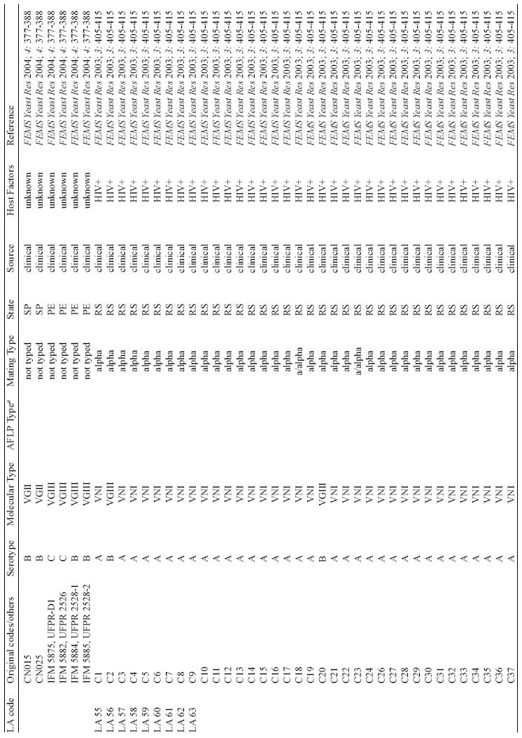
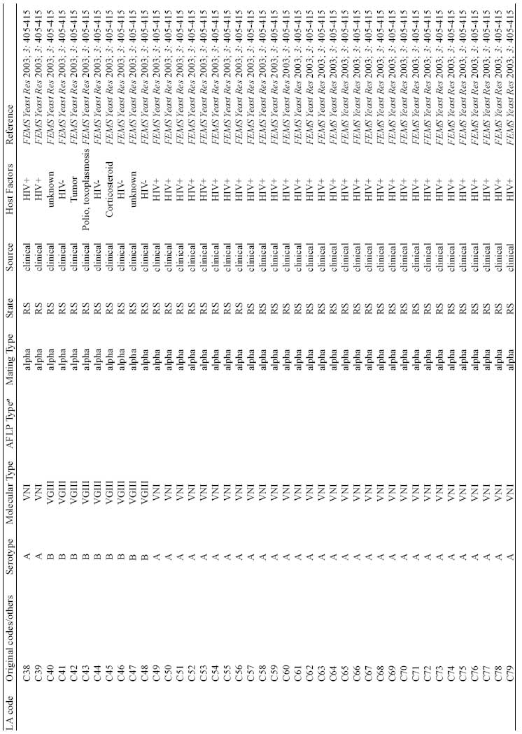
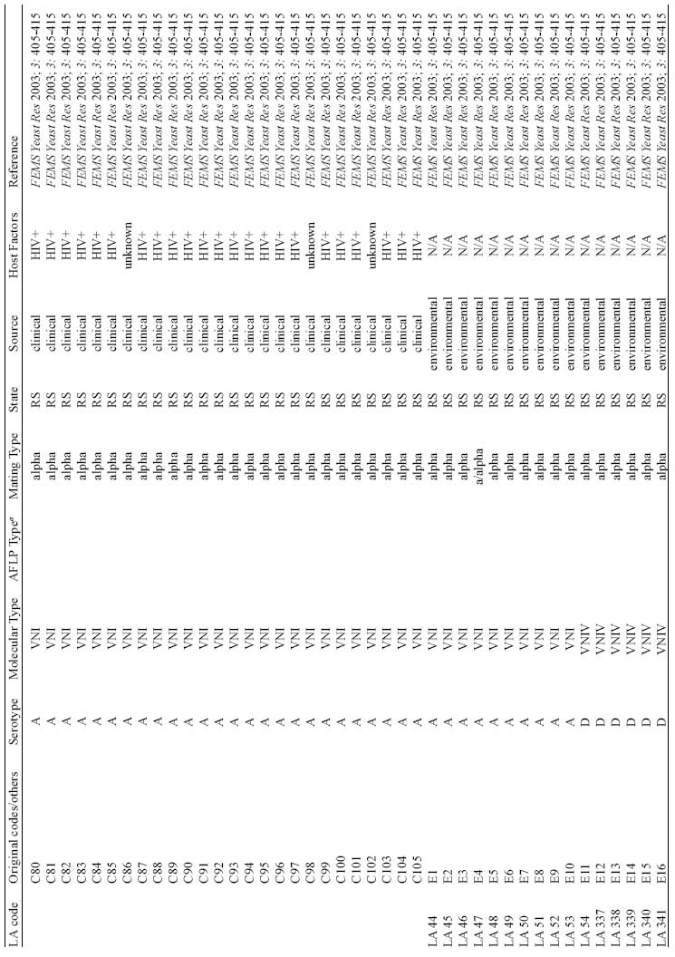
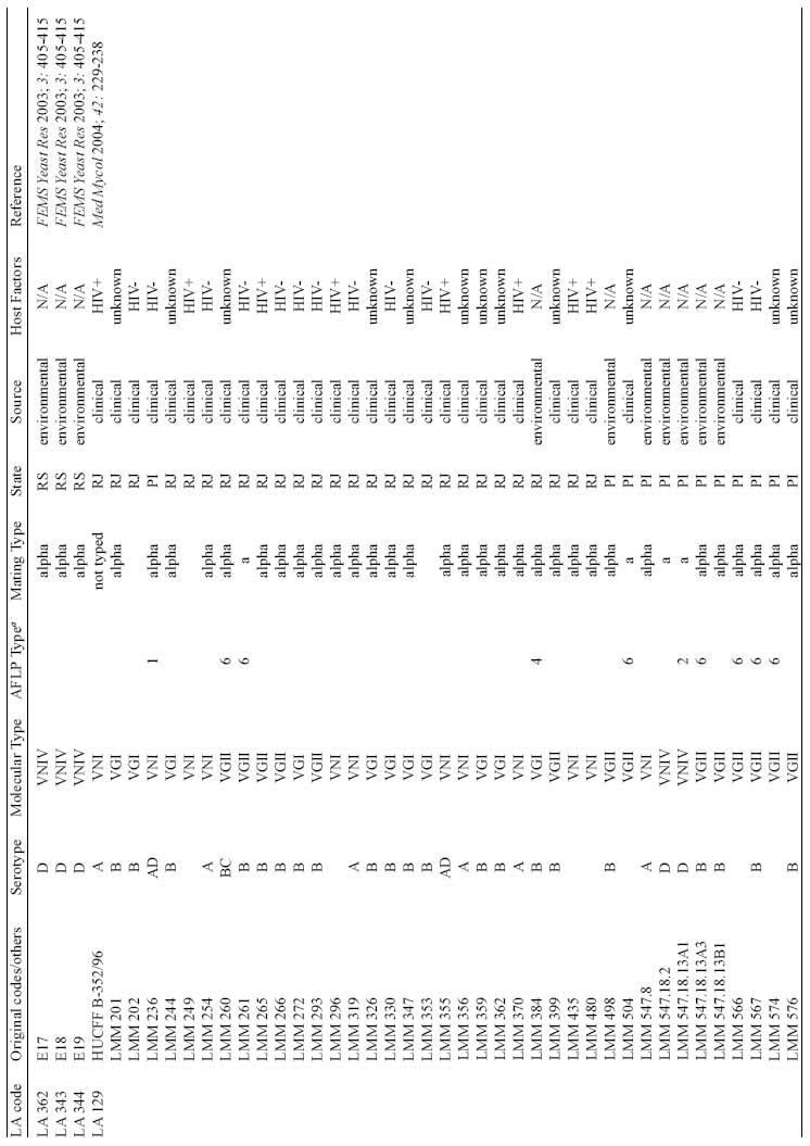
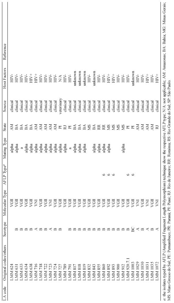
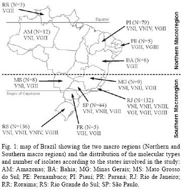
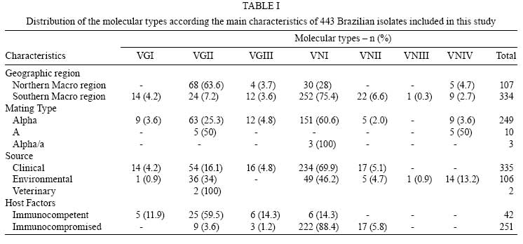
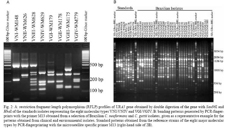
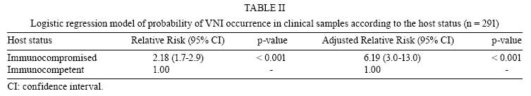
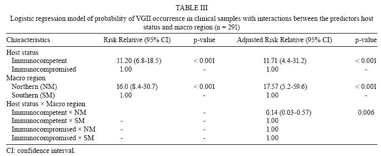
2008 Code Number: oc08080 ABSTRACT The molecular types of 443 Brazilian isolates of Cryptococcus neoformans and Cryptococcus gattii were analyzed to determine their geographic distribution within Brazil and their underlying host conditions. The following data, imported from previous epidemiological studies as well as two culture collections, were analyzed for: place of isolation, source (clinical or environmental), host risk factors, species, serotype, mating type, and molecular type. Molecular typing by PCR-fingerprinting using primers for the minisatellite-specific core sequence of the wild-type phage M13 or microsatellites [(GACA)4, (GTG)5], restriction fragment length polymorphism of URA5 gene analysis, and/or amplified fragment length polymorphism (AFLP) identified eight major genotypes: VNI/AFLP1, VNII/AFLP1A, VNIII/AFLP2, and VNIV/AFLP3 for C. neoformans, and VGI/AFLP4, VGII/AFLP6, VGIII/AFLP5, and VGIV/AFLP7 for C. gattii. The most common molecular type found in Brazil was VNI (64%), followed by VGII (21%), VNII (5%), VGIII (4%), VGI and VNIV (3% each), and VNIII (< 1%). Primary cryptococcosis caused by the molecular type VGII (serotype B, MAT) prevails in immunocompetent hosts in the North and Northeast regions, disclosing an endemic regional pattern for this specific molecular type in the Northern Brazil. Key words: Cryptococcus neoformans - Cryptococcus gattii - molecular types - epidemiology - Brazil Cryptococcosis is a life-threatening, systemic mycosis affecting humans and animals. It is acquired by inhalation of viable propagules from the environment, and the most common clinical manifestation of cryptococcosis is meningoencephalitis (Lin & Heitman 2006). Cryptococcosis is caused by two different species of the genus Cryptococcus: C. neoformans (serotypes A, D, and hybrid AD) and C. gattii (serotypes B and C) (Kwon-Chung et al. 2002). While C. neoformans infections occur worldwide and are an important cause of morbidity and mortality in immunocompromised hosts (especially AIDS-patients), C. gattii usually infects normal hosts (Kwon-Chung & Bennett 1992, Lazéra et al. 2005) and is considered to be a tropical disease. The ongoing Vancouver Island outbreak in a temperate climate, however, suggests that C. gattii can adapt to new environments (Kidd et al. 2004). In Brazil, cryptococcosis caused by C. neoformans occurs in all regions; however, C. gattii behaves as a primary pathogen infecting native immunocompetent hosts and mainly infects young people and children in the North and Northeast (NE) regions of Brazil. In this group of patients, the infection is characterized by a high lethality rate that ranges from 40.6% to 56% (Correa et al. 1999, Lazéra et al. 2005) and frequently causes incapacity (e.g., visual deficits or blindness) and hydrocephalus (Rozenbaum & Gonçalves 1994, Nishikawa et al. 2003). Brazilian epidemiological data suggest a geographic, macroregional, north-south trend in C. gattii infections in Brazil. The Northern macro region (NM), which is comprised of the states of Amazonas, Roraima, Pernambuco, Piauí, and Bahia, is endemic for C. gattii. The Southern macro region (SM), which are represented by the states of Mato Grosso do Sul, Minas Gerais, São Paulo (SP), Rio de Janeiro (RJ), Paraná, and Rio Grande do Sul (RS), show sporadic infections by C. gattii. Serotyping was largely used for epidemiological studies of C. gattii and C. neoformans, but the lack of an available commercial serotyping kits and the search for a more reliable technique led to an increased use of molecular tools. Additionally, the analysis of genotypes within a species could answer several questions that may impact management, therapy, surveillance, and prophylactic actions. An attempt to standardize a technique for a global molecular epidemiological survey of the agents of cryptococcosis identified eight major molecular types via PCR fingerprinting with the minisatellite-specific core sequence of the wild-type phage M13 or microsatellites [(GACA)4, (GTG)5]. Thus, C. neoformans was grouped into the types VNI (serotype A), VNII (serotype A), VNIII (serotype AD), and VNIV (serotype D); C. gattii was grouped into the types VGI, VGII, VGIII, and VGIV (serotypes B and C) (Meyer et al. 1999). This grouping has been confirmed by others using different techniques, such as amplified fragment length polymorphism analysis (AFLP) (Boekhout et al. 2001), restriction fragment length polymorphism analysis (RFLP) of URA5 and PLB1 genes (Latouche et al. 2003, Meyer et al. 2003), and multilocus sequence typing (MLST) (Litvintseva et al. 2006). Recently, AFLP and MLST have revealed the existence of a new molecular type of C. neoformans in Botswana (genotype VNB) that seems to be geographically restricted to sub-Saharan Africa (Litvintseva et al. 2006). Although several previous studies have analyzed the molecular types of Brazilian isolates according to the previously described typing system (VNI/AFLP1, VNII/AFLP1A, VNIII/AFLP2, VNIV/AFLP3, VGI/AFLP4, VGII/AFLP6, VGIII/AFLP5, and VGIV/AFLP7 types), they demonstrated only the presence of C. neoformans and C. gattii in certain cities or states (Casali et al. 2003, Igreja et al. 2004, Abegg et al. 2006, Matsumoto et al. 2007). Even when more than one region was analyzed, the number of isolates was limited (Trilles et al. 2003). In the current study, the main objectives were to obtain an up-to-date picture of the molecular type distribution of C. gattii and C. neoformans in Brazil and correlate the genotypes to geographic regions and host conditions by reanalyzing all of data from previously published cryptococcal strains and combining them with data obtained from new strains using statistics and descriptive analysis. MATERIALS AND METHODS Data from 356 Brazilian isolates were imported from different sources: (i) previous epidemiological studies (Boekhout et al. 2001, Casali et al. 2003, Trilles et al. 2003, Meyer et al. 2003, Igreja et al. 2004, Katsu et al. 2004, Ribeiro et al. 2006), (ii) the database of the Cryptococcal Culture Collection at Laboratório de Micologia, Instituto de Pesquisa Clínica Evandro Chagas, RJ, and (iii) the database of the Australian Medical Fungal Collection, at the Molecular Mycology Research Laboratory at Westmead Hospital, Sydney, Australia. The data collected for the current analysis included: place of isolation, source (clinical or environmental), host risk factors, species, serotype, mating type, and molecular type identified by PCR-fingerprinting, URA5-RFLP according to Meyer et al. (2003), or AFLP according to Boekhout et al. (2001). In addition to the imported data described above, we determined the mating type, serotype, molecular type, and polymorphisms of additional 87 C. gattii and C. neoformans isolates. Serotyping - Serotyping was carried out using the slide agglutination test according to the manufacturer's instructions (Crypto Check Iatron RM 304-K kit; Iatron Laboratories, Tokyo, Japan). Molecular typing - The 87 clinical and environmental Brazilian isolates were typed by URA5-RFLP and PCR-fingerprinting using the minisatellite-specific core sequence of the wild-type phage M13. The following standard strains representing each molecular type were included in the analysis: WM 148 (serotype A, VNI/AFLP1), WM 626 (serotype A, VNII/AFLP1A), WM 628 (serotype AD, VNIII/AFLP2), WM 629 (serotype D, VNIV/AFLP3), WM 179 (serotype B, VGI/AFLP4), WM 178 (serotype B, VGII/AFLP6), WM 175 (serotype B, VGIII/AFLP5), and WM 779 (serotype C, VGIV/AFLP7) (Meyer et al. 2003). The DNA was extracted as previously described (Meyer et al. 1999). The isolates were grown on Sabouraud's dextrose agar at 37°C for 48 h. The tube containing the yeast cell pellet was frozen in liquid nitrogen. The pellet was ground with a miniature pestle, and 500 µl of cell lysis solution (0.5% sodium dodecyl sulfate, 1.4% NaCl, 0.73% EDTA, and Tris-HCl 0.2M) was added to the frozen, ground cells. The tubes were incubated for 5 min at room temperature with constant shaking, and 500 µl phenol:chloroform:isoamyl alcohol (v:v:v 25:24:1) was added and mixed thoroughly for 2 min to obtain a homogenous suspension. The tubes were centrifuged for 20 min at 16,110 g. After centrifugation, the upper aqueous layer was transferred to a new tube, an equal volume of chloroform:isoamyl alcohol (v:v 24:1) was added, and the mixture was shaken and centrifuged. To precipitate the genomic DNA, an equal volume of isopropanol was added to the supernatant. The mixture was then gently shaken and incubated at -20°C for at least 1 h or overnight. The DNA pellet was washed with 70% ethanol and suspended in 200 µl sterile, deionized water at 4°C overnight. PCR of the URA5 gene was performed in a final volume of 50 µl. Each reaction contained 50 ng of DNA, 1 X PCR buffer [160 mM (NH4)2SO4, 670 mM Tris-HCI (pH8.8 at 25°C), 0.1% Tween-20 - Bioline], 0.2 mM each of dATP, dCTP, dGTP, and dTTP (Roche Diagnostics GmbH), 3 mM magnesium chloride, 1.5 U BioTaq DNA polymerase (Bioline), and 50 ng of each primer URA5 (5' ATGTCCTCCCAAGCCCTCGACTCCG 3') and SJ01 (5' TTAAGACCTCTGAACACCGTACTC 3') (Meyer et al. 2003). PCR was performed for 35 cycles in a Perkin-Elmer thermal cycler (model 480) at 94°C with a 2 min initial denaturation, 45 s denaturation at 94°C, 1 min annealing at 61°C, 2 min extension at 72°C, and final extension cycle for 10 min at 72°C. A total of 30 µl of PCR products were double digested with Sau96I (10 U/µl) and HhaI (20 U/µl) for 3 h, and the fragments were separated by 3% agarose gel electrophoresis at 100 V. RFLP patterns were assigned visually by comparison with patterns obtained from standard strains (VNI-VNIV and VGI-VGIV). PCR-fingerprinting reactions were carried out in a volume of 50 µl containing 25 ng genomic DNA, 10 mM Tris-HCl, pH 8.3, 50 mM KCl, 1.5 mM MgCl, 0.2 mM each of the dATP, dCTP, dGTP, and dTTP (Roche Diagnostics GmbH, Mannheim, Mannheim, Germany), 3 mM magnesium acetate, 30 ng primer (5' GAGGGTGGCGGTTCT 3'), and 2.5 U Amplitaq DNA polymerase (Applied Biosystems, Foster City, CA). PCR was performed for 35 cycles in a Perkin-Elmer thermal cycler (model 480) with a 20 s denaturation at 94°C, 1 min annealing at 50°C, 20 s extension at 72°C, and final extension cycle for 6 min at 72°C. Amplification products were concentrated to a volume of approximately 15 µl and separated by electrophoresis on 1.4% agarose gels stained with ethidium bromide in 1 X Tris-borate-EDTA (TBE) buffer at 70 V for 9 h, and they were visualized under a UV light (Meyer et al. 2003). PCR-fingerprinting profiles were visually compared to the standard strains to determine the molecular types. The genetic relationships of the isolates were analyzed using the 1D gel analysis module (BioGalaxy, BioAware, Hannut, Belgium) in BioloMICS version 7.5.30 (BioAware). Similarity coefficients were calculated by using the Dice algorithm, and cluster analyses were performed by the Unweighted Pair Group Method with the Arithmetic mean (UPGMA). AFLP analysis was performed according to the AFLP Microbial Fingerprinting Protocol of the manufacturer (Applied Biosystems), and we used MseI and EcoRI for the DNA restriction digestion (Boekhout et al. 2001). The restriction ligation was performed using 1 unit MseI, 5 units EcoRI, and 3 units T4 DNA ligase. The first PCR procedure was performed with two preselective primers (EcoRI core sequence and MseI core sequence) and the AFLP Amplification Core Mix from the AFLP Microbial Fingerprinting Kit according to the manual. A second PCR procedure used more selective primers (EcoRI-AC FAM and MseI-G). Mating type - The mating type was determined by PCR. MAT alpha-specific mating type primers were MatalphaF (5' CTTCACTGCCATCTTCACCA 3') and MatalphaR (5' GACACAAAGGGTCATGCCA 3'), and the amplification reactions were carried out according to the procedure in Chaturvedi et al. (2000). MAT a-specific mating type primers were MFa2U (5' ACACCGCCTGTTACAATGGAC 3') and MFa2L (5' CAGCGTTTGAAGATGGACTTT 3') (Fraser et al. 2003), and the amplifications reactions were performed according to the procedure in Halliday and Carter (2003). Statistical analysis - Statistical analysis of 443 C. neoformans and C. gattii isolates was performed using SPSS version 11.0 software, and a p-value < 0.05 was used to define significance. The most representative molecular types in terms of the numbers of isolates were included in the logistical analysis. Univariate analysis was performed using the Fisher's exact and chi-square tests. Odds ratio (OD) and 95% confidence intervals was calculated to assess the univariate risk of a particular molecular type or species occurring in a certain geographic area. The variables selected for the current study were source, host factors, and region (NM or SM). Host factors were analyzed according to the immunological status of the patient as either immunocompetent, HIV negative with no other conditions or immunocompromised, HIV positive or other conditions (e.g., corticotherapy and tumor). The complete data of the isolates are shown in the supplementary data (Supp 1, 2, 3, 4, 5, 6, 7, 8, 9, 10, 11). A multivariate logistic regression model was use to analyze 291 clinical samples and was calculated with SAS software version 8.0 using the Genmod procedure. The log-binomial model was used for estimating the adjusted relative risk of the most common molecular type of each species occurring in different host conditions as well as different Brazilian regions. The variables for which the Wald test showed a p < 0.05 were included in the final model. Two models were tested separately to examine the interactions between host factors and the macro region (i.e., VNI was compared to all other molecular types, and VGII was compared to all other molecular types). RESULTS A total of 443 C. neoformans (n = 320) and C. gattii (n = 123) isolates from all Brazilian regions, representing 11 Brazilian states, were analyzed (Fig. 1). Data from 356 isolates were imported from previous studies and that from 87 isolates were newly typed using the molecular tools described above. Out of the 320 C. neoformans isolates, 251 were clinical and 69 environmental isolates. Out of the 123 C. gattii isolates, 86 were clinical (84 human and 2 veterinary) and 37 were environmental isolates. Information regarding geographic origin was not available for two isolates (0.5%). Mating type data was available for 262 (59.1%) isolates, and 249 (95%) of these were of mating type alpha. Ten isolates were of mating type a (5 VNIV environmental and 5 VGII clinical isolates, all from the NM). Host risk factor data were available for 293 of the 335 (87%) clinical isolates. The distribution of the molecular types of the Cryptococcus species complex in Brazil according to geographic region, mating type, source, and host factors is shown on Table I. Overall, the most common molecular types were VNI (64%) and VGII (21%), followed by VNII (5%), VGIII (4%), VGI and VNIV (3% each), and VNIII (< 1%). The molecular type VGIV was not identified among the studied Brazilian isolates. Fig. 1 shows the distribution of the molecular types according to the Brazilian states included in the analysis. Fig. 2A demonstrates the RFLP profiles of the eight molecular types (VNI-VNIV, VGI-VGIV) resulting from the URA5-RFLP technique using the standards isolates, and Fig. 2B shows the PCR fingerprinting profiles from the eight standards isolates as well as a representative example of the Brazilian isolates. Univariate analysis of the dataset showed that the OD of an isolate being molecular type VNI or VGII (compared to all other molecular types) varied according to the region (NM or SM) when both clinical and environmental datasets were analyzed separately. Investigation of clinical (OD 9.6; p < 0.001) and environmental (OD 2.4; p < 001) samples suggests that the molecular type VGII is more likely to occur in the NM. On the contrary, VNI isolates are more likely to occur in the SM and show an OD of 1.5 for both clinical and environmental isolates (p < 0.001 and p = 0.018, respectively). C. gattii is 4.4 times more likely to occur in the NM, whereas C. neoformans is 1.6 times more likely to occur in the SM (p < 0.001). The Wald test for the VNI log-binomial model revealed that host factors were significant (p < 0.05), whereas the macro region was not important for the occurrence of this molecular type in Brazil (p > 0.05). VNI was five times more likely than the other molecular types to infect immunocompromised hosts (Table II). When the major molecular type VGII was analyzed, the interaction analysis showed a significant correlation between host factors and macro region. This reflects a demographic pattern of distribution, because the majority of immunocompromised patients (mainly AIDS patients) live in big cities. The largest Brazilian cities are located in the SM. This correlation may partially explain the predominance of VGII (C. gattii) in the NM. However, the Wald test for the VGII log-binomial model showed that the geographic region and host condition were significant predictors (p = 0.006) (Table III). Although it is slightly higher, the relative risk is quite similar to the adjusted relative risk. Immunocompetent hosts are ten times more likely to be infected by VGII than by all other molecular types. The molecular type VGII is 16 times more likely to occur in the NR (Table III). Nine out of 34 VGII (26%) clinical isolates were associated with HIV infections; two of these were from the NM and seven were from the SM. Molecular polymorphisms identified via PCR-fingerprinting using the primer M13 grouped the 87 isolates into the major molecular types of VNI, VNII, VNIV, VGI, and VGII (Fig. 4). Overall, the similarity of the C. neoformans isolates was 87%, whereas that of the C. gattii isolates was 77%. According to the banding pattern, VNI isolates (n = 25) were divided into six subtypes: one major group comprised 50% of the VNI isolates, three groups contained three or four isolates, and two clinical isolates clustered individually. The VNII isolates (n = 3) showed individual patterns. The only three VNIV isolates analyzed by PCR-fingerprinting showed a unique, identical banding pattern. The VGI isolates (n = 13) were all from RJ (SN) and were divided into one major group (10 isolates), and three clinical isolates showed individual banding patterns. The highest degree of polymorphism was observed among the VGII isolates, which showed 14 individual patterns (1 major group with 8 isolates and a second group with 6 isolates). There was no association between these subtypes and their geographical distribution. DISCUSSION In this study, two major epidemiological trends were identified in Brazil: C. gattii predominantly occurred in the NM (OD 5.4; p < 0.001), and C. neoformans predominantly occurred in the SM (OD 2.6; p < 0.001). These results are in agreement with a phenotypic study of 467 Brazilian C. neoformans isolates by Nishikawa et al. (2003), which showed serotype A (C. neoformans) associated with the Southeastern region (which is in the SM), and serotype B (C. gattii) associated with the NE region (which is in the NM) of Brazil. C. neoformans is the most common agent of cryptococcosis worldwide, and it mainly affects AIDS patients. In Brazil, over 70% of AIDS patients live in the South and Southeast regions (Brito et al. 2001). These regions contain the largest cities of the country, and deforestation and anthropic actions are more evident in them (Lazéra et al. 2005, Pedroso et al. 2007). On the contrary, C. gattii affects mainly immunocompetent hosts and has a natural habitat predominantly related to wood decay in tropical and subtropical countries. The predominance of C. gattii as the main agent of cryptococcosis in the NM of Brazil is reinforced by the similar results found during analysis of the environmental isolates (OD 2.3; p < 0.001). With regard to the molecular types of the NM and SM of Brazil, no significant differences were observed regarding the occurrence of VNI. VGII clearly occurs endemically in the NM, where it is responsible for 89% of the C. gattii infections. In the SM, only 43% of the C. gattii infections are caused by VGII. Moreover, the majority of the VGII clinical isolates in RJ were obtained from patients coming from the NE region of Brazil (Rozenbaum & Gonçalves 1994, MS Lazéra, unpublished data) who went to the large cities of the South in search of better living conditions. This explanation may also account for the VGII clinical isolates obtained in other cities (e.g. São Paulo, the most important center of internal migration in Brazil). Considering the large extent of inland migration, therefore, the molecular types of clinical origin of a certain region should be correlated with the molecular typing of the environmental isolates of that region. In the present study, 36 of the 37 C. gattii environmental isolates were isolated in the NM. All of these were VGII, which reinforces once again its predominance in that region. The only environmental C. gattii isolate obtained in the SM was molecular type VGI. However, the molecular type VGI was not detected among the clinical and environmental isolates from the NM, suggesting that this type may occur in higher latitudes of tropical or subtropical regions. Similar results were described by Escandón et. al. (2006) in Colombia, where only one of 425 isolates of the Cryptococcus species complex was identified as VGI; the majority (99.2%) of the C. gattii isolates were of the molecular type VGII. Recent molecular typing studies in Brazil have shown the presence of VNI and VNII in clinical samples and VNI, VNIV, and VGI in environmental samples in the SM (i.e. states of SP and RS) (Abegg et al. 2006, Ribeiro et al. 2006, Matsumoto et al. 2007). Studies of molecular polymorphisms in infective agents have led to a better understanding of those agents' epidemiology and may contribute to the evaluation of interventions and treatments. Martins et al. (2007) analyzed serial isolates from HIV patients using pulsed field gel electrophoresis and RAPD-PCR, and their findings suggested that patients may be infected by more than one isolate. Similarly, Almeida et al. (2007) used RAPD-PCR to analyze primarily Brazilian C. neoformans isolates. These authors observed a high correlation between a distinct genetic profile in serial samples and the tendency to become resistant to antifungal drugs. In the current study, the molecular polymorphism analysis showed a lower similarity rate (77%) among VGII than VNI isolates (87%). The VGII isolates may have a higher rate of mutation, which could produce a poorer response to antifungal therapy (Yee-Chun et al. 2000). Mat alpha cells are more virulent than Mat a cells. As in the present study, these cells predominate in clinical and environmental samples worldwide. In the NM region, VGII Mat a was identified as agent of systemic cryptococcosis in five immunocompetent patients. These findings denote an uncommon picture of the pathogenesis of cryptococcosis and warrant further study to elucidate the impact of the mating locus in C. gattii infections. The finding of both mating types in Brazilian VGII isolates is of great interest in connection with the search for the possible origin of the recent outbreak of cryptococcosis on Vancouver Island British Columbia, Canada. In this case, all isolates of the causative agent were VGII Mat alpha strains (Kidd et al. 2004). Because the VGII molecular type existed in Brazil long before the British Columbia outbreak and we have identified both mating types, Mat alpha and Mat a, geographically proximal to one another, we suggest the possibility of genetic recombination among those yeast populations in South America. The results of such recombination may spread in the environment as adapted polymorphic populations. The first VGII strain (LMM 293) identified in Brazil was isolated in RJ in 1988 from a patient coming from the NM. Brazil is a large continental country with many different geographic and demographic patterns. In the northern region, the Amazon rainforest encompasses partially preserved wild areas surrounding urban cities or settlements. In the NE region, the central semi-arid area is covered by brushwood known as "caatinga". Despite these differences, our results showed the occurrence of C. gattii VGII in the environment of both regions. It behaved as a primary pathogen of human infection in native, normal hosts and primarily caused meningoencephalitis, attaining a prevalence of 20 to 30% in children and adolescents. In accordance with the human infections by C. gattii VGII in the NM of Brazil, several genera of trees (e.g., Cassia sp., Ficus sp., Guetarda sp., Erythrina sp., and Licania sp.) can harbor this yeast in their hollows (Lazéra et al. 1998, 2000, Fortes et al. 2001). Clinical isolates are thus more likely to be found in deforestation areas, which are located mainly on the border of rainforests. A recent diagnosis (2006) of meningoencephalitis due to a VGII strain in a 5-year-old child (not included in the present analysis) who lived his entire life in RJ (SM) suggests that the VGII type may be spreading from the NM. It may be adapting to new areas in the SM due to anthropic activity and global climatic changes. It is also possible that this molecular type has long been present at a low density in the SM, thereby causing occasional human cases. Similarly, it has been suggested that C. gattii VGII has recently colonized the temperate region of Vancouver Island via unknown events and that forestry activities and the distribution of tree by-products may have facilitated the mobility of C. gattii through aerosolization and mechanical dispersal to non-endemic areas within the Pacific Northwest (Fraser et al. 2005, Kidd et al. 2007, Upton et al. 2007). This emphasizes the need for an active surveillance program of new human and animal infections by VGII strains in the SM. It is very important to note that VGII is not a rare genotype of C. gattii in South America. In fact, it behaves as a primary fungal pathogen and causes endemic cryptococcosis in immunocompetent hosts in the northern macro region of Brazil, where it is particularly well-adapted to environmental biotypes associated with wood decay. Our findings suggest that this eco-epidemiological pattern of the VGII genotype in Brazil is not a recent event and has been recognized for at least the last 20 years. REFERENCES
Copyright 2008 - Instituto Oswaldo Cruz - Fiocruz The following images related to this document are available:Photo images[oc08080t2.jpg] [oc08080s10.jpg] [oc08080s2.jpg] [oc08080t1.jpg] [oc08080s11.jpg] [oc08080s3.jpg] [oc08080s8.jpg] [oc08080s4.jpg] [oc08080s9.jpg] [oc08080s5.jpg] [oc08080s7.jpg] [oc08080f1.jpg] [oc08080f2.jpg] [oc08080s6.jpg] [oc08080f3.jpg] [oc08080t3.jpg] [oc08080s1.jpg] |
| |||||||||
