 |
| About Bioline | All Journals | Testimonials | Membership | News |
|
||||||
|
||||||
Memórias do Instituto Oswaldo Cruz, Vol. 104, No. 1, February, 2009, pp. 27-32 Efficacy of benznidazol treatment for asymptomatic chagasic patients from state of Rio Grande do Sul evaluated during a three years follow-up CD FernandesI; FM TiecherI; MM BalbinotI; DB LiarteIII; D SchollII; M SteindelII; A RomanhaIII, + IInstituto
de Pesquisa Biológica, Laboratório Central, Fundação
Estadual de Produção e Pesquisa em Saúde, Porto Alegre,
RS, Brasil Financial support: IPB/LACEN, CNPq, Fiocruz Received 4 June
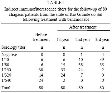
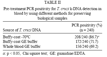
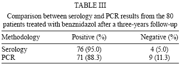
2008 Code Number: oc09004 ABSTRACT The efficacy of benznidazol on the treatment of chagasic patients from the state of Rio Grande do Sul was evaluated during a three-year follow-up. A cohort of 80 asymptomatic chronic chagasic patients or blood bank donors (49 male and 31 female) was studied. Their ages varied from 17-42 years, with a mean and a median of 30 and 35 years, respectively. The 80 patients presented positive serology, hemoculture and polymerase chain reaction (PCR). They were treated with 5 mg/Kg benznidazol twice a day for 60 days. Serological, parasitological and PCR methods were used to evaluate response. Serology was performed using commercial ELISA and indirect immunofluorescence (IFI) tests, parasitemia was monitored by hemoculture in LIT medium and PCR with primers S35/S36 was used to amplify a Trypanosoma cruzi 330 bp kDNA repetitive sequence. PCR positivity of 240 seropositive individuals was compared using DNA preparations from whole blood/guanidine EDTA (GE), buffy-coat/GE and frozen buffy-coat. Fifty non-chagasic individuals were used as negative controls. PCR positivity was 86.7% for the frozen buffy-coat, 71.7% for the GE/buffy-coat and 69.2% for the GE/whole blood. The hemocultures became negative just after treatment and remained negative during the three years of follow-up. In the third year after treatment, 9/80 (11.3%) patients presented negative PCR and, from those, four also presented negative serological tests. Furthermore, a reduction in three serological titers was observed in 27/80 (33.8%) of the patients treated. Taken together, the results show that four of the 80 (5.0%) chronic chagasic patients from the state of Rio Grande do Sul were cured after treatment with benznidazol. Key words: Chagas disease - diagnosis - polymerase chain reaction - benznidazol treatment - cure Trypanosoma cruzi, the aetiological agent of Chagas disease, is a hemoflagellate protozoan parasite that affects about 15-16 million individuals and it is estimated that 75-90 million people are exposed to infection in Latin America (Coura 2007). In Brazil, the vector control program, established in 1975 and continuing with the South Cone Initiative, eliminated the main vector, Triatoma infestans, from the country (Massad 2007). The seroprevalence of T. cruzi infection in Brazil among children aged 7-14 years fell 99.8% from 1980-1999 and the absence of seropositivity among young children (aged 0-4 years) is evidence of the interruption of vectorial transmission (Massad 2007). In Brazil, the highest prevalence of T. cruzi infection was found in the states of Rio Grande do Sul, Minas Gerais, São Paulo, Paraná, Goiás and Bahia (Silveira & Rezende 1994, Dias 1997). Human Chagas disease presents two distinct phases: the acute phase, which appears just after infection, and the chronic phase, which may last several years. After a long asymptomatic phase, around 30% of infected individuals develop chronic disease with severe damage to the heart and digestive system (Prata 2001). During the acute phase, T. cruzi trypomastigotes are usually detected by microscopic examination of fresh or stained blood-smears, as well as by xenodiagnosis and hemoculture (Luquetti & Rassi 2000). In contrast, during the chronic phase, diagnosis is based on the detection of circulating antibodies (Luquetti & Rassi 2000). However, due to the long-lasting maintenance of circulating antibodies, it is difficult to use serology as a marker for cure of the disease even after the successful treatment of T. cruzi infection (Luquetti & Rassi 2000). PCR comparative studies of whole blood were performed using traditional methods, such as xenodiagnosis and serology, for detecting T. cruzi; these showed that amplification of T. cruzi kDNA may substitute for xenodiagnosis in assessing parasitemia in chronic chagasic patients and may also be used as a complement to serological tests in blood banks (Avila et al. 1993, Marcon et al. 2002). Other studies showed that polymerase chain reaction (PCR) is a sensitive method that may detect the parasite DNA in up to 95% of blood samples from chronic chagasic patients (Britto et al. 1995a, Silber et al. 1997). Furthermore, PCR was shown to be a very useful tool for confirmation of diagnosis in patients with doubtful serology (Marcon et al. 2002). In this work, we followed up on the serological, parasitological and PCR profiles of 80 asymptomatic chagasic patients treated with benznidazol. In addition, considering that PCR sensitivity depends mainly on the target and source of the DNA, we compared different forms of biological sample preservation as sources of T. cruzi kDNA. PATIENTS, MATERIALS AND METHODS Clinical samples and procedures - A cohort of 80 asymptomatic chronic chagasic patients or blood bank donors (49 males and 31 females) from the state of Rio Grande do Sul presenting positive serology and hemo-culture was studied. Their ages varied from 17-42 years old, with a mean and median age of 30-35 years, respectively. Patients were treated with 5 mg/Kg benznidazol twice a day for 60 days. Our cohort was obtained from an initial group of 240 seropositive chagasic individuals. A negative control group, consisting of 50 individuals with negative serology, hemoculture and PCR for T. cruzi infection was included in the study. Chagasic patients were followed annually for a three-year period by serology, hemoculture and PCR. Blood collection was performed with a Vacutainer® system (Becton Dickinson, Franklin Lakes) based on the following volumes and objectives: i) 10 mL of blood for serology, ii) 30 mL of blood with heparin for hemoculture and iii) 15 mL of blood in EDTA Na2 for PCR. Five milliliters of whole blood was mixed (v/v) with 6 M guanidine hydrochloride/0.2M EDTA buffer pH 8.0 (GE buffer) immediately after collection and was stored at 4ºC until use. For the remaining 10 mL of whole blood, the buffy-coat was separated by centrifugation in a histopaque gradient® (Sigma, St. Louis), collected and divided into two tubes; one was stored at -70ºC and the other was added to 250 L of GE buffer and stored at 4ºC until use. Serological assays - The enzyme-linked immunosorbent assay (ELISA) was performed using the commercial Chagatest-Wienner kit (ELISA recombinant v. 3.0). Briefly, sera were diluted 1/20 in phosphate-buffered saline-tween 20 (0.05%) containing 1% bovine serum albumin. Two hundred microliters of diluted sera was added to microwells containing T. cruzi antigens. The plates were incubated at 37ºC for 30 min. After five washes in 0.1M phosphate buffer to remove unbound immunoglobulin, the samples were incubated at 37ºC for 30 min with a 1:40,000 dilution of peroxidase-labelled anti-human IgG conjugate. After another wash step, the plates were revealed with 60 mM hydrogen peroxide in 50mM citrate buffer, pH 3.2 and 0.01mM tetramethylbenzidine in 0.1N chloride acid. After 30 min incubation at rt, the reaction was stopped by the addition of 2N sulphuric acid. The plates were read at a wavelength of 450-620 nm in a microplate reader (Anthos Labetec Instruments, Austria). The mean absorbance of the negative controls plus 0.3 OD was used for cut off determination according to the manufacturer's instructions. Indirect immunofluorescence (IFI) assays were performed using the (IFI)-Chagas Bio Manguinhos Kit, FIOCRUZ, RJ, according to the manufacturer's instructions. Serum dilutions (1/40-1/1280) in phosphate buffer saline, pH 7.2 (PBS), were incubated for 30 min at 37ºC with total antigen pre-adsorbed onto the slide surface. Unbound immunoglobulins were removed by washing the slides twice with PBS. Following incubation with fluorescein-labeled anti-human IgG conjugate for 30 min at 37ºC, unbound conjugate was removed by two washes with PBS. Slides were mounted with buffered glycerine, pH 9.5, and observed under a fluorescence microscope (Labophot Nikon, Japan). Positive chagasic and negative sera were used as controls. The cut off value for IFI was 1/40. Hemoculture - Hemoculture was performed as described by Galvão et al. (1989). Briefly, 30 mL of heparinized venous blood was collected from each individual in a vacutainer system (Vacuum II) and centrifuged at 600 g for 30 min at 4ºC. Plasma was collected and stored at -20ºC. The pellet was resuspended in 15 mL of LIT (liver infusion tryptose) medium and centrifuged as described above. After removal of the supernatant, the pellet was resuspended in 30 mL of LIT and distributed into six tubes (18 x 150 mm), which were maintained at 28ºC and examined monthly until the fourth month. Only patients with positive hemocul-tures were treated. They received 5 mg/kg benznidazol twice a day for 60 days, followed by yearly serology, hemoculture and PCR analyses over the three years. DNA extraction and PCR - The buffy coat was added to 200 µL of extraction buffer (50 mM-Tris HCl, pH 8.0/50 mM EDTA/100 mM NaCl/0.5% SDS) and incubated with 20 µg/mL proteinase K (Sigma) for 2 h at 45ºC. Following a phenol/chloroform extraction and ethanol/3M sodium acetate precipitation, the DNA was washed twice with 70% ethanol, resuspended in 50 µL of TE buffer (Tris-HCl 10 mM and EDTA 1 mM), pH 8.0, and incubated with RNAse A (10 µg/mL) at 42ºC for 1 h. Blood lysates were boiled for 15 min for kDNA decatenation (Britto et al. 1993) and a 0.5 mL sample was subjected to phenol/chloroform extraction as already described. Primers S-35 (5´ -AAA TAA TGT ACG GGT GAG ATG CAT GA-3´) and S-36 (5´ -GGG TTC GAT TGG GGT TGG TGT-3´) (Sturm et al. 1989) were used to amplify a 330 bp fragment corresponding to the T. cruzi minicircle. Amplification reactions were performed in a final volume of 10 µL containing 1 U Taq DNA polymerase (Biotools), 200 µM of each dNTP, 1.5 mM MgCl2, and 10mM Tris-EDTA, pH 9.5, 5 pmoles of each primer and 2 µL of DNA preparation diluted 1/10 in TE buffer. DNA amplification was carried out in an Eppendorf Mastercycler using the following temperature profile: an initial step at 95ºC/for 5 min, followed by 34 cycles at 95ºC/1 min, 60ºC/1 min and 72ºC/1 min, and a final extension step at 72ºC/5 min. Positive controls consisted of 10 pg of T. cruzi DNA (Y strain) and 2 µL of an artificial mixture of human + T. cruzi DNA (diluted 1:10). Negative controls consisted of 2 µL of non-chagasic human DNA (diluted 1:10). The amplified products were subjected to 1.5% agarose gel electrophoresis, visualized under UV light after ethidium bromide staining and digitally recorded in a Digidoc-it UVP® system (Cambridge, England). Forward HuHGPRT1 (5' ATGGCGACCCGCAGCCCTGG 3') and HuHGPRT2 reverse (5' GAATGGATCTATCACTATTT 3') primers were used to amplify a 280 pb DNA fragment corresponding to the human gene sequence # gi164518913. The thermal conditions and reagent concentrations were the same as above except that the number of cycles was 30 and DNA preparation dilution was 1:100. Ethics - Informed written consent was obtained from all individuals enrolled in this study. This work was approved by the Ethical Committee IPB/LACEN/FEPPS (protocol n. 05/2003) and fulfills resolution number 196/1996 from the Brazilian National Health Council for research involving human beings. RESULTS From the initial cohort of 240 seropositive asymptomatic chagasic patients, 57.5% were male and 42.5% were female with ages ranging from 17-53 years old (mean age 38 years). Anti-T. cruzi IgG antibodies were confirmed by both ELISA and IFI tests. Antibody titration by IFI varied from 1/40-1/640. A serological titer of 1/40 was found in 27.5%, 1/80 in 20.0%, 1/160 in 23.3%, 1/320 in 12.1% and 1/640 in 17.1% of patients, respectively. From those, a group of 80 individuals, 49 men and 31 women with positive serology, hemoculture and PCR, aged 17-42 years with a mean of 30 years and a median of 35 years, received treatment with benznidazol. They were followed annually by serology for a period of three years (Table I). At the third year after treatment, a decrease of three, two and one, titer in the IFI test was observed in 27 (33.8%), 33 (41.2%) and 12 (15.0%) patients, respectively. Four (5.0%) patients remained with the same title and four (5%) became negative (data not shown). The last four patients (one in the 2nd year and three in the 3rd) presented negative serology by both IFI and ELISA tests, showing evidence of being cured. The hemocultures became negative after treatment and remained negative throughout the three years of follow-up. As little as 0.1 fg of pure T. cruzi DNA could be detected by PCR. In artificial mixtures, T. cruzi k-DNA was specifically detected even in the presence of an excess of 106-fold human DNA. Pre-treatment PCR positivity for T. cruzi k-DNA detection in blood by using different methods for preserving biological samples is shown in Table II. The results show that preservation of buffy coat at -70°C provides the highest PCR positivity (86.7%). All 50 individuals used as negative controls were PCR negative. A comparison between serology and PCR results from the 80 patients treated with benznidazol after three years of follow-up is shown in Table III. In the third year after treatment, nine patients with positive PCR before treatment presented negative PCR and, from those, four presented negative serology as well. All the patients who were PCR-negative for T. cruzi DNA presented the specific band (280 bp) for the human hypoxanthine phosphorybosil transferase (HGPRT) gene, excluding the possibility of PCR reaction inhibition. DISCUSSION Different tools have been used to diagnose chronic T. cruzi infection. Most of the serological commercial kits are highly sensitive for the detection of anti-T. cruzi antibodies, but in Latin American countries where other parasites such as Leishmania spp. and Trypanosoma rangeli are found, false-positive results have been reported (Saldaña & Sousa 1996, Caballero et al. 2007). In the state of Rio Grande do Sul, asymptomatic infection by Leishmania Viannia parasites in humans has been recently reported (Fagundes et al. 2007). In the present study, 240 chronic chagasic patients with an indeterminate form of Chagas disease were evaluated by serology, hemoculture and PCR methods. According to the WHO proposal, at least two distinct tests should be performed for a final serodiagnosis conclusion. In our study, 100% concordance between IFI and ELISA assays was found. However, distinct antibody titers varying from 1/40-1/640 were found among these patients. It is well established that the T. cruzi taxon presents two major lineages (TcI and TcII) that may influence parasite antigenic composition and, consequently, patient antibody responses (Di Noia et al. 2002, Buscaglia & Di Noia 2003). This fact must be considered when low or borderline antibody titers are found since most of the commercial kits use the T. cruzi TcII lineage as the antigen. Gutierrez et al. (2004) compared four different serological tests and reported discordant results for 15 out of 94 sera from Colombian chagasic patients. According to the authors, an in-house ELISA using an autochthonous T. cruzi TcI as the antigen shows the highest sensitivity, probably due to the fact that the TcI genotype is the most prevalent in that region. The TcII lineage is found most frequently in both acute and chronic chagasic patients in the south cone countries (Miles et al. 1980, Yeo et al. 2005, Steindel et al. 2008). In Brazil, distinct zymodemes are associated with acute and chronic Chagas disease (Luquetti et al. 1986). The state of Rio Grande do Sul showed one of the highest prevalence rates (8.84%) of T. cruzi infection in humans (Camargo et al. 1984). Despite this high infection prevalence, it is noteworthy that manifestations of cardiac or digestive disease are rare in the state of Rio Grande do Sul (Vinhaes & Dias 2000). It has been speculated that this apparent difference may be due to the characteristics of the T. cruzi populations circulating there. In the southern cone regions where the TcII genotype is most frequently found in humans, chagasic mega syndromes are common. In comparison, they are absent in the North of the Amazon (Miles et al. 1981). It seems that the TcII genotype is more virulent than TcI (Di Noia et al. 2002). Studies of 29 T. cruzi strains isolated from chagasic patients from the state of Rio Grande do Sul showed that these strains exhibit low virulence patterns in mice (Fernandes et al. 1997). According to the Brazilian Ministry of Health, treatment of T. cruzi infection is recommended during the acute phase, congenital infection, early chronic phase (children under 15 years old and elderly patients with evidence of early chronic infection) and reactivation of the infection in individuals infected with HIV. At the Centro de Cardiologia do Rio Grande do Sul, specific treatment for T. cruzi infection is offered to all patients having a positive hemoculture. In the present study, 80 patients with positive hemoculture received treatment with benz-nidazol (5 mg/kg of body weight for 60 days taken twice a day) and were followed up for a three-year period by means of serological, parasitological and PCR methods. Differences in the sensitivity of hemoculture have been reported. Luz et al. (1993) found 90% positivity among untreated chronic chagasic patients from the state of Minas Gerais. Fernandes et al. (1999) reported 76% positivity among children from the state of Rio Grande do Sul. Lower rates of sensitivity for chronic patients have been shown by other authors (Chiari et al. 1989, 55.8%, Gomes et al. 1999, 36.5%, Lages-Silva et al. 2006, 53.0%). Such differences may represent distinct levels of parasitemia that may depend on the phase of the disease, the parasite strain and the host immune response. In the present study, 80/240 (33.3%) positivity by hemoculture was found in the chronic chagasic patients with positive serology. Longitudinal studies in endemic and non-endemic areas have shown that anti-T. cruzi antibodies persisted in infected individuals for many years (Coura et al. 1996, Gomes et al. 1999, Francolino et al. 2003). On the other hand, serology became negative after successful parasitological treatment of both acute and chronic infection, indicating cure from the infection (Luquetti 1999). A negative parasitological test alone does not represent treatment success; however, its positivity after chemotherapy represents therapeutic failure. In a study of 27 chronic chagasic patients from the state of Goiás submitted to chemotherapy with benznidazol, 11.1% of the patients presented positive hemoculture, demonstrating treatment failure (de Castro et al. 2006). In the present study, none of the 80 treated patients showed positive hemocultures in the three-year follow-up. Seventy-six out of the 80 (95.0%) patients showed positive serology with decreasing titers during the follow-up and four patients (5.0%) revealed negative serology by both methods - evidence of being cured. One of those patients negated the serology in the second year after treatment and the other three negated the serology in the third year. Due to the long persistence of anti-T.cruzi antibodies after chemotherapy and the low sensitivity of parasitological methods, PCR has been suggested to be a very useful tool for treated patients' follow-up (Britto et al. 1995b). In our study, we analyzed the performance of PCR for the detection of T. cruzi k-DNA in the blood of two panels of patients using three distinct sample preservation methods. PCR performed with the frozen buffy coat as the T. cruzi DNA source showed a higher positivity than the GE-preserved buffy coat or whole blood. The higher PCR positivity on buffy coat preparations is due to similar density between leucocytes and T. cruzi. Therefore, the bloodstream parasites concentrate on that zone after differential centrifugation. It is well known that hemoglobin is a strong PCR inhibitor. In our experiments using primers for the detection of the human HGPRT gene, we observed the expected band in all PCR-negative patients, revealing that the PCR reaction was not inhibited. The interpretation of the PCR results in the diagnosis of Chagas disease is controversial since a positive result may reflect the detection of the DNA of either an intact or a lysed parasite (Britto et al. 1995b, 2001). During the recent outbreak of Chagas disease in the state of Santa Catarina, 100% PCR positivity was found among the 18 patients evaluated (Steindel et al. 2008). In the present study, nine out of the 80 (11.3%) patients negated the PCR three years after treatment with benznidazol. Considering that antibody titers can be detected for several years, the negative PCR results suggest the process of healing. One may speculate that these patients will be the next to be cured. Taken together, our results suggest that four out of the 80 asymptomatic chagasic patients submitted for chemotherapy with benznidazol were cured of the infection after a three-year follow-up. REFERENCES
Copyright 2009 - Instituto Oswaldo Cruz - Fiocruz The following images related to this document are available:Photo images[oc09004t2.jpg] [oc09004t1.jpg] [oc09004t3.jpg] |
| |||||||||
