 |
| About Bioline | All Journals | Testimonials | Membership | News |
|
||||||
|
||||||
Memórias do Instituto Oswaldo Cruz, Vol. 104, No. 1, February, 2009, pp. 33-36 Prostatic paracoccidioidomycosis: differential diagnosis of prostate cancer Daniel Lima LopesI; Stanley de Almeida AraújoII, +; João Paulo Lemos da Silveira SantosII; Ana Claudia LyonII; Diogo Vieira DantasII; Bernardo Sgarbi ReisIII; Alfredo Miranda de GóesIII; Ênio Roberto Pietra PedrosoII IHospital
do Instituto de Previdência dos Servidores do Estado de Minas Gerais,
Belo Horizonte, MG, Brasil Received 3 September
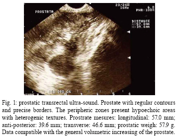
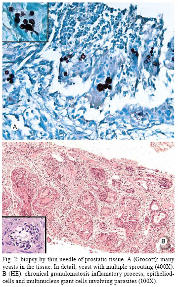
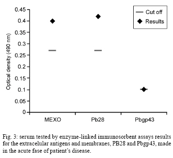
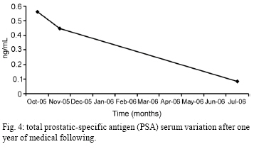
2008 Code Number: oc09005 ABSTRACT Symptomatic prostatic paracoccidioidomycosis (PCM) is a very rare condition; however, it may express as a typical benign prostatic hyperplasia or a simulating prostatic adenocarcinoma. This case report presents PCM mimicking prostatic adenocarcinoma. The purpose of this paper is to call the general physician's attention to this important differential diagnosis. Key words: Paracoccidioides brasiliensis - paracoccidioidomycosis - prostatitis - prostate cancer Paracoccidioidomycosis (PCM) is an important deep mycosis in Latin America. It is caused by the dimorphic fungus Paracoccidioides brasiliensis, whose spores enter the body via the respiratory tract and evolve either asymptomatically or in a sub-acute or chronic manner. The parasite is able to provoke several clinical presentations due to its capacity to disseminate itself from the lungs through the lymphatic system or via the blood stream to any organ or system. PCM mainly affects adult males, preferentially in the lungs, mucosa, skin and phagocytic-monocytic system (Paniago et al. 2003, Prado et al. 2005). On a smaller scale, it reaches the nervous, musculoskeletal and suprarenal systems (Paniago et al. 2003). Its clinical incidence in the urinary tract, especially in prostatic injuries, is not well known. In a few necropsy studies involving disseminated PCM cases, its incidence varies from 2.7-9% (Salfelder et al. 1969, Begliomini et al. 1993). Prostatic diseases have become more common due to the increase in the population's age. Generally, they appear in males over 40-years-old, being more often bacterial and viral infections, benign hyperplasia and adenocarcinoma. Prostatic adenocarcinoma is the most fearful lesion due to its malignant and metastatic potential, and it is seen clinically in 10% of cases. Its diagnosis must always be set apart because its mortality is 3%, making it the greatest cause of morbi-mortality among men (Crawford 2003). Fungal prostatitis is unusual, having as its most common agents Coccidioides immitis, Candida albicans, Aspergillus sp., Cryptococcus neoformans and Blastomyces dermatitidis, which mainly affect patients who are immune-suppressed. The symptomatic attack of the prostate by PCM has rarely been described. Its potential severity justifies the reporting of this case. In this paper, infection of the prostate by P. brasi-liensis is described, mimicking cancer with urethral obstruction and urgent expansion of the bladder. Case study AAF, a 54-year-old married rural worker who was born in and is a current resident of São Sebastião do Maranhão, Minas Gerais, Brazil, presented in December 2003 with palate stomatitis associated with dysphagia and odynophagia. A biopsy of this injury revealed P. brasiliensis. He was treated with Sulfametoxazol (800 mg) and Trimethoprim (160 mg) daily in an irregular way for 20 months. In December 2004, he started to complain of dysuria, polyuria and urinary urgency and frequency, which culminated after one year in prompt urinary retention. Concomitantly, a stomatitis with moriforme injury of the palate appeared. A digital rectal exam revealed an increased prostate volume, induration, precise limits and a nodule on the right lobe. It was thought to be prostate cancer. His PSA level was measured at 0.563 ng/mL and transrectal ultrasonography demonstrated a prostrate with irregular contours, with peripheral zones containing hypoechoic areas with heterogenic texture (Fig. 1). There was a volumetric increasing of the gland, which weighed 57.9 g. The prostatic biopsy revealed a diffuse chronic granulomatous inflammatory process showing epithelial and multinucleated giant cells, neutrophils and eosinophils involving the P. brasiliensis (Fig. 2 A, B). A complete blood count showed a total counting of leukocytes of 12,940/mm3 (neutrophils 9,614/mm3, lymphocyte 2,057/mm3, eosinophils 349/mm3, basophils 116/mm3, monocytes 660/mm3). The urine exam revealed hemoglobin +/4+, urobilinogen 0.2 mg/dL, leukocytes +/4+, piocytes 20/field, red blood cells 10/field, bacterian microbiota +++/4+, rare grouped piocytes and the presence of renal epithelium. Gram staining did not reveal any bacteria and serum biochemistry did not show any abnormalities. A chest X-ray showed localized micro-reticulo-nodule alterations, mainly on the hilum and in the pulmonar basis bilaterally. Abdominal ultrasonography did not show any alteration and the other urinary organs were apparently normal. Collected blood samples were positive in anti-P. brasiliensis serologic tests using the antigens PB28, Pbgp43 and membrane and extracellular antigens (MEXO) (Fig. 3). The initial treatment was done with Itraconazol (100 mg per day), which was replaced after 10 days with Sulfametoxazol (800 mg) and Trimetoprim (160 mg every 12 h) because the former was not available at public health care centres in Brazil. After six months, the patient was asymptomatic and without deficits or clinical or radiological residual injuries. The total PSA level remained low despite the intense inflammation (Fig. 4). After one year of ambulatory observation, there was no recurrence of the infection or side effects from the treatment. DISCUSSION We report this case due to the rare presentation of PCM in the prostate, which may be a serious symptomatic condition if untreated and due to the importance of the differential diagnosis with prostate adenocarcinoma, which is a very severe disease, with lethality and me-tastatic potential whose possibility of treatment is only surgical. Depending on the extension of the neoplastic tissue and on the chosen technique, prostatectomy may lead to minor or serious complications such as erectile problems urinary incontinence and damage to the urethra or rectum (Wilt & Brawer 1999). Thus, a proper histopathological exam may demonstrate an infectious prostatitis and avoid an unnecessary surgical intervention and its related risks. PCM has special importance in Latin America, where it is endemic among infecto-parasitarian diseases due to the severity and the peculiarity of some of its clinical forms. This infection is not commonly isolated in the urogenital tract. PCM in the prostate is not considered a primary disease, affecting only 1.59% of patients in the disseminated form (Brito & Caprini 1959, Ciconelli et al. 1969, Begliomini et al. 1993). All of the urogenital tract may be affected by the disease, including the epididymis, testicles, prostate, ureters, penis, urethra and kidneys. The first case of prostatic involvement was described by Brito and Caprini (1959); since then, only a few other cases have been described (Melo et al. 1992, Severo et al. 2000). Because of the general symptoms, the diagnosis of PCM is frequently done later than expected. Patients with prostatic involvement may present obstruction symptoms of the lower urinary tract. Cancer and chronic inflammation always constitute the differential diagnosis for complaints related to prostatic disease symptoms. When the prostate is involved in PCM infection, differential diagnosis with cancer is difficult due to the variety of damage and absence of specific signs. The diagnosis of prostatic injuries is suggested by the clinical aspects, but it often requires confirmation by a histopathological exam. A biopsy is mandatory due to the necessity for cancer exclusion (Stillwell et al. 1987).Differential diagnosis for PCM of the prostate is divided into specific, caused by bacteria (Treponema pallidum, Mycobacterium tuberculosis, Brucella sp.), fungi (Candida sp., Aspergillus sp., C. neoformans, Histoplasma capsulatum, Actinomyces sp., B. dermatitidis and C. immitis), viruses or parasites, and non-specific, derived from xantogranulomatousis, allergies, post-TURP or post-biopsies. Granulomatous prostatitis is a rare disease that frequently simulates the development of prostatic carcinoma due to the formation of nodules and the increase of the gland by rectal touch (Stillwell et al. 1987, Begliomini et al. 1993). A histopathological exam of the granulomatous prostatitis, however, showed infiltrated lympho-plasmocyte, pseudotubercules, epithelioidic cells and the absence of signs of atypical cellular proliferation, which characterize the neoplasia. In coccidioidomicosis and North American blastomicosis, the involvement of the genitourinary tract is frequent, being common in prostatic attack in the disseminated form (Brito & Caprini 1959, Stewart 1964, Eickenberg et al. 1975). There have been a few cases of transmission of B. dermatitidis from men with prostatic involvement to their female sexual partner (Faber et al. 1968). PCM has no record of inter-human transmission despite its potential for sexual transfer by contaminated prostatic secretion. Tuberculosis or other mycotic diseases that affect the genitourinary tract are other important differential diagnoses and the microbiologic study of urine and cultures are fundamental for its discernment (Stillwell et al. 1987). Since P. brasiliensis is not coloured by Gram stain, its diagnosis may not be noticed in routine exams. Induration of the prostate may persist upon physical examination for years and scarring and fibrosis have been observed in a third of the patients (Paniago et al. 2003). The case described here presented sexual dysfunction after the treatment and the effects tobacco use, aging process and adverse effect of the treatment with Sulfametoxazol-Trimetoprim or Ketoconazole, which were concomitant conditions in this case, could be potential factors, in addition to prostate fibrosis induced by PCM (Brito & Carpini 1959). There is no report of alterations in serum levels of the prostatic-specific antigen PSA during the treatment. In granulomatous prostatitis, both increasing and decreasing serum levels of PSA may be observed. A significant reduction in PSA, as in this case, suggests that cytokines derived from the activated macrophages and T-lymphocytes may have a regulatory effect on the cells' secretions and cause the destruction of the prostatic epithelia (Dhundee & Maciver 1991). Current serum tests are highly sensitive and specific for diagnosis and useful for the clinical follow-up of people affected with PCM. Normal serum results are extremely rare with evidence of the disease's histopathology. Serum tested by enzyme-linked immunosorbent assays (ELISA) for the extracellular antigens and membranes - MEXO - from P. brasiliensis presents a specificity of 96.6% compared to control groups and 81.2% in relation to groups with diverse infections (Reis et al. 2005). A purified protein with a molecular weight of 28-kDa of MEXO, called Pb28, is the most specific antigen in the humoral immunological response of PCM. This protein reacts and has a specificity of 100%. It may be used as an alternative antigen to the serum diagnostic method. ELISA, either for MEXO or for Pb28, is a strong reactor in serum analysis when collected during the acute period of the disease (Fig. 4). However, one glycoprotein with a molecular weight of 43 Kda, known as Pbgp43, which is an important extracellular component released by P. brasiliensis during its pathogenic phase, is used in the detection of the serum antibodies in patients with PCM and in epidemiologic studies through the intradermal reaction (Kalmar et al. 2004). It presents a sensitivity of 95.1% and a specificity of 97.5%. In this case, it was reactor-moderated (Fig. 4). Such serum data is in accordance with those reported by Reis et al. (2005) because Pb28 has proven to be more sensitive and specific for PCM and Pbgp43 presented irregular results. PCM is diagnosed by the identification of the yeasts in direct exams of clinical samples (biopsy, mucous, scratched) or cultures. The yeast has specific microscopic aspects, including a bi-refringent wall, reproduction by multiple-sprouting and originating blastospores that do not detach from the mother cell, which gives them a unique appearance among the pathogenic fungi (Fig. 2A). The granuloma is formed with the objective of restricting the parasite and avoiding its dissemination (Fig. 2B). Thus, the more efficient the cellular immunologic response is the better the efficiency of the granulomatos reaction and the lesser the severity of the disease. The histopathological response of the host caused by P. brasiliensis resembles tissue invasion by B. dermatitidis and C. immitis. Injuries of the oropharyngeal mucosa are frequently observed. The most characteristic aspect is moriform ulcerous stomatitis. PCM is a disease that lacks standards for cure and a standard treatment protocol. There are no specific treatments for the urogenital form. Prolonged medication is, in many cases, necessary to prevent recurrence, which exhausts the patient and impairs his/her adequate compliance with the proposed treatment. Interruption of the medication or irregular or inadequate use may drive the emergence of resistance, making the problem worse and producing several side-effects (Prado et al. 2005). Once a diagnosis of prostatic PCM is made, a specialist in infectious diseases should be consulted. Finally, the patient needs to be observed for many years for prevention and occasional treatment of disease recurrence. PCM is a systemic disease with the potential for involvement of all organs and it is necessary to pay attention to all complaints presented by the patients, even those not associated with the most frequent localizations of the disease. In South America, especially in Brazil, prostatic PCM is an important differential diagnosis to prostatic adenocarcinoma and can evolve into a serious symptomatic condition if left untreated. REFERENCES
Copyright 2009 - Instituto Oswaldo Cruz - Fiocruz The following images related to this document are available:Photo images[oc09005f1.jpg] [oc09005f4.jpg] [oc09005f3.jpg] [oc09005f2.jpg] |
| |||||||||
