 |
| About Bioline | All Journals | Testimonials | Membership | News |
|
||||||
|
||||||
Memórias do Instituto Oswaldo Cruz, Vol. 104, No. 3, May, 2009, pp. 451-455 ARTICLES The IFN-γ+874T/A gene polymorphism is associated with retinochoroiditis toxoplasmosis susceptibility Maíra Cavalcanti de AlbuquerqueI; Ana Luisa Quintella do Couto AleixoII; Eliezer Israel BenchimolII; Ana Cristina Câmara S LeandroIII; Leandro Batista das NevesI; Regiane Trigueiro VicenteI; Maria da Glória Bonecini-AlmeidaIII, +; Maria Regina Reis AmendoeiraI ILaboratório
de Toxoplasmose, Instituto Oswaldo Cruz-Fiocruz, Rio de Janeiro, RJ, Brasil Financial support: IOC, IPEC/Fiocruz, MCA fellowship from IOC/Fiocruz Received 12 August
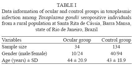
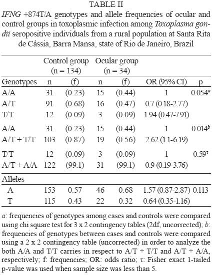
2008 Code Number: oc09071 ABSTRACT Toxoplasmosis is a worldwide zoonosis that generally produces an asymptomatic infection. In some cases, however, toxoplasmosis infection can lead to ocular damage. The immune system has a crucial role in both the course of the infection and in the evolution of toxoplasmosis disease. In particular, IFN-γ plays an important role in resistance to toxoplasmosis. Polymorphisms in genes encoding cytokines have been shown to have an association with susceptibility to parasitic diseases. The aim of this work was to analyse the occurrence of polymorphisms in the gene encoding IFN-γ (+874T/A) among Toxoplasma gondii seropositive individuals, including those with ocular lesions caused by the parasite, from a rural population of Santa Rita de Cássia, Barra Mansa, state of Rio de Janeiro, Brazil. Further, we verified which of these polymorphisms could be related to susceptibility to the development of ocular toxoplasmosis. This study included 34 individuals with ocular toxoplasmosis (ocular group) and 134 without ocular lesions (control group). The differences between A and T allele distributions were not statistically significant between the two groups. However, we observed that a higher frequency of individuals from the ocular group possessed the A/A genotype, when compared with the control group, suggesting that homozygocity for the A allele could enhance susceptibility to ocular toxoplasmosis in T. gondii infection. Key words:toxoplasmosis - interferon-gamma - chorioretinitis - single nucleotide polymorphism Toxoplasmosis is a worldwide zoonosis caused by Toxoplasma gondii, an intracellular protozoan. The prevalence of this infection ranges from 15-85%, depending on social habits, geographic and weather distribution (Amendoeira et al. 1999, Garcia et al. 1999, Sobral et al. 2005). Cats and other felids from the genera Lynx and Felis are the definitive hosts of the parasite and warm-blooded animals, including humans, are intermediate hosts (Krick & Remington 1978, Amendoeira 1995, Garcia et al. 1999). T. gondii is a lifelong persistence parasite and almost 90% of infected individuals are asymptomatic. In some cases, however, infection can cause destructive inflammation that targets multiple organs, including the brain and eyes. Ocular toxoplasmosis can originate from acquired or congenital toxoplasmosis, causing necrotizing retinitis or retinochoroiditis (Lynfield & Guerina 1997, Garcia et al. 1999, Montoya & Liesenfeld 2004). Although most cases of human eye infection are believed to be congenital in origin, acquired acute toxoplasmic retinochoroiditis may be more prevalent than has been recognised (estimates of prevalence range from 0.6-17.7%) (Smith & Ganley 1972, Melamed 1988, 2009, Glasner et al. 1992, Abreu et al. 1998, Holland 2009). Immune mechanisms against T. gondii ocular infection have not yet been fully clarified. However, it has been suggested that a strong T helper 1 response, with gamma interferon secretion, is crucial for protection in a murine model (Gazzinelli et al. 1994) and for controlling parasite growth in chronically infected hosts (Suzuki et al. 1989). Norose et al. (2005) observed that IFN-γ knockout mice showed mild inflammation and sludging of blood in the retina and choroid, demonstrating the role of this cytokine in controlling ocular disease. In addition, the relationships between polymorphisms in genes encoding cytokines and resistance or susceptibility to several diseases have been studied (Barret et al. 2003, Mac Lean et al. 2004, Rodriguez-Pérez et al. 2005, Spriewald et al. 2005, El-Shabrawi et al. 2006, Martinez-Pomar et al. 2006). These studies suggest that these polymorphisms may influence the course of a parasitic disease. In toxoplasmosis infection, single nucleotide polymorphisms (SNPs) in IL10 -1082G/A (*124092 INTERLEUKIN 10; IL10), TNF -308G/A (*191160 TUMOUR NECROSIS FACTOR, ALPHA; TNF), IL1A -889C/T (*147760 INTERLEUKIN 1-ALPHA; IL1A) and IL1B +3954C/T (*147720 INTERLEUKIN 1-BETA; IL1B) have been previously described in retinochoroiditis (Cordeiro et al. 2008a-c). However, there is no data in the literature regarding the role of the IFN-γ +874T/A SNP in either toxoplasmosis infection or ocular outcome. We conducted a population case-control study to investigate the role of the (*147570 interferon, gamma; IFN-γ) IFN-γ +874T/A SNP in retinochoroiditis susceptibility. PATIENTS, MATERIALS AND METHODS Study population - This study was performed in a rural area called Santa Rita de Cássia, Barra Mansa, located 176 miles from city of Rio de Janeiro, state of Rio de Janeiro (RJ), Brazil. Several cases of visual impairment had been reported by the local Health Service as suspected ocular toxoplasmosis. Serological anti-T. gondii tests and fundoscopy were performed on 1,071 individuals, comprising about 82.4% of community population of Santa Rita de Cássia. Among these, 706 (65.9%) were seropositive for T. gondii. Ocular lesions were defined by the presence of IgG antibodies against T. gondii and retinochoroiditis with active or cicatrized lesions. T. gondii seropositive community controls (n = 134) were recruited and matched by sex and age. T. gondii seropositive individuals were excluded if they were related to either toxoplasmosis ocular cases or controls. Written informed consent was obtained from all subjects and the study was approved by the Human Research Ethics Committee of Instituto de Pesquisas Clínicas Evandro Chagas-Fiocruz, RJ, Brazil, on 07/07/2006 (CEP: 0039.0.011.099-06). Toxoplasmosis diagnosis - Indirect immunofluorescence (IIF) and Enzyme Linked Immunosorbent Assays (ELISA) were performed as described by Coutinho et al. (1970) and Uchôa et al. (1999), respectively. Serum was considered positive if it showed titer >1/16 (IIF) or an ELISA score of 1.4-fold higher than the ELISA cut-off. Genomic DNA extraction - Five mililiter of blood was collected in EDTA tubes for genomic DNA extraction, using a DNA blood mini kit (QIAgen), following the protocol described by the manufacturer. All genomic DNA extractions were subjected to 2% agarose gel electrophoresis, visualised with ethidium bromide and observed on an ultraviolet transilluminator to confirm their integrity. IFN-γ polymorphism - The IFN-γ T/A polymorphism typed in this study was a SNP at the +874 position. The SNP typing protocol was described by Pravica et al. (2000). The T and A polymorphism sequences were identified using a specific single stranded oligonucleotide, synthesised to cover a 24-bp region for each allele. An amplification refractory mutation system by polymerase chain reaction (ARMS-PCR) was performed in a total volume of 20 µL containing 1 µL of generic primer (100 pmol/µL; 5´ -TCA ACA AAG CTG ATA CTC CA -3´), 1 µL of specific A primer (100 pmol/µL, 5´ - TTC TTA CAA CAC AAA ATC AAA TCA -3´) or 1 µL of specific T primer (100 pmol/µL, 5´ - TTC TTA CAA CAC AAA ATC AAA TCT -3´), 1 µL of internal control 1 (10 pmol/µL, 5´ - GCC TTC CCA ACC ATT CCC TTA -3´), 1 µL of internal control 2 (10 pmol/µL, 5´ - TCA CGG ATT TCT GTT GTG TTTC -3´), 4 µL of dNTP (2 mM), 2.4 µL of MgCl2 (25 mM), 3 µL of buffer (10X), 0.5 µL of Taq DNA polymerase (5 U/µL), 4.1 µL of sterile MiliQ H2O and 2 µL of genomic DNA. ARMS-PCR was performed in a thermocycler (Applied Biosystems) with the cycles: 95ºC (3 min), 10 cycles of 95ºC (15 s), 65ºC (50 s) and 72ºC (40 s), followed by 20 cycles of 95ºC (20 s), 55ºC (50 s) and 72ºC (50 s), 72ºC (7 min) and 4ºC (hold temperature). ARMS-PCR amplified products were subjected to 1.5% agarose gel electrophoresis, stained with ethidium bromide and visualised on an ultraviolet transilluminator. Statistical analysis - Genotypes and allele frequencies were compared between cases and controls by Chi Square analysis for a 3 x 2 contingency table, using the Epi Info 2000 software (CDC, Atlanta, GA, USA); p values were corrected for multiple comparisons (age and sex). A corrected p < 0.05 was considered to be statistically significant. RESULTS We identified 41 (5.8%) individuals with ocular lesions caused by T. gondii infection from all T. gondii seropositive individuals (n = 706). Seven individuals were excluded from the study. Of these, four were related, two had moved to another city and one refused to participate in the study. Among the remaining ocular group, 10 (29.4%) were male and 24 (70.6%) were female and ages ranged from 16-88 (44 ± 20.9 SD) years. We included 134 control individuals from the T. gondii IgG positive individuals, matched by sex and age, for a case:controls ratio of 1:4 (Table I). All 168 individuals were tested by ARMS-PCR at the IFNG +874T/A polymorphic site. Forty six (27.3%) showed A/A homozygocity, 107 (63.7%) were A/T heterozygous and 15 (9%) were T/T homozygous. Genotype frequency analysis of the control group showed that A/T heterozygotes (68% of 134 genotypes) were the largest group. However, the ocular group showed a similar frequency of A/A and A/T genotypes. The T/T genotype was present in the same frequency among case and control groups. No statistically significant difference was observed (p = 0.054) (Table II) between the cases and controls. However, A/A homozygous individuals showed a 1.62-fold higher risk than other genotypes of developing retinochoroiditis during T. gondii infection (p = 0.014; df = 1) (Table II). The distribution of alleles did not differ between the ocular and control groups (p = 0.113) (Table II); however, the presence of a T allele in heterozygous individuals was sufficient to confer protection against retinochoroiditis when the A/T heterozygous individuals were compared with the A/A homozygous subjects (p = 0.01, df = 1; uncorrected X2 = 6.21, OR = 0.36; 0.15-0.88). Since the frequency of retinochoroiditis during T. gondii infection was higher in female (n = 24; 71%) than male (n = 10; 29%) individuals in our community case-control study, we adjusted the p value by gender. The effect remains invariable, since the odds ratio was not affected; thus, the p values remain the same. DISCUSSION Ocular toxoplasmosis can cause visual alterations, retinochoroiditis and, in some cases, blindness. T. gondii infection induces a response by T helper 1 cells, which produce IFN-γ, a cytokine that is involved in resistance to toxoplasmosis (Suzuki et al. 1989, Scharton-Kersten et al. 1996). Resistance to ocular toxoplasmosis is associated with the ability to produce IL-12 and IFN-γ against parasitic antigens (Vallochi et al. 2002). High levels of IFN-γ have been described in both asymptomatic and symptomatic T. gondii seropositive individuals, compared to negative controls (Fatoochi et al. 2006). However, only a slight increase was reported in T. gondii asymptomatic individuals compared with individuals afflicted with acquired ocular toxoplasmosis (Yamamoto et al. 2000). Polymorphisms in the genes encoding cytokines, including pro-inflammatory cytokines such as TNF-α, IL-6, IL-12 e IFN-γ and anti-inflammatory cytokines such as IL-10 and TGF-β, have previously been shown to be associated with several diseases (Barret et al. 2003, MacLean et al. 2004, Spriewald et al. 2005, El-Shabrawi et al. 2006, Martinez-Pomar et al. 2006). Some studies have demonstrated an association between IFN-γ production levels and the polymorphism at the IFN-γ +874T/A position. The T/T genotype associates with high levels of IFN-γ production, A/T with medium levels and A/A with lower levels (López-Maderuelo et al. 2003, Daí et al. 2006, Henao et al. 2006). Recently, Cordeiro et al. (2008a-c) described an association between the IL10 -1082A allele and retinochoroiditis. This study also found that the TNF -308G/C, IL1A -889C/T and IL1B +3954C/T alleles did not show any association with retinochoroiditis. To our knowledge, the +874T/A polymorphism in the gene encoding IFN-γ has not previously been described in toxoplasmosis. The overall distribution of this SNP has previously been described in Brazilian populations. Our results were proportionally similar to those described by Laguila Visentainer et al. 2005. They observed 30.3% of the population possessed the A/A genotype, 55% possessed the A/T genotype and 14.7% with the T/T genotype. Similarly, Matos et al. (2007) reported a 38.4%, 45% and 16.6% genotype distribution and Visentainer et al. (2008) identified a profile of 31.8%, 54% and 14.2% of each genotype. Interestingly, Amim et al. (2008) described a different profile of genotype distribution in their population (45%, 32% and 23%). The polymorphism in IFN-γ at the +874T/A position has been previously described to associate with several diseases. The A/A genotype has been shown to be associated with hepatitis B in China, Yu et al. 2006, Helicobacter pylori gastritis in Italy, Zambon et al. 2005, tuberculosis in Spain (López-Maderuelo et al. 2003, type 2 diabetes mellitus in Greece (Tsiavou et al. 2005 and Wegener's granulomatosis in Germany (Spriewald et al. 2005). The A/T and T/T genotypes have been shown to associate with breast cancer in Iran (Kamali-Sarvestani et al. 2005, hepatitis C in Taiwan (Daí et al. 2006) and Hashimoto's disease in Japan (Ito et al. 2006). Our results showed that the A/T genotype is the most frequently in both the control and ocular groups (47% of individuals in the ocular group and 67.9% in the control group). We observed a statistically significant difference in the frequencies of the A/T and A/A genotypes. This result suggests that homozygocity for the A allele is associated with susceptibility to development of ocular toxoplasmosis. It is know that the resistance to development to ocular toxoplasmosis involves the production of IL-12 and IFN-γ against parasitic antigens (Vallochi et al. 2002). Individuals with the A/A genotype produce less IFN-γ than individuals with other genotypes. Together, these facts suggest a possible mechanism that could explain the relationship between the A/A genotype and susceptibility to ocular damage leading to retinochoroiditis. We conclude that the A/A genotype can be associated with susceptibility to ocular toxoplasmosis. Further studies involving polymorphisms in other cytokines should be performed to understand the role of the immune system in the course of T. gondii infection. REFERENCES
Copyright 2009 - Instituto Oswaldo Cruz - Fiocruz The following images related to this document are available:Photo images[oc09071t3.jpg] [oc09071t1.jpg] [oc09071t2.jpg] |
| |||||||||
