 |
| About Bioline | All Journals | Testimonials | Membership | News |
|
||||||
|
||||||
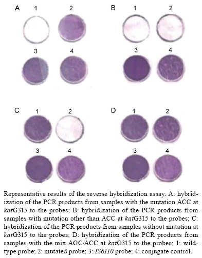
Memórias do Instituto Oswaldo Cruz, Vol. 104, No. 5, August, 2009, pp. 710-714 ARTICLES In house colorimetric reverse hybridisation assay for detection of the mutation most frequently associated with resistance to isoniazid in Mycobacterium tuberculosis Mirela VerzaI, II; Raquel de Abreu MaschmannI, II; Márcia Susana Nunes SilvaI, III; Elis Regina Dalla CostaI, IV; Marta Osório RibeiroI; Franciele RossoI, II; Philip Noel SuffysV; Enrico TortoliVI; Fiorella MarcelliVI; Arnaldo ZahaII; Maria Lucia Rosa RossettiI, III, + ICentro
de Desenvolvimento Científico e Tecnológico, Fundação
Estadual de Produção e Pesquisa em Saúde, Av. Ipiranga
5400, 90610-000 Porto Alegre, RS, Brasil Financial support: CNPq, FINEP, FEPPS Received 19 December
2008 Code Number: oc09159 ABSTRACT Mutations in the katG gene have been identified and correlated with isoniazid (INH) resistance in Mycobacterium tuberculosis isolates. The mutation AGC→ACC (Ser→Thr) at katG315 has been reported to be the most frequent and is associated with transmission and multidrug resistance. Rapid detection of this mutation could therefore improve the choice of an adequate anti-tuberculosis regimen, the epidemiological monitoring of INH resistance and, possibly, the tracking of transmission of resistant strains. An in house reverse hybridisation assay was designed in our laboratory and evaluated with 180 isolates of M. tuberculosis. It could successfully characterise the katG315 mutation in 100% of the samples as compared to DNA sequencing. The test is efficient and is a promising alternative for the rapid identification of INH resistance in regions with a high prevalence of katG315 mutants. Key words: drug resistance - isoniazid - katG315 - reverse hybridization assay - tuberculosis Tuberculosis (TB) is the only infectious disease that has been considered a global emergency by the World Health Organization as one third of the world's population is latently infected with Mycobacterium tuberculosis (WHO 2008). The emergence and spread of multidrug-resistant (MDR) strains of M. tuberculosis are serious threats to the control of TB and represent an increasing public health problem. Patients infected with MDR strains, namely those resistant to at least rifampicin (RIF) and isoniazid (INH), are difficult to cure and likely to remain as sources of infection for a longer period than patients with drug-susceptible strains (Zumla & Grange 2001). The recently observed spread of extensively drug-resistant TB, which is defined as MDR-TB with additional resistance to a fluoroquinolone and a second-line injectable drug, is often incurable (WHO 2008). The early identification of resistant strains is essential for efficient treatment and control of MDR-TB. Faster application of effective chemotherapy also prevents further spreading of drug-resistant isolates (Parsons et al. 2004). INH is one of the most effective and specific antibiotics available for the treatment of TB and is also widely used to treat latent M. tuberculosis infections (Espinal et al. 2001). This antibiotic is a prodrug that requires activation by the catalase-peroxidase enzyme encoded by the katG gene (Hazbon et al. 2006). Activated drug appears to disrupt the synthesis of essential cell wall mycolic acids (Rawat et al. 2003). In the majority of isolates, resistance to INH is mainly achieved through mutations in the katG gene and in the inhA promoter region. Mutations in other genes, such as ahpC and ndh, have been implicated in INH resistance, but their roles have not yet been proven (Hazbon et al. 2006). In various studies, the frequency of INH-resistant strains containing a mutation in codon 315 of the katG gene ranged from 50-100% (Musser et al. 1996, van Soolingen et al. 2000, Silva et al. 2003, Hillemann et al. 2005, Zhang et al. 2005, van Doorn et al. 2006, Dalla Costa et al. 2009). In most cases, one amino acid substitution at codon 315 (AGC→ACC/Ser→Thr) was reported to be the most frequent (Mdluli et al. 1996, Musser et al. 1996, van Soolingen et al. 2000, Abate et al. 2001, van Doorn et al. 2001, Mokrousov et al. 2002, Silva et al. 2003, Zakerbostanabad et al. 2008, Dalla Costa et al. 2009). Mutations in katG315 may be favoured because mutations at this location appear to decrease INH activation without abolishing catalase-peroxidase activity, a potential virulence factor (Saint-Joanis et al. 1999, Pym et al. 2002, Wei et al. 2003, Kapetanaki et al. 2005). The katG315 mutation is associated with the development and transmission of MDR-TB, whereas other INH resistance-conferring mutations, such as inhA -15C→T, are not (van Soolingen et al. 2000, Hazbon et al. 2006, van Doorn et al. 2006). Drug susceptibility testing by conventional methods takes more than four weeks. Generally, DNA sequencing-based approaches are considered the reference assays for the detection of mutations, but have often been found to be too cumbersome for routine use. Commercial molecular hybridisation tests for the detection of resistance to INH, such as GenoType MTBDR (Hain Lifescience, Nehren, Germany), are sensitive and specific, but the high cost and need for validation hamper the widespread application in geographic areas where it is most urgently needed (Makinen et al. 2006). We therefore developed a molecular assay based on reverse hybridisation (RHA) and colorimetric detection for the characterisation of katG315 in M. tuberculosis. The results of this procedure were compared to data obtained by DNA sequencing. Additionally, some of the DNA was also analyzed with the GenoType MTBDR commercial test. MATERIALS AND METHODS M. tuberculosis isolates - The RHA was standardised with DNA of M. tuberculosis extracted from the culture collection of the Laboratório Central do Rio Grande do Sul (Brazil). The assay was performed with a total of 180 DNA samples previously sequenced for the katG gene: 60 DNA samples contain a mutation in katG315 (58 with the mutation AGC→ACC and 2 with the mutation AGC→AAC); 119 samples have no mutation in this codon; one sample contains the mix AGC/ACC. The M. tuberculosis reference strain H37Rv was used as a wild-type control (ATCC27294). Extraction of nucleic acids - Nucleic acids were extracted from M. tuberculosis cultures using the CTAB method described by van Soolingen et al. (1994). DNA sequencing of the katG gene - A 232 bp fragment of katG was amplified using the primers katG1 (CATGAACGACGTCGAAACAG) and katG2 (CGA GGAAACTGTTGTCCCAT) as described by Silva et al. (2003). Amplifications were carried out in a thermocycler Mini-Cycler-Hot Bonnet PTC-150 (MJ Research) as follows: 94ºC for 2 min, 55ºC for 1 min and 72ºC for 2 min for 30 cycles. Amplification products were analysed by electrophoresis in 1.5% agarose gels. PCR products were purified with the polyethylene glycol method (http://pubmlst.org/neisseria/mlst-info/nmeningitidis/pcr.shtml). Sequencing was performed using the Big Dye® Terminator Cycle Sequencing Kit and AmpliTaq DNA polymerase (Applied Biosystems, Foster City, CA, USA) in the ABI Prism 3100 DNA Sequencer (Applied Biosystems). The sequences obtained were analysed using the program PREGAP and GAP4 from the STADEN package 10.0. Only nucleotide sequences with a phred value higher than 20 were considered for analysis. GenoType MTBDR assay - The RHA results from 24 DNA samples extracted from cultures of M. tuberculosis (15 with the mutation ACC in katG315, 8 with no mutation in this codon and 1 isolate with the mix AGC/ACC) were compared with the results of the GenoType MTBDR assay (Hain Lifescience, Nehren, Germany). These samples were submitted to GenoType MTBDR, a commercial molecular test based on reverse hybridisation for the determination of mutations associated with susceptibility or resistance to RIF and INH. Experimental conditions were set up according to the manufacturer's instructions. PCR primers and hybridisation probes - Primers katG1 and katG2 (biotinylated) were used to generate a 232 bp fragment of katG. A 245 bp fragment of the insertion element IS6110 (IS) (used to confirm the presence of the M. tuberculosis complex) was amplified using primers INS1 (BIO-CGTGAGGGCATCGAGGTGGC) and INS2 (BIO-GCGTAGGCGTCGGTGACAAA), described by Hermans et al. (1990). The oligonucleotide probes were designed using the Primer Express Software v2.0 (Applied Biosystems) and included a wild-type probe (1W) (AMN-TCACCAGCGGCATCGAG) and a mutated probe (2M) (AMN-TCACCACCGGCATCGAG) for detection of the katG315 mutation ACC. The probe for IS (AMN-TTTTTTTTTTGCCCGTCCCGCCGATCTC) was designed to be complementary to an internal sequence of insertion element IS. These probes were manufactured by Invitrogen with the 5'-terminal amino group. Amplification conditions - The katG and IS fragments were generated in a single PCR reaction of 50 μL containing 200 μM of each dNTP, 10 mM Tris-HCl (pH 8), 50 mM KCl, 2 mM MgCl2, 25 pmol of primers katG1 and katG2, 10 pmol of primers INS1 and INS2, 2.5 units of Taq DNA Polymerase (Cenbiot/UFRGS, Brazil) and 100 ng of genomic DNA. The amplification reactions were carried out in a Mini-Cycler-Hot Bonnet PTC-150 thermocycler (MJ Research) as follows: 95ºC for 3 min; 35 cycles of 95ºC for 1 min, 60ºC for 1 min, 72ºC for 1.5 min and 72ºC for 4 min. Each experiment included a negative (water instead of template DNA) and a positive control (M. tuberculosis H37Rv DNA, 100 ng). The PCR products were analysed by electrophoresis in a 10% polyacrylamide gel. Immobilisation of probes on the membrane - Four circles of 5 mm in diameter were drawn on a negatively-charged nylon membrane (Biodyne C, Pall Corporation), which was cut into 1.5 cm x 2 cm pieces. The membrane pieces were then activated in a freshly prepared solution of 16% EDAC [1-(3-dimethylaminopropyl)-3-ethylcarbodiimide hydrochloride, Acros Organics] for 15 min at room temperature (RT) and were washed in distilled water for 2 min at RT. Next, 10 μL of the 5'-aminated oligonucleotide probes 1W (0.02 pmol/μL), 2M (0.1 pmol/μL) and IS (1.15 pmol/μL) in 0.5 M NaHCO3 (pH 8.4) and the conjugate control (CC) (0.01 μg/μL of streptavidin-alkaline phosphatase conjugate, Invitrogen) were spotted separately in each of the four circles and incubated for 1 min at RT. The membranes were then incubated in 0.1 M NaOH for 10 min at RT and rinsed first with distilled water for 2 min at RT, second with 2 X SSC/0.1% SDS (sodium chloride-sodium citrate buffer/sodium dodecyl sulfate) for 10 min at 50ºC and finally with 20 mM EDTA (pH 8.0) for 15 min at RT. Membranes were then sealed and stored at 4ºC until further use. Hybridisation and colorimetric detection of biotinylated PCR products - Before hybridisation, membranes were rinsed with 2 X SSC/0.1% SDS for 5 min at 50ºC and blocked by incubating in 2 X SSC with 3% bovine serum albumin (BSA, Inlab) at 50ºC for 15 min. Twenty microlitres of biotinylated PCR products were diluted in 150 μL of 2 X SSC/0.1% SDS, denatured at 100ºC for 10 min, transferred to 4ºC and incubated with the membranes in 2 X SSC/0.1% SDS at 62ºC for 45 min. After hybridisation, the excess PCR products were removed. The membranes was consecutively washed twice with 2 X SSC/0.5% SDS and 0.2 X SSC/0.5% SDS at 57ºC for 10 min, incubated in TSB buffer (100 mM Tris-HCl, 150 mM NaCl, pH 7.5) containing 6% BSA for 30 min at 50ºC and in TSB buffer for 5 min at RT. The membranes were then incubated in TSB buffer containing 0.33 μg/mL streptavidin-alkaline phosphatase conjugate for 15 min at RT. Excess conjugate was removed by washing in TSB buffer for 10 min at RT and in TSA buffer (100 mM Tris-HCl, 150 mM NaCl and 5 mM MgCl•6H2O, pH 9.5) for 10 min at RT. Visualisation of hybridised amplicons was achieved through incubation with TSA buffer containing 40 μg/mL 5- bromo-4-chloro-3-indoyl phosphate and 82.5 μg/mL nitro blue tetrazolium (BCIP/NBT, Sigma) for 10 min at RT. The colorimetric revelation reaction was stopped with distilled water. The membranes were then dried at RT and photographed. The intensity of the hybridisation signals was compared with that obtained from the positive and negative PCR controls. Sensitivity and specificity tests - The analytical sensitivity of the RHA was determined by performing the procedure described above using PCR products obtained from a serial dilution of M. tuberculosis H37Rv DNA (1 pg to 100 ng). The specificity of the procedure was determined by performing the described PCR and hybridisation procedure using 100 ng of DNA extracted from Neisseria meningitidis, Pneumococcus pneumoniae, Salmonella enterica and Haemophilus influenzae obtained from the Laboratório Central do Rio Grande do Sul and Mycobacterium marinum, Mycobacterium intracellulare, Mycobacterium scrofulaceum, Mycobacterium gordonae, Mycobacterium avium, Mycobacterium smegmatis, Mycobacterium kansasii, Mycobacterium xenopi, Mycobacterium fortuitum-peregrinum and Mycobacterium phlei obtained from the Instituto Oswaldo Cruz-Fiocruz, Brazil. Statistical analysis - Statistical analysis was performed using the SPSS 12.0 statistical program (SPSS Ins. Chicago, IL, USA). Agreement between the RHA and DNA sequencing techniques was evaluated using the kappa score. RESULTS The RHA was standardised for detection of the katG315 mutation in DNA from M. tuberculosis. For this purpose, amplification products with the expected sizes of 245 bp (IS) and 232 bp (katG) were hybridised to the membrane. All 58 samples with the mutation ACC in katG315 hybridised correctly with the 2M. The positive result was visualised by a purple precipitate on the membrane's spot. The spot of the 1W appeared clear, while the spot of the IS probe and CC were visualised as purple precipitates (A in Figure). Two samples with the mutation AAC in katG315 did not hybridise with 1W and 2M probes, but hybridised with the IS probe and reacted with the CC (B in Figure). All 119 samples with no mutation in katG315 hybridised correctly with the 1W probe. The 2M spot appeared clear, while the spots of IS and CC were visualised as purple precipitates. The same results were obtained with the reference strain H37Rv (C in Figure). One sample containing both wild-type and mutated DNA sequences (AGC/ACC) in katG315 hybridised with both the 1W and 2M probes (D in Figure). The negative PCR control did not hybridize with 1W, 2M and IS probes. Complete concordance was found with the sequencing results. The RHA results of 24 of the 180 samples, which displayed complete concordance with the sequencing results, were compared with the results of the GenoType MTBDR assay. This assay could not identify four of the 24 samples; three of them (with the ACC mutation) did not hybridise with the 2M and reacted with only the CC and one sample hybridised only with the 1W despite the fact that it contained a 75/25% mixture of wild-type and mutated DNA sequences (AGC/ACC) in katG315, respectively. Hybridisation of the amplicons from serial dilutions of M. tuberculosis H37Rv showed that the sensitivity of the method was 10 pg. The PCR products from N. meningitidis, P. pneumoniae, S. enterica, H. influenzae, M. marinum, M. intracellulare, M. scrofulaceum, M. gordonae, M. avium, M. smegmatis, M. kansasii, M. xenopi, M. fortuitum-peregrinum and M. phlei did not hybridise with any probe. DISCUSSION The early identification of drug resistance would help not only to optimise the treatment of MDR-TB, but also to break chains of transmission, to identify any hot-spot regions in the country for proper implementation of TB control programs (Suresh et al. 2007) and to contribute to the reduction of the cost of total treatment (Jiao et al. 2007). Mutations in katG315 are associated with a higher minimum inhibitory concentration for INH than mutations in the inhA promoter region and therefore are probably of greater clinical significance (van Soolingen et al. 2000, Ramaswamy et al. 2003, van Doorn et al. 2006). This mutation is associated with viable and virulent pathogens (Heym & Cole 1992, Mdluli et al. 1998). A possible relationship between the mutation of katG315 and transmissibility was suggested in previous reports, in which patients infected with strains containing this mutation were associated with an increased transmission risk (or an increased progression from infection to disease) (van Soolingen et al. 2000, van Doorn et al. 2006). Part of the success of the katG315 mutation is probably due to the fact that catalase peroxidase is still active in these mutants (van Doorn et al. 2006). Recently, a study by Dalla Costa et al. (2009) characterised mutations in the katG, ahpC and inhA gene loci in 224 INH-resistant M. tuberculosis strains from three South American countries, including Brazil. Mutations in katG were observed in 181 (80.8%) of the isolates and, of those mutations, 178 (98.3%) were the katG315 mutation AGC→ACC/Ser→Thr. The data from this study suggest that genetic screening for this mutation may provide rapid information for the selection of an anti-TB regimen, the epidemiological monitoring of INH resistance and, possibly, the tracking of the transmission of INH-resistant strains. These observations indicate that the katG315 mutation is an interesting target for diagnostics and further research. The results obtained in this study using the RHA were very satisfactory. The test could successfully characterise the katG315 mutation in 100% of DNA samples from M. tuberculosis as compared to DNA sequencing. The assay also showed high analytical sensitivity and specificity. The RHA, when compared to the GenoType MTBDR assay, showed greater accuracy in detecting the mix AGC/ACC. The RHA could be an accurate and sensitive tool for the rapid identification of INH-resistant M. tuberculosis associated with the katG315 mutation. The test requires simple equipment, is inexpensive and the results are easily interpretable. The test also reveals the presence of additional mutations besides ACC in katG315 by a negative hybridisation result with the 1W. This type of test may be particularly important for some geographical locations/ethnic groups where M. tuberculosis strains with the mutation ACC in the katG315 gene occur more frequently. It will also be helpful in locations with a low prevalence of these mutations because, by adjusting treatment regimens, the emergence of MDR-TB strains can be controlled. This control is important since MDR-TB strains are more likely to acquire additional resistance (van Soolingen et al. 2000). In addition, resistance surveillance gives a good measure of the efficacy of regional control programmes; molecular epidemiological studies based on the dot-blot method could determine the frequency of specific mutations in a geographical area and monitor the spread of a cluster of drug resistant strains within a community or institution (Victor et al. 1999). It needs to be mentioned that the use of primers INS1 and INS2 for the identification of isolates of the M. tuberculosis complex could lead to false positive results in laboratories that perform fingerprinting by IS-RFLP. Therefore, the RHA was done with all the necessary precautions to avoid this contamination. The design of primers that amplify another sequence such as 16S-23S rRNA is underway. As of yet, molecular genetic testing cannot fully replace conventional phenotypic susceptibility testing, but this test might be a good alternative for a drug resistance screening of katG315 in the context of an MDR DOTS-plus strategy (van Rie et al. 2001). REFERENCES
Copyright 2009 - Instituto Oswaldo Cruz - Fiocruz The following images related to this document are available:Photo images[oc09159f1.jpg] |
| |||||||||
