 |
| About Bioline | All Journals | Testimonials | Membership | News |
|
||||||
|
||||||
Memórias do Instituto Oswaldo Cruz, Vol. 104, No. 7, Nov, 2009, pp. 970-974 ARTICLES Evaluation of HA negatively charged membranes in the recovery of human adenoviruses and hepatitis A virus in different water matrices C RigottoI, +; CK KolesnikovasI; V MorescoI; CMO SimõesII; CRM BarardiI IDepartamento
de Microbiologia e Parasitologia Financial support: CNPq, (473041/2007-3) Received 19 March
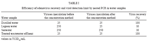
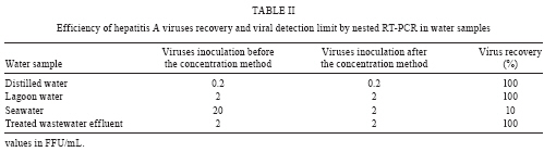
2009 Code Number: oc09202 ABSTRACT Human adenoviruses (HAdV) and hepatitis A virus (HAV) are shed in the faeces and consequently may be present in environmental waters, resulting in an increase in pathogen concentration that can affect water quality and human health. The aim of this study was to evaluate an adsorption-elution method which utilizes negatively charged membrane HA to determine the efficient recovery of HAdV and HAV from different water matrices and to combine this procedure with a qualitative molecular method (nested RT-PCR and nested PCR). The best efficiency recovery was achieved in distilled water and treated wastewater effluent (100%) for both viruses and in recreational lagoon water for HAV (100%). The efficiency recovery was 10% for HAdV and HAV in seawater and 10% for HAdV in lagoon water. The viral detection limit by nested PCR for HAV in water samples ranged between 20-0.2 FFU/mL and 250 and 25 TCID50/mL for HAdV. In conclusion, these results suggest that the HA negatively charged membranes vary their efficiency for recovery of viral concentration depending upon the types of both enteric viruses and water matrices. Key words: virus recovery - negatively charged membrane - environmental waters - hepatitis A virus - adenovirus - PCR Human enteric viruses are excreted in the faeces of infected patients in high concentrations and transmitted mainly by the faecal-oral route via contaminated food and water. Viruses are the major cause of water-related disease and have been estimated to cause about 30-90% of gastroenteritis cases worldwide (Bosh et al. 2008). Enteric viruses represent diverse and commonly studied groups of enteric viruses belonging to the families Picornaviridae [polioviruses, enteroviruses, coxsackieviruses, hepatitis A virus (HAV) and echoviruses], Adenoviridae (adenoviruses), Caliciviridae (noroviruses and saporovirus), Astroviridae (astroviruses) and Reoviridae (rotaviruses). Enteric virus groups are considered to be emerging waterborne pathogens based on their cellular and molecular structures that make them resistant to current water treatment processes (Fong & Lipp 2005). Traditionally, bacterial indicators, such as faecal coliforms, have been used as indicators of water quality; however, it has been clearly established worldwide that bacterial standards do not always reveal the presence of viruses in environmental waters (Formiga-Cruz et al. 2002, Pusch et al. 2005). This fact is due mostly to the different physical/chemical properties of viruses and bacteria (Hsu et al. 2007). In recent years, many researchers have demonstrated the presence of human enteric viruses in several sources of water samples such as raw sewage (Formiga-Cruz et al. 2005), treated sewage (Harwood et al. 2005), river water (Borchardt et al. 2004) seawater (Katayama et al. 2002) and tap water (Lee et al. 2005), using molecular amplification techniques (PCR). PCR has become a major tool for detection and various types of viruses have been isolated in surface water by PCR, including fastidious viruses. PCR can also contribute to epidemiological studies because it is capable of differentiating specific viruses and also different genotypes of the same virus through the use of specific primers (Kinsgley & Richard 2001). Because usually only a few viral particles are present in water samples, it is necessary to concentrate the viruses from a large volume of water. Different viral filtration methods, such as cartridge filters (electropositive or electronegative), associated with conventional viral isolation in cell cultures and/or molecular methods for virus detection have been developed to collect and concentrate viral particles from water samples (Fong & Lipp 2005). However, viral recoveries from various types of water are not always similar due to the fact that adsorption of viruses to the charged membranes may be influenced by salts, multivalent cations or acid conditions (Haramoto et al. 2005, 2006, 2007, Hsu et al. 2007, Victoria et al. 2009). Katayama et al. (2002) described an adsorption-elution method followed by ultrafiltration for virus detection in water samples using poliovirus as a model. This procedure included the addition of MgCl2 to water samples to adsorb viruses onto negatively charged membranes, an essential step that permits viral retention on the membrane. Victoria et al. (2009) evaluated this adsorption-elution method using different MgCl2 concentrations to achieve the best virus recovery of norovirus and astrovirus in water samples. In our study we evaluated the efficiency of recovery of HAV and human adenoviruses (HAdV) by applying the adsorption-elution method using an HA negatively charged membrane with minor modifications (Katayama et al. 2002), followed by molecular amplification (nested RT-PCR and nested PCR) for virus detection in four different kinds of waters (seawater, lagoon water, treated wastewater effluent and distilled water). MATERIALS AND METHODS Cells and viruses - FRHk-4 cells (rhesus kidney-derived cells) and Hep-2 cells (a continuous line of human oropharyngeal carcinoma) were obtained from the American Type Culture Collection (ATCC®, The Global Bioresource Center, USA). Cells were cultured in a CO2 atmosphere at 37ºC, in Eagle's minimal essential medium supplemented with 10% foetal bovine serum (GIBCO/BRL, Life Technologies do Brasil Ltda, São Paulo, SP, Brazil), streptomycin (100 μg/mL), penicillin G (100 U/mL) and amphotericin (0,025 μg/mL) (Gibco-BRL). HAV (strain HM 175) was propagated in FRHk-4 cells as reported (Cromeans et al. 1987) and HAdV5 (genogroup C, serotype 5) in Hep-2 cells (Bardell 1983). For determination of virus titers, an indirect immunofluorescence assay was used for HAV, as previously described (Barardi et al. 1999), and the TCID50 assay was the method of choice for HAdV5 (Reed & Muench 1938). Water samples - Five hundred millilitres of each water sample (seawater, recreational lagoon water, treated wastewater effluent and distilled water) were collected from Florianópolis city in South Brazil. All water samples were autoclaved at 120°C for 30 min before the standardization of the method. These matrices were selected in order to ascertain the recovery of this method in different environmental waters to further apply for the testing of field samples. Distilled water was included as the "gold standard" for the method (inhibitor free) because different inhibitory compounds can be present in environmental samples. Virus concentration method - In order to evaluate the efficiency of the method for viral recovery from different water matrices and to define the limit of sensitivity for virus detection using molecular methods, 500 mL of distilled water, lagoon water and treated wastewater effluent were spiked with HAdV (107 TCID50/mL) and HAV (3x10⁴ FFU/mL) and the seawater was spiked with with HAdV (108 TCID50/mL) and HAV (3x10⁵ FFU/mL). All water samples were spiked in triplicate, with inocula of the same viruses before and after (positive control) the method for concentration of virus. For each matrix of water, a negative control without spiking of virus was performed in order to verify the absence of any natural contaminants. Concentration of viruses in water was performed by adsorption onto an electronegative membrane and subsequent elution, as described by Katayama et al. (2002) with minor modifications. Briefly, an HA (mixed cellulose esters) negatively charged membrane (Nihon Millipore®, Tokyo, Japan) with a pore size of 0.45 μm and 142 mm diameter was placed into a vacuum pump and viruses were then adsorbed in the presence of 25 mM MgCl2 (the exception to this was seawater). The membrane was rinsed with 350 mL of H2SO4 (0.5 mM, pH 3.0) to elude the cations and subsequently treated for 10 min with 10 mL of NaOH (1.0 mM, pH 10.5) to allow the elution of viruses. The filtrate was neutralized with 50 μL of 50 mM H2SO4 and 100X TE buffer (pH 8.0) and then immediately ultrafiltered using a Centriprep Concentrator 50® system (Nihon Millipore®, Tokyo, Japan) at 1500 g for 10 min at 4ºC to obtain a final volume of 2 mL. Four hundred microliters of this 2 mL was further used for nucleic acid extraction. Nucleic acid extraction - Viral nucleic acids were extracted by a procedure described by Boom et al. (1990). This procedure is a simple and inexpensive alternative for nucleic acid extraction and it uses guanidinium thiocyanate for adsorption of the nucleic acids to silica particles. In order to reduce the presence of PCR inhibitors and verify the sensitivity of the nested PCR, RT-PCR or nested RT-PCR, prior to nucleic acid extraction, a 10-fold serial dilution of each concentrated seeded water sample was performed in a sterile (autoclaved) water sample. For instance, the seeded Lagoon water was diluted in sterile Lagoon water and so forth. The viral detection limit was considered to be the highest virus dilution that demonstrated a positive result. Nested reverse transcription-PCR and nested PCR to detect HAV and HAdV in water samples - Reverse transcription and genome amplification were performed using random and specific primers. Random hexamers primers were purchased from Promega (Brazil) and were used for cDNA synthesis. Briefly, a 5.0 μL aliquot of RNA was heated at 99°C for 5 min, followed by quick chilling on ice for 2 min. The denatured RNA was added to a mixture containing random primers, 50 mM Tris-HCl, pH 8.4, 75 mM KCl, 0.5 mM of each dATP, dCTP, dTTP and dGTP, 20U of RNAse inhibitor and 100 U of M-MLV reverse transcriptase (all reagents were purchased from Promega, Brazil), in a 25 μL total volume. Reverse transcription of viral genomic RNA was carried out at 37°C for 60 min. HAV RNA was detected in sewage sludge and wastewater samples by nested RT-PCR, using the oligonucleotide primer pairs F6 (+) and F7 (-), which amplifies a 392 bp fragment, suitable to amplify all HAV genotypes ("universal primers"). Internal primers were F8 (+) and F9 (-), which amplifies a 247 bp fragment (de Paula et al. 2004). AdV DNA was detected in samples using the oligonucleotide primer pairs hexAA 1885 /hexAA 1913 and nexAA 1893/nexAA 1905 described by Allard et al. (1992). The expected size of the PCR product was 300 bp and 142 bp for nested PCR. Amplified fragments of HAdV and HAV were visualized by standard gel electrophoresis of 10 μL of final reaction mixture in 1% agarose gels stained with ethidium bromide (1 μg/mL). Quality control - To avoid the number of false positives resulting from carryover contamination of amplified virus particles or viral nucleic acid, separate areas and equipment were used for each stage of the process. Negative controls (non spiked autoclaved distilled water) and positive controls (virus suspensions) were included with each set of test samples and used throughout the nucleic acid extraction and the nested PCR and nested RT-PCR assays. Additional blank controls containing the same reaction mixture except for the nucleic acid template were incorporated alongside all PCR assays. RESULTS Table 1 and 2 list the recoveries of HAdV and HAV, respectively, from different kinds of environmental waters. We obtained the same viral detection limit for the independently carried out triplicates for all samples. The negative control, without virus spiking, was negative by nested RT-PCR for HAV and nested PCR for HAdV. Viral recoveries were calculated based on the following statement: water samples spiked with viruses after the concentration method were considered positive controls, indicating 100% virus recovery; water samples spiked before the concentration method with the same viral inocula were the tested samples from which the recovery was calculated. The best recovery was achieved in distilled water and treated wastewater effluent (100%) for both viruses and in the lagoon water for HAV (100%). The lowest efficient recovery (10%) obtained was in seawater for both viruses and in lagoon water for HAdV. The viral detection limit by nested RT-PCR for HAV in water samples ranged between 0.2 (positive control) and 20 FFU/mL. Applying the nested PCR method, the detection limit of HAdV was 25 (positive control) and 250 TCID50/mL. DISCUSSION In Brazil, few studies have been developed in order to evaluate the presence of human enteric viruses in water samples using the adsorption-elution method with an HA negatively charged membrane (Villar et al. 2006, De Paula et al. 2007, Guimarães et al. 2008, Miagostovich et al. 2008). A highly sensitive technique with high recuperation efficiency for virus detection is needed in order to ascertain the presence of these viruses in environmental samples. The recovery efficiency of the virus concentration method based on the use of an electronegative filter was previously evaluated for poliovirus (Katayama et al. 2002), noroviruses and sapoviruses from a sewage treatment plant (Haramoto et al. 2006, 2008), HAV (Villar et al. 2006) and astrovirus and norovirus (Victoria et al. 2009) from mineral water, tap water, river water and seawater. In this study, HAdV and HAV were used as a model for enteric viruses in order to assess the recovery efficiency of an adsorption-elution procedure from distilled water, seawater, lagoon water and treated wastewater effluent, using a combination of qualitative molecular methods (nested RT-PCR and nested PCR). It should be noted that the recovery efficiencies estimated are purely for the purpose of orientation. The efficiency of a concentration method depends on many variables, such as the quantity of virus present in the sample, the nature and volume of the sample etc. (Albinana-Gimenez et al. 2009a). The recovery efficiency calculated from the method employed in this study is based on the viral detection limit of the positive control, which consisted of water samples seeded after the filtration step (100%) and water samples seeded before the filtration step. Nested PCR and nested RT-PCR results in this study showed clear differences between environmental samples for HAdV and HAV. The lowest recovery efficiency (10%) was found in seawater (Table 1, 2) for HAdV and HAV. These results are in agreement with findings of previous studies that have demonstrated a low recovery yield from seawater (Villar et al. 2006, Victoria et al. 2009). Researchers studying viral detection in seawater in previous studies did not add salts to the sample, assuming that natural salts within the sample were sufficient to adsorb viruses onto the membrane (Pallin et al. 1997, Katayama et al. 2002). Victoria et al. (2009) demonstrated a low efficiency recovery of human astrovirus and norovirus from seawater when the samples were not treated with MgCl2, achieving the highest recovery treating the samples with 25 mM or 50 mM of MgCl2. Despite the low recovery yield for seawater in this study, we were able to obtain a detection limit by nested PCR of 250 TCID50/mL and 20 FFU/mL for HAdV and HAV respectively, which is in accordance with previous studies (Katayama et al. 2002, Villar et al. 2006). We were able to achieve the same detection limit and 100% recovery efficiency for the distilled water and the treated wastewater effluent by comparing test samples with the positives controls of each sample for both viruses (25 TCID50/mL for HAdV from distilled water and wastewater and 0.2 and 2 FFU/mL for HAV from distilled water and wastewater, respectively). Molecular detection by qualitative PCR of adenoviruses, HAV and enteric viruses, when correctly applied, provides reliable data about the presence of these viruses in the environment. However, inhibition of amplification reactions by substances present in the samples, such as humic acids and metals, has been observed frequently in water samples (Albinana-Gimenez et al. 2009a). In addition, PCR is often unable to discriminate between infectious and inactivated virus (Reynolds et al. 1996, Ko et al. 2003). To improve the sensitivity of the detection methods, other molecular based protocols, such as real-time-PCR associated with virus viability, can also be used (Albinana-Gimenez et al. 2009b). Sensitive and practical methods for detecting enteric viruses in environmental waters are needed to determine the public health significance of these pathogens in the event of waterborne outbreaks of acute gastroenteritis. In this study, qualitative nested PCR and nested RT-PCR methods were capable of detecting HAdV DNA and HAV RNA in the water samples tested. In fact, improvements in the virus recovery method, such as treating seawater samples with MgCl2, must be made in order to obtain better recovery in this particular water sample. Nevertheless, the method was more efficient for the other kinds of water tested in this study. These results demonstrate the impact of the concentration method for the detection of HAV and HAdV by qualitative PCR; as a result, this method can be applied to monitor the presence of virus and the quality of environmental waters. ACKNOWLEDGMENTS To Ms. Doris Sobral Marques Souza, for the technical support in the PCR assays. REFERENCES
Copyright 2009 - Instituto Oswaldo Cruz - Fiocruz The following images related to this document are available:Photo images[oc09202t2.jpg] [oc09202t1.jpg] |
| |||||||||
