 |
| About Bioline | All Journals | Testimonials | Membership | News |
|
||||||
|
||||||
Memórias do Instituto Oswaldo Cruz, Vol. 104, No. 7, Nov, 2009, pp. 975-979 ARTICLES Fine structure of Henneguya hemiodopsis sp. n. (Myxozoa), a parasite of the gills of the Brazilian teleostean fish Hemiodopsis microlepes (Hemiodontidae) Carlos AzevedoI, +; Graça CasalII, III; Ivete MendonçaIV; Edilson MatosV IDepartment
of Cell Biology, Institute of Biomedical Sciences Financial support: Eng. A Almeida Foundation (Porto, Portugal), CNPq, CAPES Received 2 April
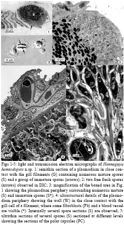
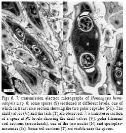
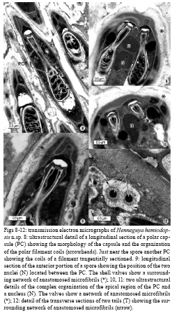
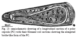
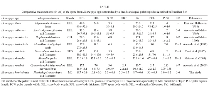
2009 Code Number: oc09203 ABSTRACT A fish-infecting myxosporean, Henneguya hemiodopsis sp. n., found infecting the gills of Hemiodopsis microlepis and collected from the Poty River near the city of Teresina, Brazil, was described based on ultrastructural studies. The parasite occurred within large whitish polysporic plasmodia (up to 200 μm in diameter) containing asynchronous developmental sporogonic stages, mainly mature spores. The spores measured 19.7 ± 0.9 μm in total length (n = 30) and the ellipsoidal spore body was 10.8 ± 0.5 μm long, 3.3 ± 0.4 μm wide and 2.5 ± 0.5 μm thick. The spores were composed of two equal shell valves adhering together along the straight suture line, with each valve having equal-sized caudal tapering tails measuring 8.7 ± 0.6 μm in length. The spores were surrounded by a thin anastomosed network of microfibrils, more evident on the tails. There were two symmetric elongated bottle-like polar capsules 3.5 ± 0.3 μm long and 1.0 ± 0.2 μm wide, each with a polar filament with five to six coils. Given the morphological and ultrastructural differences from previously described parasites and the specificity of the host species, we propose a new species, named H. hemiodopsis sp. n. Key words: ultrastructure - Henneguya hemiodopsis sp. n. - Myxozoa - parasite - gill - Brazilian fish The South American Continent contains one of the biggest hydrographic networks in the world, in which a great variety of ictiofauna species reside. Since the first description of Henneguya Thélohan, 1892 (Lom & Dyková 2006), the second largest genus of the Myxobolidae family, many species have been reported, mainly parasitising freshwater fishes throughout the world. However, the number of myxozoan parasites described from Brazil is very low, especially considering that the country has one of the most diverse freshwater fish populations in the world with about 8,000 species (Cellere et al. 2002). Thirty-six species have been described from Brazilian fauna based on light microscopy micrographs and diagrammatic illustrations (Jakowska & Nigrelli 1953, Kent & Hoffman 1984, Gioia et al. 1986, Martins et al. 1988, Gioia & Cordeiro 1996, Martins & Souza 1997, Barassa et al. 2003a, b, Eiras et al. 2004a, b, 2008, 2009, Martins & Onaka 2006, Abdallah et al. 2007). Recently, ultrastructural studies of developmental stages and mature spores supported the classification of some new species of the genus Henneguya (Rocha et al. 1992, Azevedo & Matos 1995, 1996, 2002, 2003, Azevedo et al. 1997, 2008, Casal et al. 1997, 2003, Vita et al. 2003, Adriano et al. 2005, Matos et al. 2005). In the present paper, we report light and electron microscopy-based data on the mature spores of a new parasite, designated herein as Henneguya hemiodopsis sp. n., infecting the gills of a teleostean fish of some economic importance found in a river in North-Eastern Brazil. MATERIALS AND METHODS Fifty specimens (18 males and 32 females) of the freshwater teleost Hemiodopsis microlepis Kern, 1858 (Teleostei: Characiformes: Hemiodontidae) (Brazilian common name flexeiro), endemic to this region, were recently collected from the Poty River (05º05'S 42º48'W) near the city of Teresina [state of Piauí (PI)], Brazil. The fish were lightly anaesthetised with MS 222 (Sandoz Laboratories) and dissected and the gills, containing several whitish cysts (cyst-like plasmodia), were removed from the gill lamellae and examined with a light microscope equipped with Nomarski interference-contrast (DIC) optics. For ultrastructural studies, small fragments of the gill lamellae containing cyst-like plasmodia were excised and fixed in 3% glutaraldehyde in 0.2 M sodium cacodylate buffer (pH 7.2) at 4°C for 10 h. After being rinsed overnight in the same buffer at 4ºC and post-fixed in 2% osmium tetroxide in the same buffer for 3 h at 4°C, the fragments were dehydrated through an ascending ethanol series followed by propylene oxide and embedded in Epon. Semi-thin sections were stained with methylene blue-Azure II and observed by DIC optics. Ultra-thin sections were double-stained with aqueous uranyl acetate and lead citrate and observed under a JEOL 100CXII transmission electron microscope (TEM) operated at 60 kV. RESULTS Of the 50 adult specimens of H. microlepis (Teleostei: Hemiodontidae) examined, 32 (64%) contained gills parasitised by an organism identified as follows: Phylum Myxozoa Grassé, 1970; Class Myxosporea Bütschli, 1881; Order Bivalvulida Shulman, 1959; Family Myxobolidae Thélohan, 1892; genus Henneguya Thélohan, 1892, according to the classification proposed by Lom and Dyková (2006). The morphological and ultrastructural data, mainly the shape and dimension of the spores and polar capsules (PC), as well as the polar filament coils and arrangement, indicate that this parasite is a new species, herein named H. hemiodopsis. For description of the plasmodium and mature spores, light microscopy (DIC) (Figs 1-3), TEM (Figs 4 and 5, Figs 6 and 7, and Figs 8-12) and a schematic drawing (Fig. 13) were used. H. hemiodopsis sp. n. (Figs 1-3) Description - Vegetative stages: whitish round-shaped polysporic plasmodia (cyst-like plasmodia), measuring up to 200 μm contained numerous mature and immature spores, indicating asynchronous development (Figs 3,4). The plasmodium wall was formed by a dense material that was in contact with the epithelial gill cells. The zone of contact contained several fibroblasts, collagen fibrils and some capillaries (Fig 4). The spores were ellipsoidal with a total length of 19.7 ± 0.9 μm (n = 30), body length 10.8 ± 0.5 μm (n = 20), body width (frontal view) 3.3 ± 0.4 μm (n = 20) and body thickness (side view) 2.5 ± 0.5 μm (n = 15). The two valves were symmetrical and thin and each was prolonged by a tapering tail 8.7 ± 0.6 μm (n = 20) long (Figs 2-5, 6-7). Elongated bottle-like PC localised side by side in the anterior pole of the spore were equal in size, measuring 3.5 ± 0.3 μm in length (n = 25) by 1.0 ± 0.2 μm (n = 25) in width (Figs 6-7, 8-9). The apical pore of the bottle-like PC was closed by a dense stopper and an electron-dense ring surrounded the neck of the bottle-like PC. The neck of the bottle-like PC measured 1.7 ± 0.4 μm (n = 12) in length and ~0.5 μm in diameter (Figs 8-10). Inside the PC, the isofilar polar filament was coiled with 5-6 turns oblique to the longitudinal axis (Figs 8, 9). The internal matrix of the polar filaments was denser than the surrounding matrix of the PC (Figs 8, 9). The spores presented numerous ramified and anastomosed microfibrils at the surface (Figs 9-11), more evident in the tails (Figs 6, 7, 12). Type host - H. microlepis Kner, 1858 (Teleostei: Characiformes: Hemiodontidae) (12-17 cm in length on average). Site of infection: whitish cysts (cyst-like plasmodia) were found in secondary gill lamellae. Prevalence and intensity - Thirty-two out of 50 (18 males and 32 females) adult fish (64%) were parasitised, with similar rates in both sexes. Type locality - Poty River (05º05'S 42º48'W), near the city of Teresina (PI). Type data and depository - A glass slide containing semi-thin sections of mature spores of the hapantotype was deposited in the International Protozoan Type Slide Collection at the Smithsonian Institution Washington, DC 20560, USA, with acquisition USNM 1123997. Etymology - The specific epithet derives from the generic name of the host (Hemiodopsis). Histopathology - Our observations showed that the infected gill lamellae were associated with an increased number of fibroblasts just near the zone of contact of the epithelial gill with the wall of the cyst-like plasmodia, with local atrophy and deformation of the gill lamellae (Fig. 4). DISCUSSION The morphology of the myxosporean described in the present paper has the distinguishing features of the genus Henneguya Thélohan, 1899 (i.e., ellipsoidal spore body formed by two shell valves, each with an elongated tail and two PC internally) (Lom & Dyková 2006). The morphological and ultrastructural aspects of the mature spores, as well as the host specificity and localization of the infection with this parasite, were compared to those of other myxosporidian species of the genus Henneguya from different geographical areas, mainly species that parasitise Brazilian freshwater fishes. Recently, 34 valid Henneguya species described from Brazilian fish were summarised in a table containing the spore measurements (Eiras et al. 2008). Two new species, more recently described, must be added to this number: Henneguya rondoni, described on the basis of ultrastructural data (Azevedo et al. 2008) and Henneguya corruscan, described based on light micrographs and a drawing (Eiras et al. 2009). Comparing the morphology and the dimensions of the spores of these species, we observe that H. hemiodopsis sp. n. differs mainly in the shape and size of the body, tails and PC as well as in the arrangement of the polar filament coils. All of the previously described species have different morphological characteristics and host specificity compared to the presently described species. On the other hand, the presence of an external adherent sheath surrounding the spore body wall and tails has been used as an important morphological taxonomic characteristic to distinguish Henneguya spp. Taking into account this structure, we summarise in Table the spore measurements of Henneguya spp, whose spores are surrounded by a sheath. Compared to the proposed new species (H. hemiodopsis), several differences were observed in the external sheath of other Henneguya spp that contain this structure (Table). Whereas in H. hemiodopsis sp. n. the spores were surrounded by a thin anastomosed network of microfibrils, in Henneguya theca, Henneguya adherens, Henneguya testicularis, Henneguya rhamdia and H. rondoni, the spores were surrounded by a hyaline homogenous structure and in Henneguya malabarica and Henneguya striolata, the sheath was constituted by granulo-fibrilar structures organised into a complex ramified network. This inflammatory reaction seems to be of the same type as that observed and described in other Brazilian fishes (Martins et al. 1988, Adriano et al. 2005). In conclusion, on the basis of the morphological differences in the size and shape of the spore and PC and the number and arrangement of the polar filament coils, we think that there are sufficient arguments for the establishment of a new species, named H. hemiodopsis sp. n. ACKNOWLEDGMENTS To the technical assistance of Joana Carvalheiro and João Carvalheiro. REFERENCES
Copyright 2009 - Instituto Oswaldo Cruz - Fiocruz The following images related to this document are available:Photo images[oc09203f6-7.jpg] [oc09203t.jpg] [oc09203f8-12.jpg] [oc09203f13.jpg] [oc09203f1-5.jpg] |
| |||||||||
