 |
| About Bioline | All Journals | Testimonials | Membership | News |
|
||||||
|
||||||
Memórias do Instituto Oswaldo Cruz, Vol. 105, No. 1, 2010, pp. 45-51 ARTICLES Antimycobacterial neolignans isolated from Aristolochia taliscana Rosalba León-DíazI; Mariana MeckesI; Salvador Said-FernándezII; Gloria Maria Molina-SalinasII; Javier Vargas-VillarrealII; Javier TorresIII; Julieta Luna-HerreraIV; Adelina Jiménez-ArellanesI, + IUnidad
Investigación Médica en Farmacología de Productos Naturales Financial support: Instituto Mexicano del Seguro Social (FOFOI FP-2003-009, 2005/1/I/102), CONACYT (to RLD) Received 4 June
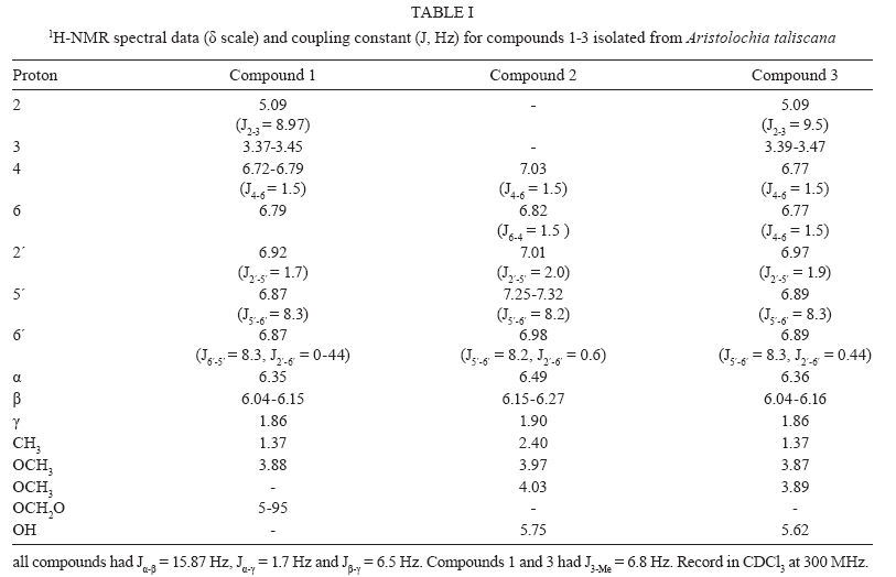
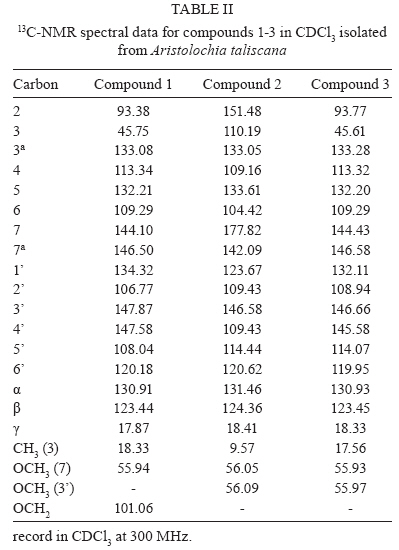
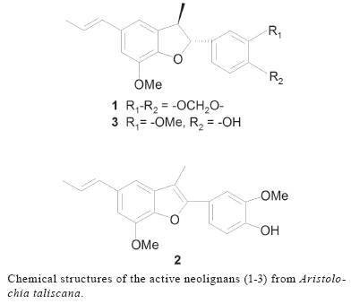
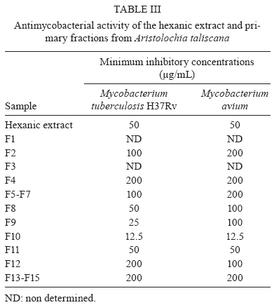
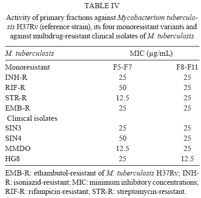
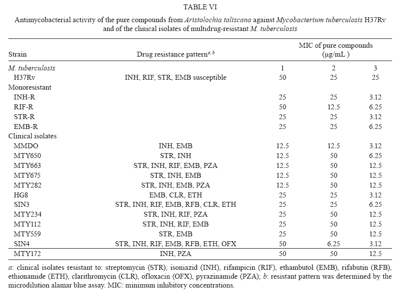
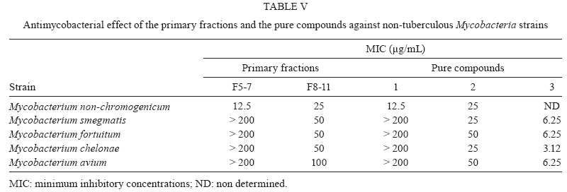
2009 Code Number: oc10006 ABSTRACT Tuberculosis (TB - Mycobacterium tuberculosis) is an ancient infectious disease that has appeared once again as a serious worldwide health problem and now comprises the second leading cause of death resulting from a single infection. The prevalence of multidrug resistance (MDR) TB is increasing and therapeutic options for treatment are not always accessible; in fact, some patients do not respond to the available drugs. Therefore, there is an urgent need to develop novel anti-TB agents. The aim of the present study was to screen extracts of Aristolochia taliscana, a plant used in traditional Mexican medicine to treat cough and snake bites, for antimycobacterial activity. The hexanic extract of A. taliscana was tested by microdilution alamar blue assay against Mycobacterium strains and bioguided fractionation led to the isolation of the neolignans licarin A, licarin B and eupomatenoid-7, all of which had antimycobacterial activity. Licarin A was the most active compound, with minimum inhibitory concentrations of 3.12-12.5 μg/mL against the following M. tuberculosis strains: H37Rv, four mono-resistant H37Rv variants and 12 clinical MDR isolates, as well as against five non-tuberculous mycobacteria (NTM) strains. In conclusion, licarin A represents a potentially active anti-TB agent to treat MDR M. tuberculosis and NTM strains. Key words: antimycobacterial neolignans - A. taliscana - M. tuberculosis H37Rv - MDR M. tuberculosis - non-tuberculous mycobacteria Medicinal plants are an important natural source of novel leads in the field of antimycobacterial therapeutics (Cantrell et al. 2001, Copp & Pearce 2007, Gutierrez-Lugo & Bewley 2008). According to ethnobotanical data, some species of Aristolochia, such as Aristolochia elegans and Aristolochia grandiflora have been widely utilized in Mexican traditional medicine to treat cough (Diaz 1976). A preliminary biological evaluation of the hexanic extract from Aristolochia taliscana Hook roots showed that it possessed an in vitro antimycobacterial effect against Mycobacterium tuberculosis H37Rv and Mycobacterium avium [minimum inhibitory concentrations (MIC's) = 50 μg/mL]. The plant is commonly known in Mexico as guaco or raíz de guaco and neolignans with antiprotozoal activity have already been identified in the species (Enriquez et al. 1984, Abe et al. 2002). In recent years, the number of patients with tuberculosis (TB) has increased rapidly due, in part, to the appearance of multidrug-resistant (MDR) and extensively drug-resistant (XDR) strains in both developing and developed countries. One-third of the world's population is currently infected with M. tuberculosis and approximately 10% of these cases will develop clinical manifestations, particularly those patients with compromised immunological systems. The AIDS/HIV pandemic has contributed to the worsening of the problem; in fact, about 30% of registered mortality has been associated with TB, especially in developing countries (Jain & Mondal 2008, Rivers & Mancera 2008). Patients with AIDS are also susceptible to becoming infected with non-tuberculous mycobacteria (NTM) such as M. avium (Rodriguez et al. 2006). It is estimated that the worldwide prevalence of MDR-TB is about 3.2% and that 6.6% of these cases are XDR-TB (Rivers & Mancera 2008). The emergence of XDR-TB strains constitutes a serious health problem because, at present, there is no pharmaceutical alternative for treating patients infected with such strains (Tomioka 2006, Zager & McNerney 2008). Consequently, novel drugs to treat or prevent the disease are urgently needed (O´Brien & Spigelman 2005, Gutierrez-Lugo & Bewley 2008). The aim of the present paper was to isolate and structurally characterize A. taliscana hexane-extract compounds that possessed activity against M. tuberculosis H37Rv, mono-resistant variants of H37Rv, MDR M. tuberculosis clinical isolates and NTM. MATERIALS AND METHODS Plant materials - A. taliscana roots were purchased at a medicinal plant market in city of Mexico, Mexico. The plant material was compared with the botanical specimen deposited at the Herbarium of the Instituto Mexicano del Seguro Social and a voucher was deposited under code 1106. Extraction and isolation - Powdered air-dried roots (1.5 kg) were macerated (3 × 48 h) with 12 L of n-hexane. The extract was filtered and evaporated in vacuo to yield 33 g of the crude extract. Open column chromatography (CC) was performed employing the silica gel 60 GF254 (70-230 mesh, Merck) as the stationary phase and silica gel 60 F254 pre-coated aluminium plates (0.2 mm, Merck) for analytical and preparative thin-layer chromatography (TLC) analysis. Spots were visualized by spraying with a 10% solution of H2SO4 followed by heating the plates at 100°C. For silica gel CC, the extract (15 g) was fractionated by eluting with n-Hex: CHCl3(100→0) and CHCl3:MeOH (100→0); 72 fractions (250 mL each) were obtained. The primary fractions (F1-F15) were combined according to the results from the TLC analysis. All primary fractions were tested for antimycobacterial activity. From the active fraction F5-F7, 975 mg of white needles with a melting point (m.p.) of 82-86ºC (lit. m.p. 89-90°C) crystallized. The compound was identified as licarin B (1) and was also detected in fractions F8-F11. Fractions F8-F11 (2.5 g) were re-chromatographed on CC using silica gel (75 g) with a solvent gradient of n-Hex:CHCl3 (100→0) and CHCl3:MeOH (100→0). This process yielded 14 secondary fractions (FA-FN) of 150 mL each. Secondary fraction FD yielded 40 mg of 1 and fraction FF (400 mg) was re-chromatographed in CC and eluted with n-Hex: CHCl3(100→0) and CHCl3:MeOH (100→0) to obtain eight tertiary fractions Fa-Fh. The isolated maroon-coloured powder (80 mg) from Fc-Fe was characterized as eupomatenoid-7 (2) with an m.p. of 100-104ºC (lit. m.p. 105-106°C). The secondary fraction FJ (300 mg) was further re-chromatographed on CC utilizing n-Hex:CHCl3 and CHCl3 as elution systems. Nine 50 mL tertiary fractions (Fa'-Fi') were obtained. From Fb', 196 mg of a white product was obtained by crystallization with an m.p. of 107-110°C (lit. m.p. 133-134°C), which was identified as licarin A (3). Chemical characterization - Chemical characterization of the isolated neolignans was determined by 1H-NMR (Eclipse 300 Jeol, 300 MHz) and 13C-NMR (Variant Unity, 300 MHz) using tetramethylsilane as an internal standard in CDCl3. Electron impact-mass spectra were obtained on a Jeol AX-505 HA mass spectrometer at 70 eV. Infrared (IR) spectra on film over NaCl in a Bruker model Tensor 27 spectrometer, optical rotation in a Perkin Elmer model 345 polarimeter at 25°C using a sodium lamp (589 nm) and m.p. in a Fisher-Johns apparatus. All the spectroscopic data (1H and 13C-NMR) of each compound were compared with those previously reported in the literature (Enriquez et al. 1984) and are described in Tables I, II. Licarin B (1) - White needles soluble in CHCl3, m.p. 82-86°C, [α]D25°C = -0.262 (MeOH), IR: ½max 2900, 1600 and 1050-1200. IE-MS: m/z (rel. int.) 324 [M+ (100)], 309 (12), 293 (8), 278 (28), 202 (6), 135 (20), 121 (8), 91 (7), 77 (14) and 46(5). Eupomatenoid-7 (2) - Maroon-coloured powder, soluble in CHCl3, m.p. 100-104°C, [α]D25°C = -0.280 (MeOH), IR: ½max 3429, 2937, 2849, 1725, 1604, 1513, 1452, 1371, 1267, 1221, 1147 and 1056. IE-MS: m/z (rel. int.) 324 (100), 309 (20), 293 (15), 123 (6), 91 (9), 77 (5) and 31 (15). Licarin A (3) - White powder, soluble in CHCl3, m.p. 107-110°C, [α]D25ºC = -0.15 (MeOH), IR: ½max 3541, 2938, 1673, 1608, 1496, 1269 and 1143. IE-MS: m/z (rel. int.) 326 (100), 311 (20), 308(7), 295 (5), 202 (10), 123 (8), 91 (10), 77 (8) and 31 (25). Mycobacterium strains - The following mycobacteria from the American Type Culture Collection (ATCC) were used: M. tuberculosis H37Rv (27294); mono-resistant strains: H37Rv isoniazid-resistant (35822), H37Rv streptomycin-resistant (35820), H37Rv rifampicin-resistant (35838) and H37Rv ethambutol-resistant (35837); M. avium (35717) and Mycobacterium smegmatis (35798). In addition, drug-resistant M. tuberculosis clinical isolates (12 strains) obtained from Mexican patients with pulmonary disease were also tested. Drug-resistant M. tuberculosis clinical isolates were selected based on their drug susceptibility patterns against antimycobacterial drugs employing the microdilution alamar blue assay (MABA) test. In addition, the following clinical or environmental non-TB mycobacteria isolates were included: Mycobacterium chelonae, Mycobacterium fortuitum and Mycobacterium non-chromogenicum. The strains were cultured in Middlebrook 7H9 broth supplemented with 10% OADC enrichment (Becton Dickenson, USA) at 37°C until a logarithmic growth phase was achieved. M. tuberculosis and non-TB mycobacteria were diluted in 7H9 at the ratios of 1:20 and 1:50, respectively. Bacterial suspensions were fresh when utilized in the assays. Antimycobacterial assay - Extracts, fractions and the pure compounds were evaluated by the previously described MABA assay (Jimenez-Arellanes et al. 2003, 2007). Briefly, samples were dissolved in dimethyl sulfoxide (DMSO) (20 mg/mL) under sterile conditions. Serial two-fold dilutions of each sample (range, 100-3.12 μg/mL) were prepared to a final volume of 100 μL with 7H9 broth and 100 μL of each mycobacterium suspension was added to 96-well sterile microplates (Nunc). For M. tuberculosis, plates were incubated at 35°C during five days, whereas non-TB mycobacteria were incubated for two days. MIC is expressed as the lowest concentration of the compound that causes 99% inhibition of mycobacterium growth. All assays were run in duplicate and streptomycin (0.5 μg/mL, Sigma), isoniazid (0.06 μg/mL, Sigma) and rifampicin (0.1 μg/mL, Sigma) were utilized as positive controls. Cytotoxicity assay - The assay was carried out in a J774A.1 murine macrophage cell line (ATCC HB-197) using the trypan blue exclusion test. Briefly, purified neolignans were dissolved in DMSO at a concentration of 20 μg/mL. Cells were grown in 24-well plates using DMEM supplemented with 10% foetal bovine serum (FBS) and antibiotics. Immediately prior to testing, monolayers were washed with warm Hanks' balanced salt solution. Serial two-fold dilutions of each compound were prepared in DMEM supplemented with 10% FBS (1-1/16 of MIC against M. tuberculosis H37Rv) and 1 mL/well of each dilution was added. To evaluate cell viability, controls were included in the microplate by adding DMEM media with DMSO; cell viability was determined after a 24-h incubation period. Trypan blue solution was added and the percentage of viable cells was calculated to determine the cytotoxic index (IC50). The assay was run in triplicate. Acute toxicity in mice - Male Balb/c mice (22 ± 2.2 g) were used to determine the acute toxicity parameter following the methodology previously described by Lorke (1983) and according to the guidelines of the local Ethical Committee for Experimentation in Animals. Animals were maintained under standard environmental conditions at 12-h light/dark photoperiods with free access to food and water. Mice were randomly divided into five groups of three animals each. Group 1 received the control vehicle (Tween 20:H2O 2:8), while Groups 2-5 were treated orally with the crude extract at doses of 0.6, 1.0, 1.6 and 2.9 g/kg. The same design was employed to test the most active primary fraction (F8-F11) and the pure compound (licarin A). All samples were solubilized in Tween 20:H2O (2:8) and were intragastrically administered in a volume that was less than 10 mL/kg of body weight. Treatment response was monitored at 1, 2, 4, 6 and 24 h and daily for 14 days, registering any signal of toxicity. At the end of the experimental period, the animals were sacrificed in a CO2 chamber to obtain the internal organs (lung, kidney, heart, spleen and liver) for pathological analysis. RESULTS Chemical characterization of the isolated neolignans - The three neolignans were characterized by comparing spectral data (Tables I, II) with those previously reported in the literature (Enriquez et al. 1984) and the respective molecular structures of the compounds are illustrated in Fig 1. Biological evaluation - As shown in Table III, a MIC of 50 μg/mL was determined for the hexanic crude extract against M. tuberculosis H37Rv and M. avium. Primary fractionation yielded F8-F11 as the most active fractions, with MIC's of 12.5-50 μg/mL against M. tuberculosis H37Rv strains and 12.5-100 μg/mL against M. avium. These fractions, as well as F5-F7, were active against all tested mono-resistant strains of H37Rv and MDR M. tuberculosis clinical isolates (SIN3, SIN4, MMDO and HG8) and the MIC values obtained ranged from 12.5-50 μg/mL (Table IV). In addition, fractions F8-F11 inhibited the growth of NTM as follows: M. non-chromogenicum (MIC = 25 μg/mL) and M. smegmatis, M. chelonae and M. fortuitum (MIC = 50 μg/mL); the fractions were less active against M. avium (MIC = 100 μg/mL). By contrast, fractions F5-F7 were highly active against M. non-chromogenicum (MIC = 12.5 μg/mL) (Table V). Antimycobacterial activity of the pure isolated compounds is shown in Table VI. Licarin B (1) was moderately active against H37Rv and against mono-resistant variants (MICs, 25-50 μg/mL), but was highly active against the majority of MDR M. tuberculosis clinical isolates tested (with MIC values ranging from 12.5-50 μg/mL). Eupomatenoid-7 (2) was active against H37Rv strains (MIC = 25 μg/mL), the four mono-resistant variants of H37Rv and three of the MDR clinical isolates tested (MIC values ranging from 12.5-25 μg/mL). The most clinically relevant activity of this compound (MIC = 6.25 μg/mL) was against an M. tuberculosis clinical isolate (SIN4) that is resistant to first- and second-line drugs (Table VI). Finally, while licarin A (3) exhibited moderate activity against M. tuberculosis H37Rv (MIC = 25 μg/mL), this compound was highly active against all mono-resistant and MDR M. tuberculosis strains tested, with MIC's ranging from 3.12-12.5 μg/mL (Table VI). Clinical isolates with highest sensitivity to this compound included MMDO, HG8 and SIN4. In addition, licarin A (3) inhibited the NTM M. avium, M. smegmatis, M. fortuitum (all with MIC = 6.25 μg/mL) and M. chelonae (MIC = 3.12 μg/mL) (Table V). Cytotoxicity assay of the pure neolignans on murine macrophage J774A.1 cell line yielded values of IC50 = 6.25 μg/mL for licarin A and B and IC50 = 3.12 μg/mL for eupomatenoid-7. The acute toxicity of the crude extract, F8-F11 and its most active component, licarin A, determined in mice was > 1.706 mg/kg. DISCUSSION TB is a severe global health problem and the search for novel therapeutic molecules is a necessity due to the appearance of resistance to the anti-mycobacterial drugs currently in use (Cantrell et al. 2001, O'Brien & Spigelman 2005, Tomioka 2006, Gutiérrez-Lugo & Bewley 2008). Medicinal plants comprise a promising natural source for the discovery of anti-TB drugs and the in vitro activity of several secondary metabolites has already been recognized. At present, 12-demethylmulticauline isolated from Salvia multicaulis (MIC= 0.46 μg/mL), micromolide from Micromelum hirsutum (MIC= 1.5 μg/mL) and (E)-phytol from Leucas volkensii (MIC= 2 μg/mL) are the most highly active compounds reported against M. tuberculosis H37Rv (Cantrell et al. 2001). Unfortunately, little information is available concerning the activity of natural compounds against MDR M. tuberculosis strains (Newton et al. 2002, Gibbons et al. 2003, Luna-Herrera et al. 2007). While the use of Aristolochia species has been discussed extensively because of its content of aristolochic acid (Chen et al. 2007), this toxic compound was not detected in A. taliscana-root hexane extract. Moreover, the LD50 for the hexanic extract determined in mice was > 1706 mg/kg. When evaluating this extract against M. tuberculosis H37Rv and M. avium, moderate in vitro activity (MIC = 50 μg/mL) was determined. Activity against M. avium is of interest because currently, there is a high frequency of TB cases associated with this species in HIV/AIDS cases. Bioguided fractionation of the extract led to the isolation of the previously identified neolignans licarin B, eupomatenoid-7 and licarin A (Enriquez et al. 1984, Abe et al. 2002). While several biological effects (antibacterial, antioxidant, anticancer, trypanocidal, neuroprotective, insecticidal and anti-inflammatory) of these compounds have been reported, to our knowledge, this is the first report on their anti-TB activity (Tsai et al. 2001, Abe et al. 2002, Lee et al. 2004, Ma et al. 2004, 2005, Park et al. 2004, Saleem et al. 2005). Licarin A (LD50 > 1706 mg/kg) displayed the most potent effect against all tested mono-resistant strains of H37Rv and MDR clinical isolates of M. tuberculosis (MIC's ranging from 3.12-12.5 μg/mL). Likewise, licarin A was active against the non-TB mycobacteria M. avium, M. chelonae, M. fortuitum and M. smegmatis (MICs ranging from 3.12-6.25 μg/mL). A drug that is able to inhibit MDR M. tuberculosis and M. avium growth, such as licarin A, would be of extremely high value in the clinic, particularly in cases of HIV/AIDS and MDR/XDR. Lignans are well-known secondary metabolites because of the cytotoxic effect they produce in several cell lines (Tsai et al. 2001, Park et al. 2004, Kong et al. 2005). The cytotoxic activity of licarin A has also been reported against P-388, KB16 and HT-29 cell lines (Tsai et al. 2001) and this activity for licarin B (100 μM) has been described against the human promyelocytic leukaemia HL-60 cells, as it is the compound inactive for caspase-3 activation. Meanwhile licarin A induces an apoptotic effect by means of caspase-3 activation (Park et al. 2004). On the other hand, Lee et al. (2004) reported that licarin A is a potent inhibitor of phospholipase Cγ1 (PLCγ1) with an IC50 = 15.8 ± 1.3 μM and that it exerts antiproliferative effects against three human cancer cell lines [A-549 (lung), MCF7 (breast) and HCT-15 (colon)], suggesting the use of licarin A as a cancer chemotherapeutic and chemopreventive agent (Lee et al. 2004, Park et al. 2004). The cytotoxicity of A. taliscana-isolated neolignans on murine macrophages was IC50 = 3.25-6.25 μg/mL; these values were similar to those determined for the MIC parameter. The results obtained here permit us to suggest further biological evaluation of the effect of licarin A against macrophages infected with MDR M. tuberculosis, to determine the compound's intracellular activity. In conclusion, a low-toxicity neolignan was isolated from the hexane extract of A. taliscana roots, structurally identified as licarin A and shown to be the most active compound against all mycobacteria tested. Licarin A is a new prototype molecule that exerts a relevant biological effect against the mycobacteria responsible for MDR-TB, a pandemic that is increasing at present and represents a serious health problem worldwide. In vivo experimental studies are in progress to establish the anti-TB potential of this compound. REFERENCES
Copyright © 2010 - Instituto Oswaldo Cruz - Fiocruz The following images related to this document are available:Photo images[oc10006t4.jpg] [oc10006t6.jpg] [oc10006t3.jpg] [oc10006t1.jpg] [oc10006f1.jpg] [oc10006t2.jpg] [oc10006t5.jpg] |
| |||||||||

/photo/oc10006t5.jpg)