 |
| About Bioline | All Journals | Testimonials | Membership | News |
|
||||||
|
||||||
Memórias do Instituto Oswaldo Cruz, Vol. 105, No. 1, 2010, pp. 73-78 ARTICLES Comparison of HPV genotyping by type-specific PCR and sequencing Nara de Oliveira CarvalhoI, +; Dora Méndez del CastilloI; Carlos PeroneI; José Nélio JanuárioI; Victor Hugo de MeloII; Geraldo Brasileiro FilhoIII INúcleo
de Ações e Pesquisas em Apoio Diagnóstico Financial support: NUPAD, Faculdade de Medicina - UFMG Received 19 July
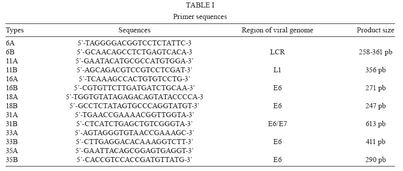
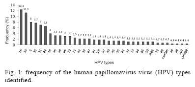
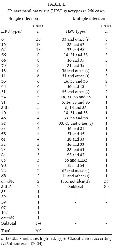
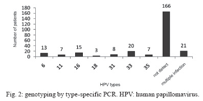
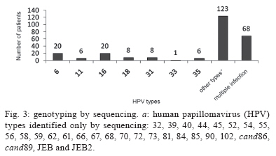
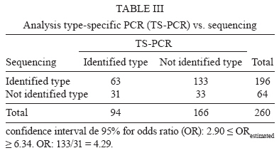
2009 Code Number: oc10011 ABSTRACT Human papillomavirus (HPV) infection is the most common sexually transmitted disease worldwide and there is a strong link between certain high-risk viral types and cervical carcinogenesis. Although there are several typing methods, it is still unclear which test is the best. This study compared the effectiveness of type-specific PCR (TS-PCR) and sequencing, with a focus on their clinical application. A total of 260 cervical samples from HPV-positive patients were tested for types 6, 11, 16, 18, 31, 33 and 35 using TS-PCR and sequencing. The genotype was identified in 36% of cases by TS-PCR and in 75% by sequencing. Sequencing was four times more likely to identify the viral type in positive samples than TS-PCR (p = 0.00). Despite being more effective for virus genotyping, sequencing was unable to identify viral types in multiple infections. Combining both techniques resulted in highly sensitive detection (87% of cases), showing that they are complementary methods. HPV genotyping is an important step in HPV management, helping to identify patients with a higher risk of developing cervical cancer and contributing to the development of type-specific vaccines. Key words: HPV - PCR - sequencing - typing Human papillomavirus (HPV) is the most common sexually transmitted virus in young and sexually active people of both sexes (Sanjose et al. 2007). Anogenital HPVs, which are primarily mucosotropic, are classified as high and low risk, according to their relationship with benign or malignant proliferative lesions (de Villiers et al. 2004). Based on the viral DNA sequence, more than 230 HPV types are known (Haws et al. 2004); 118 genotypes are well-characterised according to biological niche, oncogenic potential and phylogenetic position (de Villiers et al. 2004). Approximately 40 HPV types infect the anogenital region and 15 of them - 16, 18, 31, 33, 35, 39, 45, 51, 52, 56, 58, 59, 68, 73 and 82 - are considered oncogenic or as high risk because they are associated with high grade squamous intraepithelial lesions or cancer. Types 26, 53 and 66 are likely to be carcinogenic, whereas types 6, 11, 40, 42, 43, 44, 54, 61, 70, 72, 81 and candHPV89/Cp6108 are considered to be low risk (Munoz et al. 2003). HPVs have been associated with many proliferative lesions, with condyloma acuminatum being the most common, as well as with different types of cancer, including cervical, vaginal, vulvar, penile, anal, oropharyngeal, buccal cavity and larynx (Bosch et al. 2002, Montaldo et al. 2007). Among them, uterine cervix carcinoma is particularly important due to its high incidence and its high mortality rate. In most cases, tumours evolve slowly and can be prevented by identifying precursor lesions in the cervical epithelium as early as possible, allowing for effective treatment before local invasion and spread of the disease (Bezerra et al. 2005). A strong association between HPV and cervical cancer stimulated the development of several diagnostic tests, particularly those based on molecular biology. There are currently two main approaches for molecularly detecting HPV: PCR with generic primers to amplify part of the L1 gene of the viral capsid, which is highly conserved among anogenital HPVs and the hybrid capture test (HC2), which detects the main types of HPV by forming DNA-RNA hybrids (Iftner & Villa 2003, Giovannelli et al. 2004, Carestiato et al. 2006). As it is more sensitive, PCR has been largely used worldwide (Gravitti et al. 2000, Hubbard 2003, Kosel et al. 2003). The diversity of virus types and the incidence of multiple infections have made it necessary to develop reliable methods to identify the different genotypes, for epidemiological studies as well as for the patient follow up (Sotlar et al. 2004). As no test has officially been approved for HPV genotyping (Meijer et al. 2003), several methods have been used to identify different virus types, including PCR with generic primers (Gravitti et al. 2000), RFLP (Astori et al. 1997), hybridisation with specific probes (Mendez et al. 2005), reverse hybridisation line probe assay - HPV-LiPA (Kleter et al. 1999), reverse line-blot hybridisation (Mendez et al. 2005), nucleotide sequencing (Verteramo et al. 2006, Fontaine et al. 2007, Lee et al. 2007, Montaldo et al. 2007) and DNA Chip (Choi et al. 2005). PCR with specific primers (TS-PCR) for each virus type is another approach and is based on polymorphisms, mainly E6 and E7. This is a highly sensitive method that is easy to interpret and can characterise virus types in cases of multiple infection (Hubbard 2003, Sotlar et al. 2004, Carestianto et al. 2006, Fontaine et al. 2007, Lin et al. 2008). Selecting virus types to be tested should be based on epidemiological and prevalence studies, as there is a wide variation in the genotype distribution in different regions around the world. Over the last few years, virus genotyping has become an important way to approach cervical cancer. Several groups have searched for an effective genotyping test for HPV, due to its great contribution in the diagnosis of infections and to a better understanding of the relationship of HPV with carcinogenesis, in addition to contributing to the development of type-specific vaccines. This study compared two methods of HPV genotyping (TS-PCR and sequencing) to find an effective strategy for virus genotyping in clinical samples. SUBJECTS, MATERIALS AND METHODS Study population - The present study was part of a multicentric research project entitled Multicentric Programme for Controlling and Preventing High Degree Cervical Lesions and Cervical Uterine Cancer in HIV-positive Women. Samples came from HIV-infected women attended at the Centro de Treinamento e Referência em Doenças Infecciosas e Parasitária Orestes Diniz, in Belo Horizonte or at public gynaecology outpatient clinics in other cities (Betim, Barbacena, Divinópolis and Conselheiro Lafaiete), all of which were in state of Minas Gerais. From February 2006-February 2008, 463 samples were analysed; 260 were included in this study as they were HPV-positive and 203 were excluded (187 samples were HPV negative and 16 samples were globin negative). The research study was approved by the Research Ethical Committee of the Universidade Federal de Minas Gerais; all participants gave written consent. Sample collection and processing - Cervical cells were obtained with an Ayre's spatula and placed in a sterile tube containing 2 mL of physiological saline solution (NaCl, 0.09%); samples were sent to the laboratory within 24 h. Cytological results of the Papanicolaou test were not included. DNA was extracted using Chelex 100 chelating resin (BioRad), according to the manufacturer's protocol (Walsh et al. 1991). To control for DNA quality, the globin gene was amplified (Duggan et al. 1994) in all samples. PCR was performed in a final reaction volume of 50 μL, containing 10 μL of DNA, 5 μL buffer 10 x [100 mM Tris-HCl (pH 8,8) 500 mM KCl], 3 μL MgCl2, 1 μL dNTPs [200 μM], 2,5 μL of each primer at 10 pmol/μL and 2.5 U of Taq DNA polymerase. The PCR conditions were as follows: preheating for 1 min at 94ºC was followed by 30 cycles of 30 sec at 90ºC, 2 min at 54ºC and 1 min at 72ºC and a final extension of 10 min at 72ºC. HPV detection by PCR was carried out in a nested-PCR system, using the primers MY09/11 (Manos et al. 1989) and GP5+/6+ (de Roda Husman et al. 1995). For the first reaction, the same conditions were used as those for globin gene. Nested-PCR was performed in a final volume of 50 μL, containing 1 μL of the first reaction, 5 μL buffer 10 x [100 mM Tris-HCl (pH 8,8) 500 mM KCl], 3 μL MgCl2, 1 μL dNTPs (200 μM), 2,5 μl of each primer at 10 pmol/μL and 2,5 U of Taq DNA polymerase. The PCR conditions were as follows: preheating for 4 min at 94ºC was followed by 40 cycles of 30 sec at 94ºC, 1 min at 45ºC and 1 min and 30 sec at 72ºC and the final extension of 10 min at 72ºC. TS PCR - DNA was amplified with specific primers for the following HPV types: 6, 11, 16, 18, 31, 33 and 35 (Arndt et al. 1994, Duggan et al. 1994) in independent reactions. PCR was performed in a final reaction volume of 50 μL, containing 5 μL of DNA, 5 μL buffer 10 x [100 mM Tris-HCl (pH 8.8), 500 mM KCl], 3 μL MgCl2, 1 μL dNTPs (200 μM), 2.5 μL of each primer at 10 pmol/μL and 2.5 U of Taq DNA polymerase. Amplification conditions were the same as those for globin gene, except for annealing temperatures, which were as follows: for HPV types 16, 31 and 35: 2 min at 54ºC; for HPV type 6: 2 min at 56ºC; for HPV type 11: 2 min at 61ºC; for HPV type 18: 2 min at 58ºC; for HPV type 33: 2 min at 50ºC. All PCR products were submitted to agarose gel electrophoresis in a 2% gel, treated with ethidium bromide and analysed under UV light. Primer sequences and fragment sizes are shown in Table I. Direct sequencing - Approximately 30 μL of the nested-PCR product of each sample was purified and sequenced using the BigDye Terminator kit version 3.1 (Applied Biosystems) and Gp6+ primer (4 pmol/μL), according to the manufacturer's instructions. Sequences were read on a 3100-Avant Genetic Analyser ABI Prism sequencer (Applied Biosystems). Each sequence obtained was edited by selecting a segment of 30 nucleotides. The size and location of the L1-amplified region segment were chosen based on the degree of polymorphisms and according to published HPV sequencing methods (Feoli-Fonseca et al. 1998, Lee et al. 2007). Sequences of 30 nucleotides were aligned using the Bioedit programme (version 7.0) (Hall 1999) with HPV reference sequences obtained from the ICTVdB database (http://www.ictvdb.rothamsted.ac.uk/). A complementary analysis of sequences obtained from Blast was performed (http://www.ncbi.nlm.nih.gov/blast). Data analysis - Data were tabulated using Microsoft Office Excel 2007. The Mcnemar statistical test was used to compare the effectiveness of both methods at identifying the virus type. Differences were considered significant at p < 0.05. RESULTS Using the two strategies (TS-PCR and sequencing), the HPV genotype was identified in 227 (87%) of the 260 samples. In 33 cases (13%), it was not possible to identify the virus genotype due to the presence of non-screened types in the panel investigated by the TS-PCR and/or to the occurrence of multiple-type infection, which cannot be typed by sequencing. In Fig. 1, the frequency of the 35 types identified in the study are shown, with HPV 16 (12.4%), HPV 33 (11.3%) and HPV 6 (8%) being the most frequent types identified. Table II shows the types identified for each case. Using TS-PCR for the seven types investigated, it was possible to genotype the virus in 94/260 (36%) cases. More than one virus type was identified in 21 of the cases. However, the virus type was not identified by TS-PCR in 166/260 (64%) cases, as shown in Fig. 2. Direct sequencing of the amplified product identified the virus type in 196/260 (75%) cases. The types were distinct from those included in the panel surveyed by TS-PCR in 123 of the cases. The presence of more than one virus type, characterised by overlapping sequences, was seen in 68/260 (26%) cases, making it impossible to identify the types present in the great majority of cases; the types were identified in only four of these cases. Therefore, sequencing failed to identify the virus genotype in 64/260 (25%) cases. In addition to the seven types investigated by TS-PCR (types 6, 11, 16, 18, 31, 33 and 35), 28 other HPV types were identified by sequencing (32, 39, 40, 44, 45, 52, 54, 55, 56, 58, 59, 62, 61, 66, 67, 68, 70, 72, 73, 81, 84, 85, 90, 102, cand86, cand89 and two types that still have not been classified taxonomically, the isolated JEB and type JEB2). These results are shown in Fig. 3. Regarding multiple infections (n = 86), TS-PCR identified more than one virus type in 21/86 (24%) cases, but it failed to identify multiple types in seven of the cases, recognising only one of the types present in the sample. In contrast, sequencing identified multiple infections in 68/86 (79%) samples and it was possible to identify multiple types or at least one virus type in only 4/86 (4.6%). Considering the results of both techniques together, multiple types or at least one type were identified in 53/86 (62%) cases of multiple infection. The typing result was different between the two methods employed in 12 cases. In the 19 cases of discordance between TS-PCR and sequencing (7 in which sequencing failed to identify multiple types and 12 in which typing was different in both methods), we considered that more than one virus type was present in the sample because sequencing may fail to identify multiple types. When the effectiveness of both methods to identify HPV types present in samples was compared, sequencing identified the genotype in more cases than TS-PCR for the seven types studied at the significance level. The estimated odds ratio showed that sequencing had a 4.2-fold greater chance of identifying the virus type present in a positive sample than TS-PCR for the seven types investigated (Table III). DISCUSSION Molecular tests may accurately identify different types of HPV (of low and high cancer risk) in cells from cytological screening of cervical lesions and, due to their high sensitivities, have been the focus of attention of many studies (Gravitti et al. 2000, Hubbard 2003, Kosel et al. 2003). It is not always possible to identify the infecting virus type using PCR as a diagnostic method. The use of MY09/11 and GP5+/6+ primers in the nested-PCR system was proposed as a way to reduce this limitation, in addition to enhancing detection sensitivity, with a positivity rate 38.8% higher than that when MY09/11 was used alone; in this system, HPV can be detected in samples containing a low number of viral DNA copies (Pannier-Stockman et al. 2008). In the present study, the MY09/11 and GP5+/6+ strategy was employed and HPV90 and HPVcand86 were amplified. According to Terai and Burk (2002), these HPV types do not amplify using MY09/11 alone. Speich et al. (2004) compared the genotyping results obtained when the MY09/11 and GP5+/6+ primers were used alone. They verified that MY failed to amplify types 30, 42, 43, 51, 59, 67, 74, 92 and 91 and that GP was unable to amplify types 61 and 62. In this study, types 59, 67, 90, 61 and 62 were amplified and type 62 was the sixth most prevalent. A total of 35 virus types were identified in this study, two of which have not yet been taxonomically classified: isolated JEB and type JEB2. Furthermore, 16 high risk genotypes were identified, two of which (HPV67 and HPV70) were not included in the set investigated using the HC2 test. This data indicates that a large variety of virus is present in clinical samples, demonstrating the importance of PCR and sequencing as helpful tools for providing relevant information on the HPV infection. It was not possible to correlate the SiL/CIN grade of the lesion with the HPV DNA found in this study because its main goal was to compare the effectiveness of TS PCR (TS-PCR) and sequencing, with focuses on their clinical application. TS-PCR for the seven HPV types examined (6, 11, 16, 18, 31, 33 and 35) identified multiple types or at least one type in only 94/260 (36%) of cases. These types were chosen because types 16, 18, 31, 33 and 35 are among the eight most prevalent types in cervical cancer worldwide (Clifford et al. 2006) and therefore of great importance and types 6 and 11 were chose because they are of low risk, as they are found in up to 95% of cases of condyloma acuminatum. The low effectiveness of this method for genotyping may be attributed to the small number of types investigated in addition to the great variety of types present in these patients. Sequencing identified virus types in a larger number of cases (196/260, 75%) and recognised 28 types absent from the panel investigated by TS-PCR. Nevertheless, it was disadvantaged at identifying genotypes in samples with multiple infections, in which viral sequences overlap and it is not possible to distinguish the various types, a finding also reported by others (Vernon et al. 2000, Serrano et al. 2003, Choi et al. 2005). In this study, sequencing was able to identify types in only four cases of multiple infection, which were those having only two virus types in the sample. By sequencing, 25% of cases were not genotyped. The greatest advantage of TS-PCR was its ability to identify multiple virus types or at least one virus type in cases of multiple infections (53/86, 62% of the samples in this series). However, this procedure requires several reactions for each sample and is more laborious, an opinion also shared by Lin et al. (2008). According to these authors, several reactions are required to investigate the great number of virus types, which makes this strategy non-viable for large-scale studies. The results of the two methodologies were discrepant in 19 cases. In 10 of these cases, TS-PCR identified type 33 and sequencing identified types 58 (4 cases), 67 (4 cases), 62 (1 case) and 56/58 (1 case); in two cases, TS-PCR identified type 35 and sequencing identified types 54 (1 case) and JEB2 (1 case); in seven other cases, sequencing identified only one virus type in the sample, whereas TS-PCR identified more than one type. Such discordances may be attributed to favouring the amplification of these types in the PCR reaction, a phenomenon that can occur when there is more than one virus type; in these cases, the type or types amplified are those existing in larger amounts in the sample. Kado et al. (2001) compared typing using sequencing (n = 107) with five different primers (GP17/18, MY09/11, L1C1/L1C2+L1C2M, pU1M-L/pU-2R and pU1M-L/pU-2R-N) and they identified a different genotype in five cases, indicating a multiple infection. According to the authors, different types of HPV were preferably amplified depending on the primer used. Although this possibility is plausible, more studies are necessary to explain such differences. As already reported in other studies (Serrano et al. 2003, Fontaine et al. 2007) and in spite of its limitations, sequencing has been considered the gold standard for HPV genotyping, due to the possibility of identifying virtually all virus types without mistaken classifications through cross-reactions among similar types, which can occur using tests based on hybridisation. This study demonstrated that sequencing was more effective in recognising types of HPV, having identified 4.2 more cases than TS-PCR for the seven types studied and that both methods have advantages and disadvantages. Therefore, the best approach is the combination of both methods. Based on our results, we suggest that both methods be employed as a genotyping strategy for HPV in clinical practice because they have been shown to be complementary methods. Due to its great genotyping effectiveness, sequencing should be used in research studies or in those cases of recurrent/persistent/untreatable infections not typed by TS-PCR. When choosing types to be investigated by TS-PCR, a panel should include the most prevalent high risk types and those with greater clinical relevance. Some studies (Chow et al. 2000, Fontaine et al. 2007, Capra et al. 2008, Pannier-Stockman et al. 2008) have proposed to use TS-PCR after sequencing to identify HPV DNA, with consideration of the great variety of virus types, which are different in biological properties and carcinogenic risk. ACKNOWLEDGMENTS To Prof. Arminda Siqueira Campos and Jacqueline Tibúrcio, for their help with statistics. REFERENCES
Copyright © 2010 - Instituto Oswaldo Cruz - Fiocruz The following images related to this document are available:Photo images[oc10011t2.jpg] [oc10011t1.jpg] [oc10011f1.jpg] [oc10011t3.jpg] [oc10011f3.jpg] [oc10011f2.jpg] |
| |||||||||
