 |
| About Bioline | All Journals | Testimonials | Membership | News |
|
||||||
|
||||||
Memórias do Instituto Oswaldo Cruz, Vol. 105,
No. 2,
2010, pp. 174-178
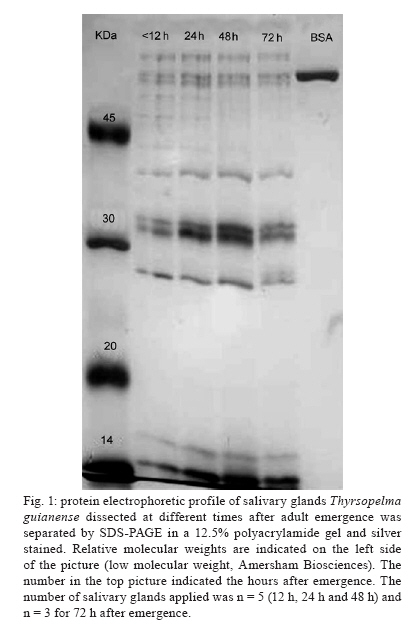
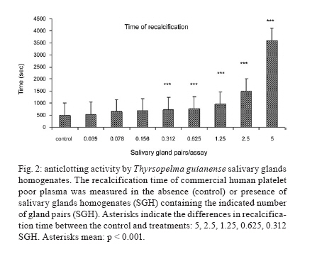
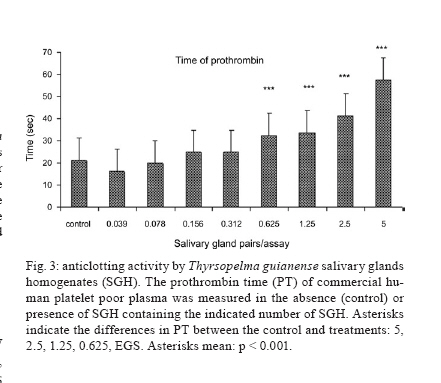
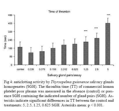
Anticoagulant activity in salivary gland homogenates of Thyrsopelma guianense (Diptera: Simuliidae), the primary vector of onchocerciasis in the Brazilian Amazon Andrezza Campos ChagasI, +; Jansen Fernandes MedeirosII; Spartaco Astolfi-FilhoIII; Victor Py-DanielIV ILaboratório de Entomologia Médica, Instituto de Pesquisa René Rachou-Fiocruz, Belo Horizonte, MG, Brasil Received 8 August 2009 Financial support: FAPEAM, CNPq (Msc scholarship for ACC) Code Number: oc10030 ABSTRACT In this study, anticoagulant activity was detected in salivary gland homogenates (SGHs) of Thyrsopelma guianense (Diptera: Simuliidae). The SGH yielded 1.07 μg ± 0.03 (n = 15) of total soluble protein per pair of glands. In addition, following SDS-PAGE (12.5% gel) and silver nitrate staining, 12 polypeptides with molecular weights ranging from 14-69 kDa were detected in all physiological ages analyzed (12 h, 24 h, 48 h and 72 h following emergence). Coagulation bioassays showed that the SGHs had activities that interacted at all levels of coagulation (the intrinsic, extrinsic and common pathways), by extending the plasma recalcification time, prothrombin time, thrombin time. This is the first report on the activity of salivary gland proteins from the main vector of onchocerciasis in Brazil. We also suggest detailed studies on the morphology and function of the salivary glands in order to understand the role of these proteins in host/vector interactions. Key words: black flies - salivary glands - anticoagulant - onchocerciasis Human onchocerciasis (river blindness) is caused by the nematode Onchocerca volvulus (Nematoda: Onchocercidae), which is transmitted through the bite of female black flies (Diptera: Simuliidae). This disease is endemic in 34 countries, 27 of which are in Africa, one is in the Arabian Peninsula and six are in Latin America. Recently, all six countries in the Americas with endemic onchocerciasis introduced programs providing treatment in all 13 foci and all six have exceeded the target coverage of treating 85% of the eligible population twice a year (Sauerbrey 2008). In Brazil, black fly saliva was determined to be responsible for the wild fire disease (Pemphigus foliaceus, also known as bulbous autoimmune disease) (Auad 1972, Lombardi et al. 1992). It was also named as the cause of death in immigrants suffering from Altamira hemorrhagic syndrome (Pinheiro et al. 1974), characterized by hemorrhage of the skin around the bites (multiple petechiae and ecchymosis), bleeding mucosa and anemia. The ability to hematophage evolved independently in different groups of arthropods (Ribeiro 1995). New compounds arose which allowed these organisms to avoid the hemostasis of the vertebrate host, making the saliva a rich source of antihemostatic molecules (Ribeiro & Francischetti 2003, Champagne 2004, 2005). These include a great diversity of vasodilators (Lerner et al. 1991, Law et al. 1992, Champagne & Ribeiro 1994, Cupp et al. 1994, 1998), inhibitors of platelet aggregation (Cupp et al. 1993, 1995, Moreira-Ferro et al. 1999, Valenzuela et al. 2001) and inhibitors of blood coagulation (Jacobs et al. 1990, Abebe et al. 1995, 1996, Capello et al. 1996, Stark & James 1998, Valenzuela et al. 1999, Cupp et al. 2000, Horn et al. 2000, Zang et al. 2002). Past studies of black flies' salivary glands are limited to the species Simulium vittatum (= Psilozia vittata Zetterstedt) due to the absence of colonies. The first report described morphological aspects of the salivary glands for this species (Hungerford 1913, Cox 1938, Rubtzov 1955) and only recently have oriental black fly species been investigated (Jariyapan et al. 2006). The salivary glands have been suggested as a tool in the identification of different species of black flies (Bennett 1963). Recently, the salivary glands of black flies have been investigated due to their important roles in blood feeding and in the transmission of filarial parasites to man (Cupp & Cupp 1997). The female black flies exhibit pool feeding behaviour where their salivary proteins are injected at the injury site and maintain the blood meal in a fluid state due to the antihemostatic properties present in the saliva. These proteins include anticoagulants (Jacobs et al. 1990, Abebe et al. 1994, 1995, 1996), vasodilators (Cupp et al. 1994), modulators of platelet aggregation as mediated by apyrase (Cupp et al. 1993, 1995) and modulators of the mouse cellular immune response (Cross et al. 1993). Andersen et al. (2009) uncovered for the first time the sialo-transcriptome of a member of the Simuliidae family and identified several new protein families with pharmacological or antimicrobial activities that might serve as epidemiological markers of Simulium exposure or as anti-disease vaccines (Cupp & Cupp 1997). In this paper we describe the anticoagulant activity and the electrophoretic protein profile from Thyrsopelma guianense (= Simulium guianensis Wise) salivary glands. This represents the first data on the salivary glands of a black fly involved in the transmission of O. volvulus in Brazil. MATERIALS AND METHODS Black flies - Nulliparous females were collected as pupae attached to plant material from breeding sites near the 40 Ilhas waterfall (0°28'48"N/60°29'49"O) in Pitinga village, municipality of Presidente Figueiredo, state of Amazonas, Brazil. The collections were made in two months (March and September) of 2004. Black flies were maintained on a glucose solution (honey Karo®) and given water ad libidum to promote accumulation of salivary secretions. The salivary glands were dissected at 12 h, 24 h, 48 h and 72 h following emergence and processed immediately as described below. Salivary gland dissection - The salivary glands of the adult black flies were dissected individually using fine entomological needles under a stereoscopic microscope in 0.15 M NaCl pH 7.4. Each pair of glands was stored in a microcentrifuge tube with a small volume of NaCl at -80°C, pending later analysis. Salivary gland homogenates (SGHs) - Salivary glands were centrifuged at 10.000 g, 4°C for 10 min after which supernatants were removed. Supernatants were pooled and referred to as SGHs. Serial dilutions of SGHs were tested in coagulation assays. Protein quantification - The soluble proteins were quantified according to Bradford (1976), using the dye Coomassie Brilliant Blue G-250. The protein concentration was determined based on a bovine serum albumin standard curve. SDS-PAGE and silver staining - SDS-PAGE was carried out on a 12.5% gel according to Laemmli (1970) and the proteins were stained with silver nitrate. Low molecular weight protein markers (Amersham Biosciences) were used. The electrophoretic profiles were determined with a pool of three SGHs at different physiological ages (12 h, 24 h, 48 h and 72 h after emergence). Coagulation assays - The assays were conducted as described by Valenzuela et al. (1996) with some modifications. Human citrated plasma as well as all the kits used to measure thrombin time (TT - thrombin activity assay) and prothrombin time (PT - extrinsic pathway assay) were provided by HUMAN GmbH (Germany) and were used to determine the anticoagulant activities of black fly SGHs. All assays were undertaken using the SGH of a newly emerged female (12 h after emergence). Recalcification time (RT) - A Biotrak II Plate Reader (Amersham Biosciences) with a kinetic module was used to measure clotting activity by the RT procedure. Briefly, citrated human plasma (20 μL) was incubated with 20 μL of 150mM NaCl, pH 7.4, either with or without the sample to be tested (SGH). Samples were mixed in the wells of a 96-well flat-bottom plate and then incubated at 37°C for 5 min. The coagulation cascade was triggered by addition of 20 μL of pre-warmed 20mM CaCl2. The plate was placed in the microplate reader, mixed and heated at 37°C to take absorbance readings at 620 nm every 10 sec. Data were recorded using the "time to selected absorbance" option in the microplate reader's Softmax software. All assays on fibrin clot formation were performed with eight replications of all dilutions. The other assays were carried out in the same way except for the substitution of 20 μL of pre-warmed Thromboplastin SI reagent (lyophilized thromboplastin from rabbit brain with the addition of calcium chloride); or 20 μL of RGT reagent (lyophilized bovine thrombin, 10 NIH U/mL) for the PT and TT assays, respectively. After 3 min of incubation at 37°C, 20 μL of pre-warmed 20mM CaCl2 was added to each sample to initiate the reaction in the plate reader as cited above. Statistical analysis - The results were expressed as mean ± SEM. For statistical analysis, a one-way ANOVA and a Tukey test were used. Values were considered to differ significantly at p < 0.001. RESULTS Female SGHs contained 1.07 μg ± 0.03 (n = 15) of total soluble protein per pair of glands (isolated from a single fly). The protein profiles of female salivary glands up until the third day following adult emergence are shown in Fig. 1. The protein profile did not change with age proteins present on the first day were present throughout the stages tested. The analysis of the female salivary gland proteins by SDS-PAGE revealed the presence of 10-12 main polypeptides. The majority of the bands are between 14-69 kDa in mass. This profile (Fig. 1) showed nine bands (69, 64, 41, 39, 33, 31, 28, 16, 15 kDa), all of which are abundant and present up to 72 h. The salivary glands of T. guianense show anticoagulant activity increasing RT (Fig. 2). The activity of the intrinsic, extrinsic and common pathways was determined using recalcification, PT and TT assays, respectively (Figs 2, 3, 4). The time of normal human plasma clotting was higher than one hour (the latest time point observed) when tested using homogenates from five pairs of salivary glands. In addition, clotting time was significantly delayed (p < 0.001) by as little as 0.312 of SGH in the recalcification assay (Fig. 2) indicating an inhibitory effect on the intrinsic pathway. The inhibition of the extrinsic pathway was measured using the PT assay (Fig. 3) and it showed a statistically significant difference at 0.625 SGH as that used on the previous assay. The inhibition of the common pathway was measured using the TT assay (Fig. 4) where 1.25 SGH was required to produce inhibition with equally significant statistical difference (p < 0.001). DISCUSSION In this work, we report for the first time the presence of anticoagulant activity in SGH of the main vector of onchocerciasis in Brazil. As a preliminary study, only salivary glands of newly emerged females were studied, due to the lack of biological knowledge on this species and the subsequent difficulty of obtaining and maintaining this species under laboratory conditions. Recently, due to increased interest in the salivary glands of this species, efforts have been made to rear this fly. Several salivary glands of hematophagous invertebrates have been intensively studied because they possess a variety of substances that are involved in counteracting the homeostatic systems and inflammatory reactions of the vertebrate host (Ribeiro 1987, 1995, Andrade et al. 2005). In black flies, the study of salivary secretion function has been limited to the species S. vittatum (Jacobs et al. 1990, Cross et al. 1993, Cupp et al. 1993, 1994, Abebe et al. 1994, 1995, 1996, Andersen et al. 2009). T. guianense is the Neotropical vector species responsible for onchocerciasis transmission in the Yanomami area (Brazil). We show that the salivary glands of this species begin synthesizing proteins soon after adult emergence, similar to the protein expression observed in mosquitoes. The levels of soluble proteins present in the salivary secretions are comparable to other anthropophilic species already studied, such as Simulium metallicum and S. ochraceum (Cross et al. 1993, Abebe et al. 1994). The protein level reflects the size of the salivary glands of each species, according to Jariyapan et al. (2006), who studied salivary protein levels in four oriental black fly species. The electrophoretic profile of T. guianense is shown to be more similar to other black fly species already studied with regard to the number of polypeptides (10-12 bands) and the fact that their expression changes with salivary gland maturation. Cross et al. (1993) analyzed four black flies species and found that zoophilic species had more bands (19-20 molecular bands) than anthropophilic species (11-12 molecular bands). This observation suggested that the difference in salivary protein composition may represent different evolutionary adaptations in anthropophilic species to aid feeding from human hosts. In addition, the differences in protein level at different developmental stages, as visualized by SDS-PAGE, reflect an increase in the amount of protein in the glands of older black flies. This observation may be associated with the fact that efficient blood feeding occurs mainly during the time of peak maximum salivary secretions - at 48 h following emergence for this black fly species (Chagas, unpublished observations). In sand flies, which are pool feeders like black flies, the number of protein components gradually increases with age and depends not only on sex but also on the physiological state in the female (Volf et al. 2000). In contrast, our results showed that the protein concentration of the black fly salivary glands did not vary qualitatively during the first three days of female adult life. Jariyapan et al. (2006) show that all black fly species studied had a small number of major proteins and that some of the major proteins differed in molecular mass between species. Our results show that the proteins responsible for inhibiting the coagulation cascade are secreted soon after emergence. It is possible that the anti-coagulation activity may be even greater than that shown here if the flies are measured at the peak of salivation. It was not possible to investigate the targets of the inhibitory saliva proteins. In this study, we show that the PT was prolonged with less efficiency than the RT, but our results suggest that both pathways are affected. We believed that this may be due to factors localized in common pathways, as factors Xa and thrombin from T. guianense SGH are able to inhibit all pathways (extrinsic, intrinsic and common). The antihemostatic molecules present in salivary glands reflect the blood meal strategy used by each species. The data for black flies shows that the different strategies are associated with the meal mode (zoophilic or anthropophilic) and that these key components reflect the vector competence of the black fly species (Cupp & Cupp 1997). In the Yanomani area, four species were found to be responsible for the transmission of O. volvulus (Py-Daniel 1997), however is not yet known what factors are making T. guianense the main vector in this area. Therefore, more investigations need to be undertaken into the antihemostatic molecules present in the salivary glands of this species in order to determine the vector competence. ACKNOWLDEGMENTS To Dr. Mary Cupp, for critical review manuscript. REFERENCES
Copyright © 2010 - Instituto Oswaldo Cruz - Fiocruz The following images related to this document are available:Photo images[oc10030f1.jpg] [oc10030f3.jpg] [oc10030f2.jpg] [oc10030f4.jpg] |
| |||||||||
