 |
| About Bioline | All Journals | Testimonials | Membership | News |
|
||||||
|
||||||
Memórias do Instituto Oswaldo Cruz, Vol. 106, No. 3, 2011, pp. 322-329 ARTICLES Efficiency of diagnostic biomarkers among colonic schistosomiasis Egyptian patients Manal Abdel Aziz HamedI, +; Samia Abdel Aziz AhmedI; Hussein Moustafa KhaledII ITherapeutic
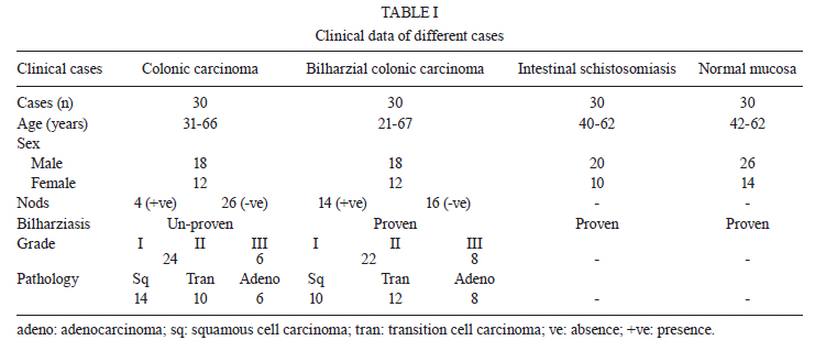
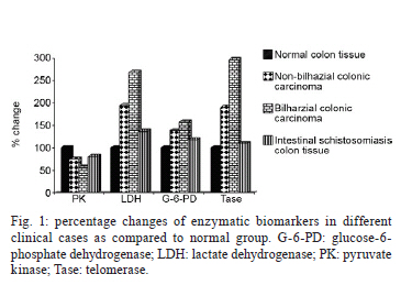
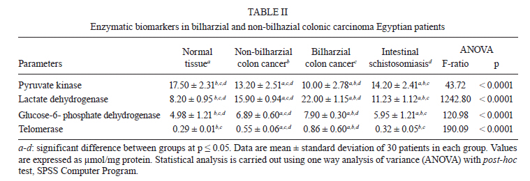
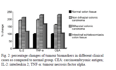
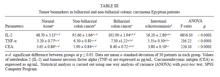
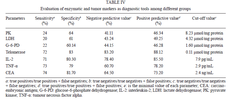
Chemistry Department, National Research Center, Dokki, Cairo, Egypt + Corresponding author: manal_hamed@yahoo.com Received 11 October 2010 Code Number: oc11053 ABSTRACT The schistosomal parasite plays a critical role in the development of malignant lesions in different organs. The pathogenesis of cancer is currently under intense investigation to identify reliable prognostic indices for disease detection. The objective of this paper is to evaluate certain biochemical parameters as diagnostic tools to efficiently differentiate between colonic carcinoma and colonic carcinoma associated with schistosomal infection among Egyptian patients. The parameters under investigation are interleukin 2 (IL-2), tumour necrosis factor alpha (TNF-α), carcinoembryonic antigen (CEA) levels, tissue telomerase, pyruvate kinase (PK), glucose-6-phosphate dehydrogenase (G-6-PD) and lactate dehydrogenase (LDH) enzyme activities. The results revealed a significant elevation in the level of the tumour markers IL-2, TNF-α and CEA as well as the activities of LDH, telomerase and G-6-PD among non-bilharzial and bilharzial colonic cancer groups, with a more potent effect in bilharzial infection-associated colonic cancer. A significant inhibition in PK activity was recorded in the same manner as compared to normal tissues. The efficacy of this biomarker was also evaluated through detecting sensitivity, specificity, negative and positive predictive values. In conclusion, schistosomal colonic carcinoma patients displayed more drastic changes in all parameters under investigation. The combination of the selected parameters succeeded in serving as biomarkers to differentiate between the two malignant types. Key words: colon - cancer - intestinal Schistosoma - biomarkers - tumour marker Colonic carcinoma is one of the most frequent malignancies in many tropical countries, where much evidence has suggested that schistosomiasis is implicated as an etiologic agent in colon cancer development (Li et al. 2006, Cao et al. 2010). Colonic schistosomiasis is a specific acute or chronic inflammatory reaction in response to Schistosoma ova that are deposited mainly in colorectal mucosa (Cao et al. 2010, Salim et al. 2010). Most of the published data refer to Schistosoma japonicum species in the induction of colonic cancer, but the evidence linking Schistosoma mansoni to colonic carcinoma occurrence is limited (Salim et al. 2010). Among Egyptians, the incidence of colonic carcinoma is less than the incidence of bladder carcinoma. Colonic carcinoma occurs predominately in the Nile Delta region where the intermediate snail hosts of schistosomiasis are extremely abundant (Fedewa et al. 2009). Tumour markers are substances, usually proteins, that are produced by the body in response to cancer growth or by the cancer tissue itself. These substances may be detected in blood, urine and tissue samples. Some tumour markers are specific for a particular type of cancer, while others are seen in several cancer types (Parekh et al. 2007). Many investigators have focused their work on the enzymatic reactions in tumour cells and consider most of these enzymes to be tumour markers (Michael et al. 1980). Thangaraju et al. (2009) have reported that glycolytic enzymes are sharply intensified in colonic cancer, while key gluconeogenesis enzymes are sharply decreased. Some investigations have dealt with pyruvate kinase (PK) activity among different grades of malignancy in colorectal cancer (Shin et al. 2009). Glucose-6-phosphate dehydrogenase (G-6-PD) has also been reported in a human adenocarcinoma cancer cell line (Vizán et al. 2009). Telomerase is a ribonucleoprotein polymerase enzyme containing an integral RNA with a short template element that directs the de novo synthesis of telemetric repeats at the chromosomal ends. These repeats maintain the length of the telomeres and allow tumour cells to survive (Gonzalo et al. 2010). Therefore, telomeres are considered to be the clock that regulates how many times an individual cell can divide. Telomere sequences shorten each time the DNA replicates until they reach a critically short length, at which point the cell stops dividing and ages (Rampazzo et al. 2010). Under normal circumstances, telomerase is active in rapidly dividing embryonic cells and in stem cell populations, but not in terminally differentiated somatic cells. Much attention has recently focused on the hypothesis that the activity of this enzyme is necessary for cells to become immortal. This hypothesis predicts that telomerase activity should be detectable in malignant cells and tissues, but not in their normal counterparts, which slowly senesce and die (Yoshida et al. 1997). Carcinoembryonic antigen (CEA) is a glycoprotein involved in cell adhesion. CEA is normally produced during foetal development, but its production stops before birth. Therefore, CEA is not usually present in the blood of healthy adults. As a specialised glycoprotein, sialofucosylated glycoforms serve as functional colon carcinoma ligands (L-selectin and E-selectin); this function is critical to the metastatic dissemination of colon carcinoma cells (Thomas et al. 2008). Nielsen et al. (2011) determined that CEA is a valuable biomarker for the early detection of colorectal cancer. Interleukin 2 (IL-2) is one of a group of related proteins made by leukocytes, such as T lymphocytes and other cells in the body. This protein increases the growth and activity of other T and B lymphocytes and affects the development of the immune system (Garcia-Tunon et al. 2004). The high expression of IL-2 and its receptors in tumour tissue is mediated by enhancing cell proliferation or inhibiting apoptosis, together with the enhancement of the anti-apoptotic factor gene (Bcl2-family gene) (Royuela et al. 2000). Several interleukins have been shown to induce the IL-2 receptor (IL-2R) α in inflammatory processes and as this chine is necessary to form the high affinity of IL-2R, it enhances IL-2 signals (Garcia-Tunon et al. 2004). Tumour necrosis factor alpha (TNF-α), isolated 30 years ago, is a multifunctional cytokine that plays a key role in apoptosis and cell survival as well as in inflammation and immunity. Although named for its anti-tumour properties, TNF has been implicated in a wide spectrum of other diseases (van Horssen et al. 2006). In tumours, TNF-α may cause direct DNA damage, have anti-apoptotic or mitogenic activity, mediate tumour/stromal cell interactions and induce a range of cytokines and che-mokines that promote tumour development. Moreover, TNF can synergise with growth factor such as transforming growth factor beta (Balkwill et al. 2006). The aim of the present study was to identify biomarkers that could be helpful in the diagnosis and differentiation between colonic carcinoma and bilharzial-related colonic carcinoma among Egyptian patients. The efficiency of each biomarker was also evaluated. The work was extended to examine these biomarkers in S. mansoni-infected patients. PATIENTS, MATERIALS AND METHODS Clinical cases - All cases were of Egyptian colonic carcinoma and schistosomal infected patients attending in the Clinical and Medical Oncology Department, National Cancer Institute (NCI), Cairo University, Egypt. Ethics - Surgical operations, biopsies and serum samples were subject to the ethical guidelines approved by the National Cancer Institute. Pathological groups - Group 1: 30 colonic cancer patients with ages ranging from 31-66 years (18 males, ages 34-66 years, and 12 females, ages 31-58 years); group 2: 30 schistosomal colonic carcinoma patients with a median age of 35 years (18 females, ages 21-60 years, and 12 males, ages 30-67 years); group 3: 30 intestinal S. mansoni-infected patients. Their ages ranged from 42-62 years (20 males, ages 42-60 years, and 10 females, ages 49-62 years); group 4: 30 individuals (26 males and 14 females with ages between 30-70 years) served as the control for all experimental groups. Unaffected colonic tissues (mucosa) were taken at the same time during the surgical operation on the patients. Gross pathology - The surgical colonic carcinoma tissue and the biopsied intestinal schistosomiasis tissue were subjected to both pathological and histological examinations to determine the diagnosis and disease progress at the Clinical Pathology Department, NCI. The oriental cell microscopic slides were examined. The tumour grade and pathological differentiation are illustrated in Table I. Preparation of tissue homogenate for PK, lactate dehydrogenase (LDH) and G-6-PD activities - Tissues were freed of necrotic debris and connective tissue and were immediately frozen at -80ºC until used. The specimens were homogenised in 4 vol of buffer containing 0.05 mol/l tris hydrochloride, 0.10 mol/l KCl, 0.01 mol/l MgCl2, 0.1 mol/l sucrose, 10 mmol/l glucose, l mmol/l EDTA and 2 mmol/l dithiothreitol. The homogenate was centrifuged for 5 min at 13,000 g and 4ºC and the supernatant was used for enzyme assays. Preparation of tissue homogenate for telomerase activity - Tissue samples were shock frozen in liquid nitrogen and stored at -80ºC. Cryostat sections of 10-15µm thickness from frozen tissue samples were prepared. The sections were transferred into a sterile tube containing 200 µl ice-cold lysate reagent, incubated on ice for 30 min and centrifuged at 16,000 g for 20 min at 4ºC. Serum separation for tumour marker tests - A fasting venous blood sample (5 ml) was withdrawn and centrifuged. Sera were separated and stored at -80ºC until used. Enzymatic assays - PK was assayed by the method of Imamura and Tanaka (1972), where the reaction velocity is determined in a LDH coupled assay system by measuring the decrease in absorbance at 340 nm caused by the oxidation of nicotinamide-adenine dinucleotide reduced form (NADH). G-6-PD activity possesses dual coenzyme specificity, where the reaction velocity is determined by measuring the increase in absorbance at 340 nm caused by the reduction of NAD or nicotinamide-adenine dinucleotide phosphate (Haghighi & Levy 1982). LDH was assayed by the method of Babson and Babson (1973), where the reduction of NAD is coupled with the reduction of a tetrazolium salt [2-(p-iodophenyl)-3-(p-nitrophenyl)-5-phenyl tetrazolium chloride] (INT). The resulting formazan of INT was measured colourimetrically at 503 nm. Telomerase activity was measured by a polymerase chain reaction (PCR)-enzyme-linked immunosorbent assay (ELISA) technique using the telomerase repeat amplification protocol according to Gelmini et al. (1998). This method is based on the use of a sensitive fluorochrome that selectively binds double-stranded DNA. The measurement of DNA concentration in post-PCR samples can be considered quantitatively related to telomerase activity. Protein concentration was determined colourimetrically by the method of Bradford (1976), where bovine serum albumin was used as a standard protein and the developed colour was read colourimetrically at 595 nm. Tumour markers assays - The measurement of serum IL-2 employed the quantitative sandwich enzyme immunoassay technique. A monoclonal antibody specific for IL-2 was used. Standards and samples were pipetted into the wells and any IL-2 present was bound by the immobilised antibody. An enzyme-linked polyclonal antibody specific for IL-2 and tetramethylbenzidine as a substrate were added. The developed colour was measured at 450 nm using a microtiter plate reader (Yang et al. 1991). The measurement of serum TNF-α employed the quantitative enzyme-linked immunosorbent technique. A monoclonal antibody specific for human TNF-α was pre-coated onto a microplate. Standards and samples were pipetted into the wells and TNF-α present in a sample bound to the wells through the immobilised antibody. Anti-human TNF-α antibody and the tetramethylbenzidine substrate were added. The developed colour was measured at 450 nm using a microtiter plate reader (Bonavida 1991). The measurement of serum CEA was based on the principle of a solid phase ELISA. The assay system utilises a monoclonal antibody directed against a distinct antigenic determinant on the intact CEA molecule. The test sample is allowed to react simultaneously with two antibodies, resulting in the CEA molecules being sandwiched between the solid phase and enzyme-linked antibodies. Tetramethylbenzidine was added as a substrate and the developed colour was measured at 450 nm (Zamcheck & Martin 1981). Statistical analysis - Data from 30 patients in each group were expressed as the mean ± standard deviation. The statistical analysis was performed using one way analysis of variance accompanied by a post-hoc test in the SPSS Computer Program. The least significance value between groups was at p < 0.05. RESULTS AND DISCUSSION The incidence of colon cancer among Egyptians is less than the reported incidence of bladder cancer (Fedewa et al. 2009). Colonic carcinoma associated with bilharzial lesions was 19% of all colonic cancer (EL-Bolkainy et al. 1982). Intestinal schistosomiasis occurs as a result of the deposition of Schistosoma ova in submucosa producing a granulomatous reaction, mucosal edema, haemorrhage and ulceration in the bowel wall. In the advanced stage, a thickened bowel wall, polyps or enteric cavity stricture can be detected (Yosry 2006). In our study and according to the pathological data received from Clinical and Medical Oncology Department, Schistosoma ova were deposited with infiltration of lymphocytes and plasma cells in the submucosa and lamina propria. In addition, atrophy of the intestinal mucosa epithelium, reduction of intestinal glands and different degrees of fibrosis were also observed in the intestinal schistosomiasis group. In colonic schistosomiasis patients, massive egg deposition, an intensive degree of fibrosis, hyperplasic polyps, canalicular and tubulovillous adenoma with intraepithelial neoplastic change were detected. In contrast, different types of papillary, mucinous and tubular adenocarcinoma were observed in colonic carcinoma patients. Cheever and Andrade (1967) and Chassot et al. (1998) showed that schistosomiasis-related intestinal polyps are frequently seen in Egyptian cases of intestinal schistosomiasis, which is different compared to most other endemic areas. Colonic involvement has been demonstrated to be meagre in Brazilian cases, rarely exhibiting polyps, and with no signs of a particular relationship with colonic cancer. In other African countries, where S. haematobium are endemic, severe intestinal involvement is not frequent. In China, a high incidence of colorectal cancer in regions with endemic S. japonicum was recorded, where patients with chronic Schistosomiasis japonica have a more than three times greater risk of developing colon cancer than those with no previous exposure to schistosomal infection (Salim et al. 2010). Therefore, the difference in parasite strains, intensity of infection, concomitant infections (including S. haematobium) and environmental conditions may be important in determining the varying patterns of the disease (Cheever & Andrade 1967). From another point of view, the problem of bilharziasis and its various aspects needs careful clinical studies to clarify its effect on the biochemical features of tumour development (Salim et al. 2010). In the present study, LDH showed a significant elevation by 93.90%, 169.29% and 36.95 % in colon cancer, bilharzial-related colon cancer and intestinal schistosomiasis colon tissue, respectively. In contrast, PK showed a significant reduction by 24.57%, 42.85% and 18.85% in colon cancer, bilharzial-related colon cancer and intestinal schistosomiasis colon tissue, respectively (Fig. 1, Table II). These findings were in accordance with Thangaraju et al. (2009), who recorded that the intracellular levels of pyruvate in colon cancer cells are much lower than those in non-malignant cells. They attributed the decrease in pyruvate level to the expression of the LDH enzyme involved in the generation and metabolism of pyruvate. They added that cancer cells generate lactate purposely to reduce the intracellular levels of pyruvate. The conversion of pyruvate into lactate is not the only mechanism by which cancer cells manage to keep the pyruvate levels low. These cells also differentially express PK splice variants, PKM1 and PKM2, such that the activity of PK, which converts phosphoenolpyruvate into pyruvate, is low. Elevation of LDH activity in malignant and bilharzial colonic carcinoma can be used as an indicator for the stimulation of anaerobic pathways in cases of abnormal growth of human cancer cells (Thangaraju et al. 2009). Aly et al. (2004) found the same pattern of LDH promotion in bladder carcinoma and S. haematobium-associated bladder cancer. The increase in LDH activity in bilharziasis can be related to the fact that the Schistosoma parasite is a homolactate form enter, producing lactate as a sole metabolic end that can in turn increase the rate of the enzymatic activity (Nabih & el-Ansary 1992). The recorded LDH increased more significantly in cancerous tissue associated with bilharzial infection than in malignant tissue. This increase in LDH was attributed to the enhancement of anaerobic glycolysis by Schistosoma toxins, suppression of pyruvate oxidative metabolism in mitochondria (where the mitochondrial enzymes were also affected) (Hamed et al. 2010), marked disturbance in glycolytic pathways due to cancer complications and the overproduction and proliferation of cancer cells (Aly et al. 2004). Regarding G-6-PD, the present results recorded a significant increase by 38.35%, 58.63% and 19.47% in colonic carcinoma tissue, bilharzial-related colonic carcinoma and intestinal schistosomiasis, respectively (Fig. 1, Table II). In agreement with the present study, Frederiks et al. (2007) reported a massive ultrastructural localization of G-6-PD in the cytoplasm of colon carcinoma cells, whereas human colon cancer cells contained five times higher G-6-PD activity than hepatoma cells. Vizán et al. (2009) found that G-6-PD, the key enzyme of the oxidative branch of the pentose phosphate pathway (which is necessary for nucleotide synthesis), is enhanced during cell cycle progression of the human colon cancer cell line. This enhanced enzyme activity coincides with an increased ratio of the pentose monophosphate to the hexose monophosphate pool during late G1 and S phases of cell cycle, suggesting a potential role for pentose phosphates in proliferation signalling. Geraci et al. (2010) detected genetic heterogeneity among G-6-PD transcripts in colon and colorectal tumours. In case of S. mansoni infection, G-6-PD was significantly increased (Shaheen et al. 1989) due to an elevation in glycogen synthesis, which enhanced enzyme activity. In patients with bilharzial colon carcinoma, G-6-PD was markedly elevated. This elevation was attributed to the synchronised effect of both diseases, accumulation of hepatic glycogen and enhancement of cell proliferation. Schistosoma toxins and ova deposition may also act as a signal on gene expression, leading to activation of transcription of a specific DNA sequence into mRNA. Gene activation is an effective way to enhance enzyme activity (Hoek et al. 1997). Telomerase is a ribonucleoprotein that synthesises TTAGGG tandem repeats at each telomeric region to re-extend the telomere to its original length. The enzyme is active in embryonic cells and in stem cells, but its activity is undetectable in normal, terminally differentiated somatic cells (Kim et al. 1994). Cells that can overcome this limitation have the potential for prolonged survival, indefinite proliferation and tumour development. Hence, if the "telomere hypothesis" applies to human malignancy, one might predict that telomerase activity would be detectable in the initial stages of neoplasia and could therefore be a particularly useful marker for early diagnosis (Yoshida et al. 1997). The telomerase activity, in the present study, was enhanced by 89.65%, 196.55% and 10.34% for the cases of colonic cancer, bilharzial colonic cancer and in bilharzial colon tissues, respectively (Fig. 1, Table II). In agreement with our results, Yoshida et al. (1997) have detected telomerase in a wide range of malignancies, including those of the gastrointestinal tract, breast and lung. Rampazzo et al. (2010) have suggested that tel-omere shortening is a key initial event in colorectal carcinogenesis, where the extent of telomere erosion is related to the tumour origin site and may be influenced by the mismatch repair pathway. Gonzalo et al. (2010) have found that long-standing inflammatory bowel disease in patients with high risk for colorectal cancer leads to an overexpression of human telomerase reverse transcriptase in non-affected colorectal mucosa, suggesting its potential usefulness as a biomarker for the risk of malignant transformation. In contrast to the results of Gonzalo et al. (2010), Yoshida et al. (1997) have detected strong enzyme activity representing 92% of colonic carcinoma patients with no sign of activity in cases of inflammatory bowel disease, suggesting its potency as a good marker for the detection of colon carcinoma regardless of the tumour stage or the histological type of tumour. In parallel with the present finding, Shaker et al. (2009) have recorded an increase in telomerase activity in schistosomal-associated bladder cancer in comparison to non schistosomal-associated bladder cancer. The higher incidence of telomerase in bilharzial colonic cancer patients than in colonic carcinoma patients may be due to the effect of Schistosoma toxins, which act as a stimulus for activating the human telomerase reverse transcriptase protein that provokes enzyme activity in addition to the role of enzyme initiation in cancer cells. In the case of tumour markers, IL-2 displayed a significant elevation by 92.14%, 111.29% and 15.40% in non-bilharzial colon cancer, bilharzial colon cancer and intestinal schistosomiasis, respectively (Fig. 2, Table III). These data are in accordance with Berghella et al. (1994), who have stated that IL-2 levels are significantly correlated with the stage of colon cancer disease, showing an increase from stage I to stage IV. Reichert et al. (1998) have determined that carcinoma cells synchronised in the G2/M-phase of the cell cycle express significantly more intracytoplasmic IL-2. Moreover, Johnson et al. (2009) have mentioned that treatment of mice bearing colon adenocarcinoma with a tumour-specific monoclonal antibody fused to IL-2 causes greater suppression of tumour growth and enhances survival rate. Silveira-Lemos et al. (2008) and Allam (2009) have observed significant elevations of IL-2 after S. mansoni infection in human and mice. In addition, Elshazley et al. (2006) have recorded a significant increase in IL-2 among different stages of schistosomiasis; in chronic schistosomiasis with hepatitis B or hepatitis C, a higher level of significance (p < 0.001) was observed. Stavitsky and Harold (1988) explain this phenomenon according to distinct signals from granulomatous inflammation that enhances proliferation of T lymphocytes and lymphokine gene expression. These enhancements promote IL-2 production, as there is a complex interplay between IL-2 and inflammatory signals during infection (Pipkin et al. 2010). These data are in line with our data through the elevation of IL-2 in schistosomal infection and the higher significance value in schistosomal colonic carcinoma patients. Almost two decades ago, TNF was identified as a protein produced by the immune system that plays a major role in the suppression of tumour cell proliferation. Extensive research since then has revealed that TNF is a major mediator of inflammation, viral replication, tumour metastasis, transplant rejection, rheumatoid arthritis and septic shock (Aggarwal et al. 2002). In our study, TNF-α was significantly increased by 96.87%, 128.12% and 67.18% in non-schistosomal colon cancer, schistosomal colon cancer and intestinal bilharziasis colon tissue, respectively (Fig. 2, Table III). These data are in accordance with Talero et al. (2011), who have recorded the highest production of TNF-α in mice experimentally induced with colorectal carcinoma. In addition, Kemik et al. (2010) have stated that TNF-alpha was significantly increased in the serum of cachectic patients with colon cancer. The recorded high level of TNF-α in schistosomal colon cancer patients was in agreement with Torben and Hailu (2007). They attributed the increased level of this inflammatory cytokine to toxins elevated by egg deposition and granulomatous reaction, which enhanced the induction of tissue fibrosis through propelling the hepatic satellite cells. This process also indirectly induces the production of other fibrogenic factors, such IL-1 and IL- 6 (Kimura et al. 2003). Moreover, Davies et al. (2004) have reported that TNF-α essentially functions as a trophic factor for maintaining the viability of adult Schistosoma worms, in addition to its role as a mitogen and promoter of tumour development in schistosomiasis-associated carcinoma (Balkwill et al. 2006). CEA, a tumour-associated antigen, is a widely used serum biomarker for colorectal cancer. Interestingly, CEA has also been shown to correlate with the differentiation state of normal colon and in colon cancer cell lines. In the normal colonic mucosa, CEA expression showed a crypt-surface distribution. CEA expression was strong in surface epithelial cells and goblet cells of the upper crypts, while very weak in the mid crypt and at the base. Cell lines with high expression of CEA showed shuttle-shape morphologic changes with long or dendritic-like cytoplasmic processes (Ruan et al. 2010). CEA, in the present study, displayed an enhancement of 61.64%, 130.13% and 4.11% in colon cancer not associated with bilharzia, bilharzial-associated colon cancer and intestinal schistosomiasis colon tissue, respectively. Thirunavukarasu et al. (2010) have recorded a significant increase of CEA in medullary carcinoma of the colorectum, which is a new histological type of adenocarcinoma characterised by poor glandular differentiation and intraepithelial lymphocytic infiltrate. Nielsen et al. (2010) have recommended the use of a combination of plasma tissue inhibitor of metalloproteinases-1 and CEA for the detection of colorectal cancer and especially in colon cancer. Chen et al. (2010) have added that the membrane protein profile of colorectal carcinoma and neighbouring normal mucosa from colorectal cancer patients is easily differentiated by the over-expression of CEAs. Sarkar et al. (2010) have mentioned that vaccination with neem leaf glycoprotein matured CEA pulsed dendritic cells enhances antigen-specific humoral and cellular immunity against CEA and restricts the growth of CEA (+) murine tumours. Over-expression of CEA in colonic carcinoma associated with schistosomiasis may be due to the advanced stage of tumour progression related to schistosomiasis. Metastasis or the development of other carcinomas due to schistosomiasis complications may also be taken into consideration. The evaluation of enzymatic and tumour markers as diagnostic tools among different groups is clearly illustrated in Table IV. Telomerase, IL-2, TNF-α and CEA displayed potent values in the diagnosis of bilharzial and non-bilharzial colon cancer, while PK, G-6-PD and LDH to some extent succeeded in differential diagnosis. In conclusion, the selected enzymatic biomarkers along with the commonly known tumour markers play a potential role in differentiating between bilharzial and non-bilharzial colonic carcinomas. Schistosomiasis-associated colon cancer displayed drastic changes in all biomarkers compared with colon carcinoma. Detailed studies must be done among colon cancer associated with schistosomiasis infection, especially in developing countries where S. mansoni is endemic, to establish new biomarkers for early diagnosis of the disease. REFERENCES
Copyright © 2011 - Instituto Oswaldo Cruz - Fiocruz The following images related to this document are available:Photo images[oc11053t1.jpg] [oc11053f2.jpg] [oc11053t4.jpg] [oc11053t3.jpg] [oc11053t2.jpg] [oc11053f1.jpg] |
| |||||||||
