 |
| About Bioline | All Journals | Testimonials | Membership | News |
|
||||||
|
||||||
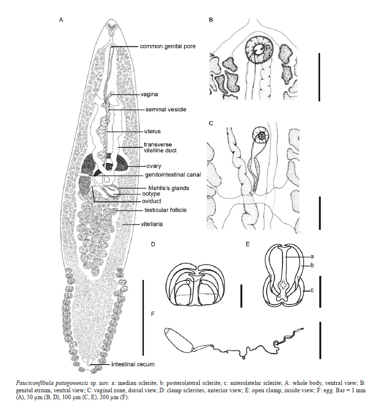
Memórias do Instituto Oswaldo Cruz, Vol. 106, No. 3, 2011, pp. 335-338 ARTICLES Pauciconfibula patagonensis sp. nov. (Monogenea: Microcotylidae) parasitizing the horsefish, Congiopodus peruvianus (Pisces: Congiopodidae), from the Patagonian Shelf, Argentina Delfina MP Cantatore+; Ana L Lanfranchi; Juan T Timi Consejo Nacional de Investigaciones Científicas y Técnicas y Laboratorio de Parasitología, Departamento de Biología, Facultad de Ciencias Exactas y Naturales, Universidad Nacional de Mar del Plata, Funes 3350, 7600 Mar del Plata, Argentina + Corresponding author: cantator@mdp.edu.ar Received 24 October 2010 Financial support: Universidad Nacional de Mar del Plata (EXA 531/10), FONCYT (PICT 02199/07), CONICET (PIP 112-200801-00024) Code Number: oc11055 ABSTRACT Pauciconfibula patagonensis sp. nov. (Monogenea: Microcotylidae), parasite of gill filaments of the horsefish, Congiopodus peruvianus (Congiopodidae) collected in the Patagonian Shelf, Argentina, is described and illustrated. The new species is characterized by having intestinal caeca not confluent and entering into the haptor, vitelline follicles extending from the genital pore to near the posterior portion of haptor, two parallel rows each comprised of 16-20 microcotylid clamps in the haptor, 25-43 testes and a fusiform egg with one very long tangled polar filament. P. patagonensis is the only member of the genus known to parasitize a scorpaeniform host and represents the first record of a representative of this genus in the southern Atlantic Ocean. Key words: Microcotylidae - Pauciconfibula patagonensis - Congiopodus peruvianus - Patagonian Shelf - Argentina Microcotylids of the genus Pauciconfibula Dillon and Hargis 1965 are parasites of inner surfaces of the operculum and gill filaments of brackish and marine fishes. At present, this genus comprises a total of six nominal species: Pauciconfibula trachini (Parona & Perugia 1889), a parasite of Trachinus radiatus Cuvier 1829 (Trachinidae), from Genoa and Bay of Naples, Italy (Dillon & Hargis 1965); Pauciconfibula draconis (Briot 1904), a parasite of Trachinus draco Linnaeus 1758 (Trachinidae), from the English Channel and the North Sea (Dillon & Hargis 1965); Pauciconfibula pogoniae (MacCallum 1913), found in Pogonias cromis (Linneaeus 1766) (Sciaenidae), from New York, USA (MacCallum 1913) and also reported from Florida, USA (Hargis 1956); Pauciconfibula euzeti (Ktari 1971), a parasite of Dentex gibbosus (Rafinesque 1810) [=Dentex filosus (Rafinesque 1810)] (Sparidae) from the Gulf of Tunis, Tunisia (Ktari 1971); Pauciconfibula gallieni (Euzet & Katari 1971), a parasite of Tylosurus acus acus (Lacepède 1803) [=Strongylura acus (Lacepède 1803)] (Belonidae), from the Gulf of Tunis and Gulf of Gabes, Tunisia (Euzet & Ktari 1971) and Pauciconfibula subsolana Chisholm, Beverley-Burton and McAlpine 1991, reported from Morone americana (Gmelin 1789) (Moronidae), from Saint John River, New Brunswick, USA (Chisholm et al. 1991). As a result of a parasitological survey of specimens of Congiopodus peruvianus (Cuvier 1829) (Congiopodidae), caught on the Patagonian Shelf, Argentina, microcotylids consistent with the diagnosis of Pauciconfibula (sensu Chisholm et al. 1991) were found on gill filaments of fishes. Their detailed study has shown that they represent a previously undescribed species, which is described herein. MATERIALS AND METHODS A total of 264 specimens of C. peruvianus, caught during research cruises on the Patagonian Shelf (41-48ºS 58-65ºW) during July 2007 and February 2009 were kept frozen until being examined for the presence of parasites. Fishes were necropsied and gill arches were removed and examined using a stereoscopic microscope. Parasites were collected and fixed in 4% buffered formaldehyde solution and then transferred to 70% ethanol for storage. Monogeneans were stained with alcoholic chlorhydric carmine, dehydrated through an ethanol series, cleared in methyl salicylate and mounted in Canada balsam. Mounted worms were studied and measured using light microscopy and drawn with the aid of a drawing tube. All measurements are based on adult specimens and given in micrometers (µm), unless otherwise specified as the mean ± standard deviation followed by the range and the number of specimens measured in parentheses. The use of ecological terms relating to parasite infection parameters follows Bush et al. (1997). The taxonomy, scientific and common name of the host are in accordance with FishBase (Froese & Pauly 2010). Type material was deposited in the Helminthological Collection of the Museo de La Plata (HCMLP), La Plata, Argentina. RESULTS Pauciconfibula patagonensis sp. nov. (Figure) Diagnosis (based on 16 mature whole-mounted worms) - Body (a in Figure) lanceolate, flattened dorsoventrally; total length 4.7 ± 0.9 (3.5-6.7; n = 16) mm, maximum body width 1.1 ± 0.2 (0.7-1.6; n = 16) mm at level of ovary. Haptor arising from ventral surface beginning at level of posterior testes; 1.6 ± 0.4 (1.0-2.5; n = 16) mm long, constituting about 35% of total length and 1 ± 0.2 (0.6-1.3; n = 12) mm maximum width; armed with two parallel approximately equal rows of 16-20 (n = 16) typical microcotylid clamps each. Clamp (d, e in Figure) bilaterally symmetrical, pedunculated; clamp sclerites include single medial sclerite with truncate anterior and posterior ends each, bearing two diverging spinelike projections, paired anterolateral sclerites bending posteriorly to reach medial sclerite and paired posterolateral sclerites; the largest clamps near midlength of each row 131 ± 12 (108-150; n = 32) wide and 95 ± 8 (83-113; n = 32) long, anterior clamp 80 ± 21 (38-113; n = 32) wide and 62 ± 18 (30-100; n = 32) long and posterior clamp 97 ± 12 (75-123; n = 32) wide and 76 ± 10 (53-95; n = 32) long. Paired prehaptoral buccal organ elliptical to subcircular in outline, septated, surrounded by minute tubercules; septum extending diagonally across its anterolateral half; buccal organ 86 ± 11 (63-100; n = 32) in maximum diameter, 75 ± 9 (55-90; n = 16) length perpendicular to maximum diameter. Mouth opening subterminal and ventral. Muscular pharynx ovoid, 110 ± 14 (68-125; n = 15) long, 81 ± 10 (50-90; n = 15) wide. Oesophagus short, without diverticula. Intestinal bifurcation immediately anterior to common genital pore; intestine obscured by vitelline follicules extending into haptor without posterior confluence; intestinal caeca subequal in length, left caecum being slightly longer. Common genital pore midventral, surrounded by weak circular muscle 29 ± 8 (18-50; n = 16) in diameter, located at 353 ± 50 (288-475; n = 16) from anterior extremity; genital atrium unarmed (b in Figure). Vagina unarmed (c in Figure), opening via single mediodorsal pore located at 989 ± 253 (720-1750; n = 16) from anterior extremity; vaginal canal muscular, short, dividing posteriorly into two lateral vaginal canals connecting with vitelline ducts. Paired vitelline ducts directing posteriorly to form vitellovaginal reservoir near level of initial portion of ovary. Vitelline follicles lateraly distributed, coextensive with intestinal caeca, extending from genital pore to near posterior portion of haptor. Ovary long, tubular, pretesticular, intercaecal, dorsal to vitelline ducts and uterus; originating on right side of body, extending anteriorly before traversing intercaecal region to left side, then looping anteriorly back to right side and finally directing posteriorly. Oviduct extending from end of ovary, receiving genitointestinal canal before vitellovaginal reservoir. Genitointestinal canal consisting of small duct connecting oviduct with right intestinal caecum. Ootype smooth walled, with Mehlis' glands surrounding proximal portion. Uterus arising at level of end of vitellovaginal reservoir, extending along body midline to genital atrium as relatively straight delicate tube dorsal to vitellovaginal reservoir and ventral to ovary and vas deferens. Testes 33 ± 4 (25-43; n = 13) in number, in post-ovarian intercaecal field occupying posterior half of body. Vas efferens not observed; vas deferens extending along body midline to male copulatory organ; long sigmoid proximal portion as a thin-walled seminal vesicle. Male copulatory organ unarmed. Egg (f in Figure) (in uterus) fusiform, operculated, 191 long, 58 wide (n = 1), with one very long tangled polar filament. Type host - Horsefish, C. peruvianus Cuvier 1829 (Scorpaeniformes: Congiopodidae). Site of infection - Gill filaments. Type locality - Patagonian Shelf (41-48ºS 58-65ºW), Argentina (June 2007 and February 2009). Type data and depository - Holotype MLP 6295 and paratypes MLP 6296 (5 whole mounted specimens) are deposited in HCMLP. Distribution - Patagonian Shelf (41-48ºS 58-65ºW), Argentina. Host-parasite data - Prevalence: 9.5% (25 fishes infected out of 264 fishes examined), mean intensity ± standard deviation (range): 4.6 ± 4.9 (1-18). Etymology - The specific name refers to the geographic area where parasites were found (Patagonia). Taxonomic discussion - Microcotylidae Taschenberg 1879 is a large monogenean family and, according to the revision made by Mamaev (1986), it comprises eight subfamilies, 39 genera and approximately 150 species. Among these subfamilies, only Protastomicrocotylinae Yamaguti 1968 and Microcotylinae Monticelli 1892 include species in which the genital pore, the vaginal pore and the male copulatory organ are all unarmed. Members of Protastomicrocotylinae are characterized by having a paired symmetrical prostatic system and by the ventromedial position of the vaginal pore (Ogawa & Egusa 1980). Consequently, the new species is assigned to Microcotylinae because it has a dorsomedial vaginal pore and lacks the prostatic system. Within Microcotylinae, the new species is accommodated in Pauciconfibula due to the lack of armament on the male copulatory organ, genital atrium and vaginal pore, the latter in dorsomedial position. Dillon and Hargis (1965) erected Pauciconfibula to accommodate two previously described species, P. trachini (Parona & Perugia 1889) and P. draconis. Later, Chisholm et al. (1991) described P. subsolana and provided an amended generic diagnosis for the genus. The authors also considered Pseudoaspinatrium Mamaev 1986 as a junior synonym of Pauciconfibula; therefore, the species previously assigned to Pseudoaspinatrium were transferred to Pauciconfibula as P. euzeti, P. gallieni and P. pogoniae. The validity of P. draconis remains uncertain, but according to Chisholm et al. (1991) this species should be considered valid pending further observations. Among these six species, only P. pogoniae and P. subsolana have intestinal caeca extending unfused into the haptor (Chisholm et al. 1991). The new species differs from the P. pogoniae by having fewer clamps (32-40 instead of 108), lower number of testes (25-43 rather than 75), by the shape of the ovary, which is longer and more convoluted in the latter, and by the extension of vitelline follicles (barely entering into the haptor in P. pogoniae). P. patagonensis sp. nov. can be distinguished from P. subsolana by the presence of one polar filament in eggs (absent in P. subsolana), by the structure of posterolateral clamp sclerites, which are not composed by two distinct sections, by having a larger number of clamps (32-40 intestead of 18-26) and by the absence of a swelling in the genitointestinal canal at its junction with oviduct. In this study, the host range of Pauciconfibula is extended to include a member of the order Scorpaeniformes. Moreover, the present finding represents the first record of this genus in the southern Atlantic Ocean. ACKNOWLEDGEMENTS To Leslie A Chisholm, for gently providing some literature used in this work, and to Claudio C Buratti and Claudia V Dato (Instituto Nacional de Investigación y Desarrollo Pesquero), for kindly providing fish samples. This work is part of the doctoral thesis of DMPC. REFERENCES
Copyright © 2011 - Instituto Oswaldo Cruz - Fiocruz The following images related to this document are available:Photo images[oc11055f1.jpg] |
| |||||||||
