 |
| About Bioline | All Journals | Testimonials | Membership | News |
|
||||||
|
||||||
Gundlachia dutrae: n. sp. from Northwest Brazil (Mollusca: Basommatophora: Ancylidae)
Sonia Barbosa dos Santos
Code Number: OC94033
Size of Files:
    Text: 19K
    Graphics: Line Drawings (Gif) - 230K
Departamento de Biologia Animal e Vegetal, Instituto de Biologia, Universidade do Estado do Rio de Janeiro, Rua Spio Francisco Xavier 524, 20550-010, RJ, Brasil
A new species of Gundlachia, Gundlachia dutrae, is described from northwest Brazil.
It is distinguishable from other congeneric species by characteristics of the shell, radula and internal organs.
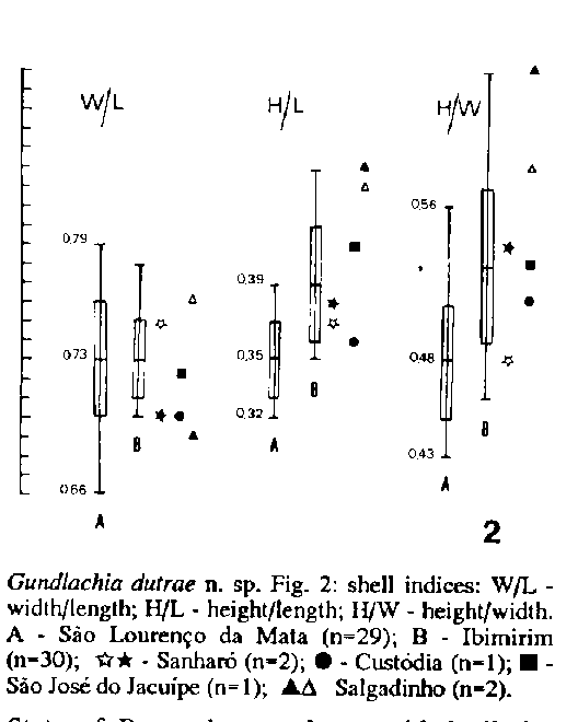
Shell relatively high. Aperture near-circular; periostracum dark brown without periostracal hairs. Apex slightly inclined to the right, projected but not hooked, with an apical depression surrounded by a sculpture of well-marked irregular punctations. Shell surface with prominent radial sculpture. No septate specimens were observed. Ratios (n= 59): shell width/shell length = 0,66- 0,79 (mean 0,73); shell height/shell length = 0,32- 0,45 (mean 0,37); shell height/shell width = 0,43- 0,63 (mean 0,51).
Body of normal ancylid type; mantle pigmentation dark brown or black, concentrated along the mantle collar.
The dorsal surface of the right anterior muscle is elongated and medially constricted. The left anterior and the posterior muscles are almost elliptical. Adhesive area is V-shaped.
Pseudobranch unpigmented bearing a very small and thin dorsal lobe.
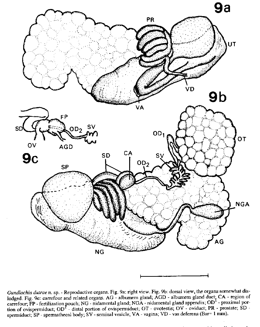
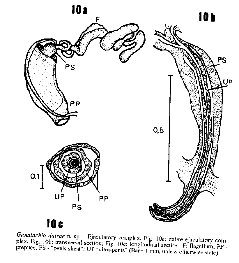
Ovotestis with more than 25 unbranched diverticula. Ovispermiduct with seminal vesicle rather developed. Elongated nidamental gland continuous with the glandular wall of the uterus. Nidamental gland appendix ending into a bulbous swelling. Spermathecal body almost rounded. Well-developed prostate with five long diverticula. Ejaculatory complex with long glandular flagellum, without a penis or true ultra-penis. "Penis sheath" developed. "Ultra-penis" projected as a tube inside the lumem of prepuce, with a slit between "ultra-penis" and "penis sheath".
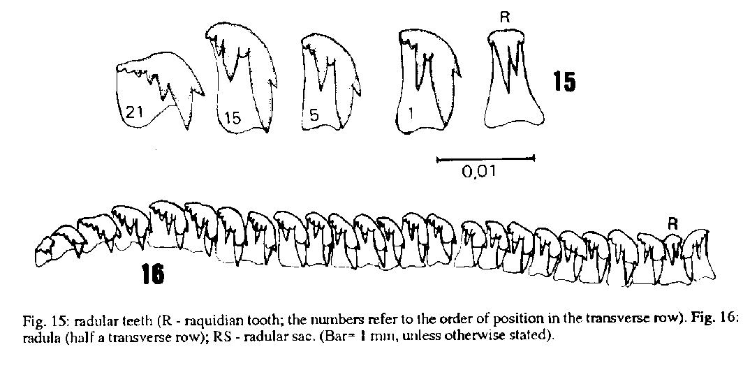
Rachidian tooth tetracuspid, with two median cusps assymmetrical and aculeated. Lateral teeth tricuspid, with a reduced endocon and a prominent mesocon. A well marked gap occurs between meso and ectocon. Marginal teeth similar to lateral ones.
Jaw T-shaped, with about 28 dorsal plates.
Key words: Mollusca - Ancylidae - Gundlachia dutrae - morphology
The ancylids are widespreading freshwater snails. They are represented in Brazil by Gundlachia Pfeiffer, 1849, Ferrissia Walker, 1903, Burnupia Walker, 1912 and Laevapex Walker, 1903.
Besides its abundance and wide distribution in South America, anatomical knowledge of ancylids is still scarce. Most species have been described on shell morphology. Anatomical des-criptions were presented by Scott (1953), Marcus and Marcus (1962), Hubendick (1964), Fernandez (1981), Lanzer (1988, 1991), and Santos (1989).
MATERIAL AND METHODS
Six samples were studied being five from the State of Pernambuco and one from the State of Bahia.
Twenty-nine living specimens from type-locality were relaxed in a 0.1% solution of nembutal for 6 hr, drawn from the shell and placed in Railliet-Henry's fixative. Five of them were dissected under the stereomicroscope.
Jaws and radulae of studied specimens were removed by means of 5% solution of KOH kept over a termic plate for 6 to 12 hr. They were mounted unstained in glicerine and some were stained with acetic carmin and mounted with Entellan.
The prepuce was pared away from the ejaculatory complex, mounted in glycerine and examined under microscope. Ejaculatory complex is used instead of penial complex because Gundlachia has no penis.
Specimens were collected at type-locality on superior surface of submersed rocks, in a small rapid stream.
Specimens were deposited in the following malacological collections: Instituto Oswaldo Cruz, Rio de Janeiro; Museu Nacional do Rio de Janeiro, Rio de Janeiro; Academy of Natural Sciences, Philadelphia; Geneve Natural Museum.
DESCRIPTION
Gundlachia dutrae n. sp.
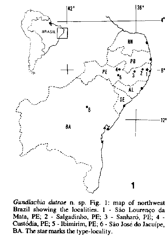
Types: Holotype: 27 C. Mol. IB-UERJ- Alto da Buchada Forest, Ecological Reserve of Tapacurbeta, Spio Louren‡o da Mata City, State of Pernambuco. S. B. Santos, N. C. Salgado & A. V. C. Dutra col. 06/90.
Paratypes: 27-A C. Mol. IB-UERJ- with the same data of holotype (25 shells and animals); DZUFPE 18- Poco da Cruz, Ibimirim, Pernambuco. A. V. Dutra col. 12/03/82 (30 shells and animals, in alcohol); DZUFPE 116- Sanharo, Pernambuco. A. V. Dutra col. 26/05/81 (4 shells and 10 animals); DZUFPE 394- Riacho Pascoal, Custodia, Pernambuco. A. V. Dutra col. 16/01/86 (1 shell and animal); FIOCRUZ 3274 - Sao Jose do Jacuipe, Jacobina, Bahia. SUCAM col. 11/07/84 (1 shell and animal).
Type-locality: Alto da Buchada Forest, Ecological Reserve of Tapacura, Spio Lourenco da Mata City, State of Pernambuco.
Other examined material: SMF 44352- Salgadinho, Recife, Pernambuco (two shells lent to Dr. Rosane Lanzer by Senckenberg Museum).
The species name was dedicated to Dr Ana Virginea C Dutra, from Federal University of the State of Pernambuco, who provided all the facilities for field work.
The map on Fig. 1 shows the localities.
The shell indices of the specimens are shown in figure 2.
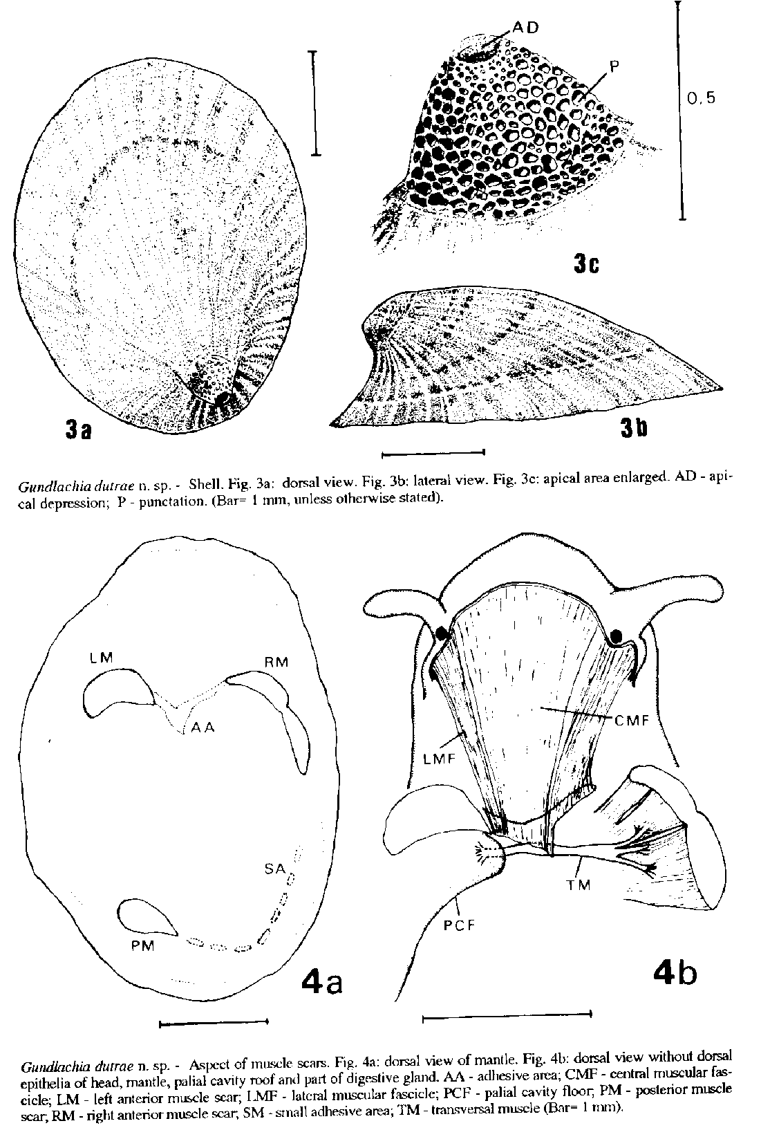
The shell aperture is near-circular, but a variation in the general shape of shell occurs. The periostracum is dark brown in color, showing various individual shades of color due to environmental impregnation. The apex is localized well-posterior in the shell, at the right of median line. It is projected but not hooked, pointing posteriorly and slightly to the right. There are a well marked apical depression surrounded by a sculpture of conspicuous irregular punctations (Fig. 3c).
A fine concentric growth sculpture and a well developed radial sculpture running from apex to margin occurs on all surface of shell (Figs 3a, b).
The highest shell, from Ibimirim, is 5.35mm in length x 3.75 mm in width x 2.00 mm in height. The shells from type-locality measure 3.10 x 2.15 x 1.10 mm to 4.75 x 3.40 x 1.70 mm.
Three muscle scars were observed, being the right anterior one the largest. It is an elongated scar, along the mantle margin, with a constriction in the middle. The left anterior muscle is elliptical, as the posterior one (Fig. 4a).
Between the two anterior muscle scars there is a V-shaped adhesive area.
Other small adhesive areas are seen between posterior muscle scar and the right anterior one. Delicate and isolated muscular fibers that come from the lateral body wall, run upward juxtaposed to the dorsal epithelia and reach these adhesive areas.
Removing carefully the head epithelia one can see, on right and left, two more evident delicate muscular fascicles formed by isolated fibers that originate on the body wall near the base of tentacles. Between these muscles there are several muscular fibers that come from the frontal region. These muscular fibers are attached on V-shaped adhesive area (Fig. 4b).
Removing the dorsal mantle epithelia, the roof of palial cavity and tearing away bit by bit the digestive gland, there is a transversal muscle running from ventral epithelia of palial cavity floor to internal wall of right anterior muscle. Just before reaching this muscle, the transversal muscle divides into three branches; the anterior one attaches the anterior fascicle of right muscle, the others the posterior fascicle (Fig. 4b).
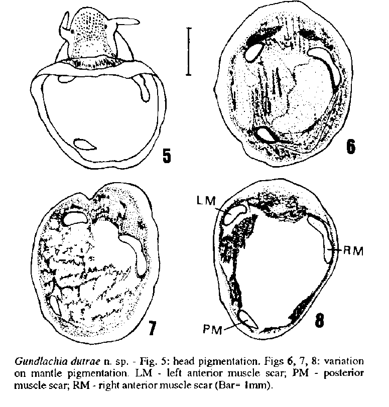
The pigmentation of mantle varies in individuals (Figs 6, 7, 8), but is generally dark brown or blackly pigmented, being more concentrated along the mantle collar and becomes lighter in the central area of mantle. Foot and tentacles are not pigmented. On the head there are scattered small blotches of pigment (Fig. 5).
The pseudobranch is rather developed; the ventral lobe has only two or three folds. The dorsal lobe is very small and thin. The pulmonary cavity is wide, reaching the midline of the animal. The excretory system has the typical serpentine shape of ancylids.
The genital system is shown in Figs 9 and 10. The ovotestis is wide, having about 35 unbranched closely pressed diverticula. It was embedded in the digestive gland, being its apex situated underneath the caudalmost intestinal loop.
The seminal vesicle is an enlargement of ovispermiduct with several digitiform-like, sometimes rounded diverticula.
The carrefour is a small structure situated between the albumen gland and the nidamental gland, represented by a short trunk that also gives rise to the oviduct, the spermiduct and receives the ovispermiduct (Fig. 9c). The spermiduct emerges from its left side, twists upward and soon downward, and runs forward giving rise to five unbranched prostatic diverticula. After, the spermiduct continues into the vas deferens, which runs forward within the lateral body wall to emerges just posterior to the attachment of the prepuce. The ejaculatory complex has a long glandular flagellum and a slitted "ultrapenis" is present.
The albumem gland is a large, almost cylindrical organ, with the posterior end twisted above and turned to the left. Its duct empties into an ovoid fertilization pouch. The oviduct emerges from right side of carrefour as a short duct that soon enlarges and reaches the nidamental gland.
The nidamental gland is a large yellowish , almost cylindrical organ, being continuous with the uterus. This organ is flattened, thin-walled and well-pigmented. Its anterior portion reaches the ejaculatory complex. At the right posterior wall of nidamental gland, pressed against the albumem gland, there is a very thin-walled pouch that gives rise to a long appendix. This appendix ends in an elongated-elliptical bulb with a metallic shine, near the posterior muscle.
The spermatheca has a roundish body. Its proximal duct is enlarged and gradually becomes more straight until it reaches the spermathecal body.
The digestive system has no features of special interest. There is only an opening for the digestive gland.
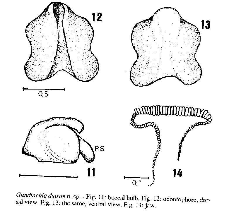
The radular sac is relatively long, about half the length of buccal bulb (Fig. 11). Figs 12 and 13 show the odontophore.
The jaw is well developed, with about 28 dorsal plates and 45 lateral plates.
The radula shows marked characteristics (Fig. 15). There are a quadricuspid raquidian tooth, being the two main cusps assymetrical and aculeated. Besides each principal cusp there is one small acessory cusp. There are three principal cusps in the lateral teeth. The mesocon is the most developed, long and aculeated; the endocon is reduced; between the meso and ectocon there is a marked gap; two or three pos-ectocon cusplets are visible, their number increasing in direction of the radular edge. The transition from lateral to marginal teeth is noted by progressive reduction of teeth size, not by form changes (Fig. 16).
REMARKS
Gundlachia Pfeiffer, 1849 , has been considered an exclusive Neotropical genus, with several nominal species, the great majority of them based exclusively on shell. A good summary about this is provided by Hubendick (1967).
Comparing shells, radula and anatomical data, it was no possible to identify our specimens as to any Gundlachia already described.
The sculpture and form of apex distinguish G. dutrae immediattelly from G. concentrica (Orbigny, 1835) where the apex is projected and hooked; from G. moricandi (Orbigny, 1837) where the apex is obtuse, and from G. radiata (Guilding, 1828) or G. ticaga (Marcus & Marcus, 1962) where the apex is rounded.
The relation length/height, the sculpture of strong radial lines and the position and inclination of the apex ressembles G. obliqua (Broderip & Sowerby, 1832) from Chile, but differs in the absence of radial lines in the protoconch.
The muscle scars are totally different from other Gundlachia. The most similar is seen in G. obliqua, where the right anterior muscle is also elongated, but never medially constricted. The genus Burnupia also has an elongated form of the right muscle scar (Brown 1961, Hubendick 1964).
The V-shaped adhesive area as well as the form of right anterior muscle scar relate G. dutrae n. sp. to G. concentrica, G. crequii (Bavay, 1904), G. obliqua and perhaps G. foncki (Philippi, 1866) according to anatomical data (Haas 1955, Hubendick 1955, 1964, 1967, Lanzer 1988) and personal observations.
Burch (1962) was probably the first author to mention the presence of additional muscle scars between the two anterior muscles, considering them as broken areas of adhesive epithelium. Lanzer (1988) observed in this area in G. crequii merely the presence of firmer tissue.
It was observed in G. dutrae n. sp. and other congeneric species (unpublished data) that the adhesive area is a true muscle scar, instead of an adhering epithelia. We believe that this character will probably be useful in taxonomic studies of ancylids.
The transversal muscle is described for the first time in ancylids.
The most similar species is G. obliqua from Chile. Apart the similarities already cited, both species present the mantle heavily pigmented, G. dutrae differing by having the foot, body wall and pseudobranchia not pigmented. The radula is similar in relation to rachidian tooth, where the median cusps are elongated and aculeated, the mesocon is the most pronounced and the endocone tends to be reduced. There are not marked differences from lateral to marginal teeth. In G. obliqua there is no gap between meso and ectocon (Pilsbry 1924) as in G. dutrae n. sp. The seminal vesicle is not so developed as in G. concentrica and G. moricandi. The ejaculatory complex is similar, both having an "ultra-penis" projected as a tube within the lumem of prepuce and with a well developed slit.
Both species are commom in lotic habitats, G. obliqua preferently on lower surface of stones (Biese 1948 and personal observations) whereas G. dutrae was found on upper surface of stones. Other congeneric species are most commom in lentic habitats on leaves, stems and remains of aquatic plants (Hubendick 1964, Indrusiak 1983, Harrison 1983, Lanzer 1988).
The shells SMF 44352 was identified by Haas (1939:267) as Burnupia (Unancylus) barilensis (Moricand, 1845) one of the synonyms of Gundlachia concentrica. It is clearly a misconception.
ACKNOWLEDGEMENTS
The author acknowledges Dr Wladimir L Paraense for permission to examine the collection of mollusks of Department of Malacology from FIOCRUZ; Dr Norma C Salgado from MNRJ and Dr Ana Virginea C Dutra from UFPE for the remarkable field work; Dr Rosane Lanzer and MSc. Fernanda Owlweiler from PUC-RS for examining SMF specimems and Dr Jose Luiz Moreira Leme from MZUSP for constant and stimulating criticism. To Biomedical Center of UERJ who provided part of financial sources for field work.
REFERENCES
Biese WA 1948. Revision de los moluscos terrestres y de betagua dulce de concha de Chile Bol Mus Nac Hist Nat Chile 24: 217-239.
Brown DS 1961. A description of Burnupia sp. cf. cafra (Krauss) (Gastropoda, Ancylidae) from Ethiopia. Ann Mag nat Hist 4 : 377-382.
Burch JB 1962. Note on the classification of freshwater limpets. Ann Rep Amer malac Un 29: 8-9.
Fernandez D 1981. Ancylidae p. 101-114. In RA Ringuelet, Fauna de agua dulce de la Republica Argentina, Vol. 15. Consejo Nacional de Investigaciones Cientificas y Tecnicas de la Republica Argentina.
Haas F 1939. Zur Kenntnis der Binnen-Mollusken NO-Brasilien. Senckenberg 21: 254-278.
Haas F 1955. Reports of Percy Sladen Trust Expedition to Lake Titicaca in 1937. XVIII. Mollusca: Gastropoda. Trans Linn Soc London 1 : 283-308.
Harrison AD 1983. Identity of Ferrissia irrorata and Gundlachia radiata, species of Ancylidae from St. Vicent, W I. Arch Moll 113: 7-15.
Hubendick B 1955. The anatomy of the Gastropoda. The Percy Sladen Trust Expedition to Lake Titicaca. Trans Linn Soc London ser. 3, 1: 309-327.
Hubendick B 1964. Studies on Ancylidae. The subgroups. Goteborgs K Vetensk -o Vitterh Samh Handl 9B: 1-72.
Hubendick B 1967. Studies on Ancylidae. The Australian, Pacific and Neotropical form groups. Acta R Soc sci litt Gothoburg 1: 5-52.
Indrusiak LF 1983. Inventbetario da fauna malacologica do Rio Ibicui-Mirim, RS. Ciencia e Natura 5: 127-134.
Lanzer R 1988. Rasterelektronenmikroskopische Untersuchungen an Gundlachia crequii (Bavay, 1904). (Basommatophora: Ancylidae). Arch Moll Frankfurt am Main 19: 205-217.
Lanzer R 1991. Duas novas especies de Ancylidae (Gastropoda: Basommatophora) para o sul do Brasil. Rev Brasil Biol 51: 703-719.
Marcus E, Marcus E 1962. On Uncancylus ticagus. Bol Fac Filos Cienc Univ S Paulo Spio Paulo, (Zool. 24), 216: 217-254.
Pilsbry HA 1924. South American land and freswater mollusks: notes and descriptions. II- The South American genera of Ancylidae. Proc Acad Nat Sci Phil 76: 54-61.
Santos SB 1989. On the morphology of Laevapex vazii n. sp. from Brazil. (Mollusca: Pulmonata: Basommatophora: Ancylidae). Mem Inst Oswaldo Cruz 84, Supl. IV: 467-473.
Scott MIH 1953. Notas sobre morfologia de Gundlachia Pfr. (Ancylidae). Physis 20: 467-473.
Copyright 1994 Fundacao Oswaldo Cruz - FIOCRUZ The following images related to this document are available:Line drawing images[oc94033a.gif] [oc94033c.gif] [oc94033g.gif] [oc94033f.gif] [oc94033d.gif] [oc94033h.gif] [oc94033b.gif] [oc94033e.gif] |
| |||||||||
