 |
| About Bioline | All Journals | Testimonials | Membership | News |
|
||||||
|
||||||
The Use of Ferromagnetic Dacron as Solid-phase in Enzyme ImmunoassaysAna Maria dos A Carneiro Leao (/***), Luiz B Carvalho Jr (*/***/), Elizabeth Malagueno (**/***)
Code Number: OC94039
Size of Files:
    Text: 20K
    Graphics: Line Drawings (gif) - 39K
Departamento de Morfologia e Fisiologia Animal, Universidade Federal Rural de Pernambuco * Departamento de Bioquimica ** Departamento de Medicina Tropical *** Laboratorio de Imunopatologia Keizo Asami (LIKA), Universidade Federal de Pernambuco, Cidade Universitaria, 50670-420, Recife, PE, Brasil
Ferromagnetic dacron is proposed as an alternative solid-phase for magnetic enzyme immunoassays. Human serum albumin (HSA) was covalently immobilized onto ferromagnetic dacron and an enzyme immunoassay was developed using anti-HSA rabbit sera. Peroxidase, o- phenylenediamine (OPD) and hydrogen peroxide were used as the enzymatic label and substrates, respectively. Best results were observed when particles of 63-100 um (diameter) and 10 ug of immobilized antigen were used. Positive reactions were detected until dilutions of 1:51200 of immune sera. Its reproducibility was similar to standard ELISA. Disruption of the immunocomplexes formed and recuperation of the immobilized antigen in other immunoassays also proved to be reliable.
Key words: magnetic ELISA - ferromagnetic dacron - human serum albumin
Immobilized biosystems have been widely employed as described in extensive and recent reviews (Coughlan et al. 1988, Kennedy et al. 1989, Powell 1990). Heterogeneous immunoassays require the use of antigen or antibody immobilized on a solid phase. The most common solid phases used in immunology are different kinds of plastics, such as PVC, nylon, glass and polystyrene (Guesdon & Avrameas 1981a, Kemeny & Challacombe 1988). Magnetic supports were firstly used in immunology by Hersh and Yaverbaum (1975) employing silanized magnetite as solid phase in a radioimmunoassay for digoxin. Lately other magnetic supports were proposed like polyacrylamide-agarose (Guesdon & Avrameas 1977, 1981b, Camargo et al. 1984, 1986), Sepharose (Mosbach & Anderson 1977), polystyrene (Robinson et al. 1985) and cellulose (Paus & Nustad 1989).
Among a lot of supports described in literature, Dacron has been successfully used in enzyme immobilization (Weetall 1970, Goldstein et al. 1977). It is a polyester widely employed as a plastic in several industrial applications. Lately, an easier method was proposed to immobilize enzymes on dacron consisting in thee steps: (1) Hydrazynolysis, (2) Conversion of Hydrazide Groups to Azide and (3) Immobilization of Protein (Carvalho Jr et al. 1986, 1987, Oliveira et al.1989). Its application has been extended to other proteins such as antigen from Yersinia pestis (Montenegro et al. 1991, 1993).
Recently, ferromagnetic Dacron was proposed as a matrix to immobilize proteins covalently (Carneiro Leao et al. 1991). It presents good mechanical properties, is resistant to solvents and to microbial attack, and is easily recovered from the medium using a magnetic field, replacing centrifugation or filtration.
In this work, antigen (human serum albumin) has been immobilized on ferromagnetic dacron and magnetic ELISA has been carried out successfully.
MATERIALS AND METHODS
Proteins and reagents - Dacron (R) was produced by Rhodia do Brasil SA. Human serum albumin (HSA) fraction V and bovine serum albumin (BSA) were purchased from Sigma Chemical Co. FeCl3.6H2O and FeCl2.4H2O were acquired from Reagen SA. NUNC Immunoplates 96 wells were purchased from Intermed, Denmark. All other reagents were analytical grade obtained from Merck SA.
Sera and immunization procedure - Normal and anti-HSA sera were obtained in our laboratory. Two New Zealand white three months rabbits were inoculated with two injections of HSA proximal to the lymphonode (10 mg with complete Freund adjuvant/each) on days 1st and 21st. On days 40th, 47th and 54th the animals were inoculated with alumen precipitated HSA (5 mg/ml) i.m. On day 59th rabbits were bleeded. The pool of sera contained 2.47 mg/ml of specific antibodies as determined by immunoprecipitation (Hudson & Hay 1989). Pig anti-rabbit IgG conjugated to peroxidase was bought to Dako (Denmark).
Activation and magnetization of dacron - This stage was proceeded according to Carneiro Leao et al. (1991). Hydrazide-dacron was freeze-dried and the particles were separated into 63-100 um (small particles) and 100-250 um (large particles) sizes though appropriate sieves. The resulting ferromagnetic-hydrazide-dacron was converted to ferromagnetic-azide-dacron as described previously.
Immobilization of HSA ferromagnetic dacron - HSA (5 ml of a 5.0 mg/ml solution prepared in 0.15 M NaCl) was incubated with the ferromagnetic-azide-dacron ( 0.5 g ) for 3 hr at 4 oC under mild stirring. Then, the ferromagnetic powder of HSA-dacron was successively washed with 0.15 M NaCl (100 ml), 1.0 M NaCl (100 ml) and 0.15 M NaCl (100 ml), and stored at - 20 oC in 0.15 M NaCl until use.
Antibody assay - A suspension of the ferromagnetic HSA- dacron particles was adjusted to a protein content of 250 ug/ml in 0.15 M NaCl and aliquots of 50 ul were withdrawn to the assay tubes. Skinned milk (1% w/v) prepared in 0.01 M phosphate buffer, pH 7.2, containing 0.15 M NaCl (PBS) was used as a blocking buffer during 1 hr at 25 oC. Incubation with anti-HSA serum and with swine anti- rabbit IgG conjugated to peroxidase (diluted 1:5,000) were carried out at 37 oC during 1 hr. Washing between incubations were proceeded using PBS Tween 0.05 % v/v (PBS/Tween) by precipitation of the ferromagnetic HSA-dacron particles under a magnetic field (6,000 Oe) and discarding the supernatant. The development of the reaction was carried out using 0.026% v/v H2O2 and 0.17 M o-phenylenediamine (OPD) prepared in 0.3 M Tris/citrate buffer, pH 6.0 as substrates during 15 min and read at 492 nm. Protein contents were determined according to Lowry et al.(1951), using a BSA standard curve.
Classical ELISA - It was done following the general procedure described by Voller et al. (1977). In brief: 5 ug/ml (0,25 ug/well) of HSA were adsorbed to a NUNC immunoplate using carbonate buffer pH 9.0 overnight. First antibody was incubated during 1 hr at 37 oC. The rest of the reaction followed some general procedure described previously to ferromagnetic-dacron. Anti-rabbit IgG conjugated to peroxidase was diluted at 1:5000 after checkerboard titration.
Recuperation of immunocomplexes - The final immunocomplexes obtained from an antibody assay using ferromagnetic- dacron were washed in 500 ul of PBS/Tween for five times. Magnetic field was used to recover the magnetic modified immobilized antigen preparation. Subsequently, this derivative was vigorously stirred in 1,000 ul of 0.05 M glicine/HCl buffer, pH 2.3, by using a vortex mixer, and kept resting for 60 min at 28 oC, with similar stirring each 15 min intervals. Then, Tris/HCl buffer (1,500 ul of 0.05 M solution, pH 8.6) was added to the magnetic preparation, with stirring. Finnaly, the material was washed with PBS/Tween for five times. The recovered material in the last washing was used for another antibody assay as described above.
RESULTS AND DISCUSSION
Ferromagnetic-dacron using amyloglucosidase as a model has been previously tested as a suitable matrix for protein immobilization (Carneiro Leao et al. 1991). The ferromagnetic dacron derivative was completely recovered by using a magnetic field and no enzymatic activity was detected in the supernatant. Else, the presence of magnetite did not interfere with the hydrazide/azide conversion step and with the catalytic property of the immobilized enzyme.
In order to obtain larger particles (powder), making recovery by magnetic force more efficient, incubation time of the dacron with hydrazine hydrate was reduced from 72 to 48 hr (Carvalho Jr et al. 1986, 1987).
The relationship between offered HSA for the immobilization onto ferromagnetic-azide-dacron and fixed protein was determined. A linear correlation was observed until concentration of 5.0 mg/ml (10 ug of protein are fixed onto 1 mg of support per each mg of offered protein). A maximum value of about 120 ug of fixed protein per g of ferromagnetic-dacron was calculated if protein concentration of about 200 mg/ml would be used during immobilization. Here, protein concentration of 5.0 mg/ml (50 ug of fixed protein per mg of support) was proposed since it should be advisable to avoid antigen overloading that can yield steric hindrance for the antigen- antibody complex formation. This preparation was used to calculate the optimum amount of protein per assay.
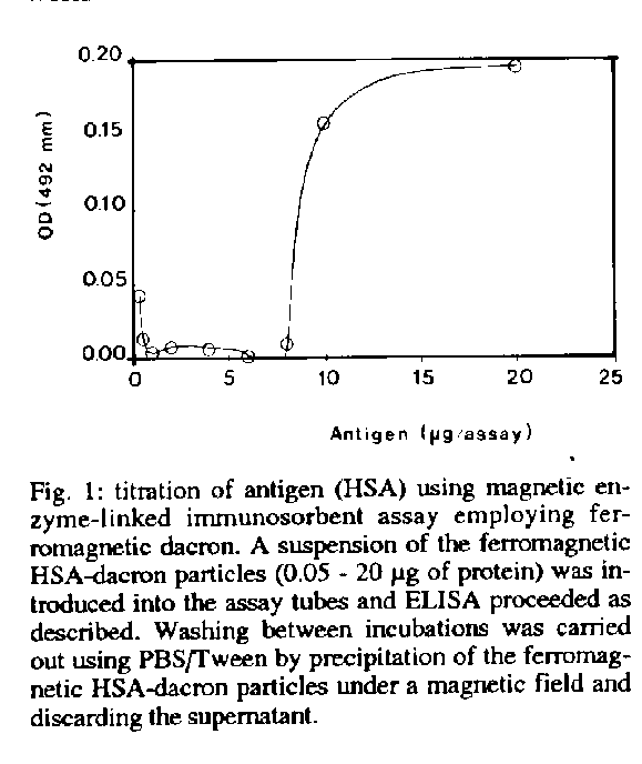
Figure 1 presents the relationship between the amount of antigen (ug of HSA-ferromagnetic-dacron derivative) used in the proposed assay and the absorbance at 492 nm by using ELISA procedure. It was compared with the standard procedure employing polystyrene plates. The amount of 12.5 ug of protein per assay used thoughout this work were selected from these results. Optimization of this technique would improve its performance so that lower amount of protein is required.
TABLE
Titration of rabbit anti-HSA serum using magnetic enzyme-linked immunosorbent assay and the recuperation of this derivative
===============================================================
                                    Sera
                      -----------------------------------------
Assay                 Normal (a)        lmunized (a)
                      1:100   1:100     [F]   1:1.600      [F]
                      (O.D.)  (O.D.)           (O.D.)
---------------------------------------------------------------
First assay           0.059   0.463     7.85   0.309       5.24
After recuperation    0.118   0.835     7.08   0.687       5.82
Second recuperation   0.162   0.956     5.90   0.684       4.22
===============================================================
a: these values represent the means of duplicates of OD at 492 nm. [F]: correlation factor Ferromagnetic HSA-dacron particles (63-100 Um; 12.5 Um of immobilized protein) were introduced into the assay tubes and antibody assay proceeded as described. The used HSA-ferromagnetic- dacron derivative was washed in PBS/tween and kept overnight. Subsequently, it was treated with glicine/HCl buffer and Trisl/HCl buffer, successively. Finnaly, the material was washed with PBS/tween and used for another assay as described above. A second reuse was proceeded. The ferromagnetic HSA-dacron particles were recovered by using magnetic field.
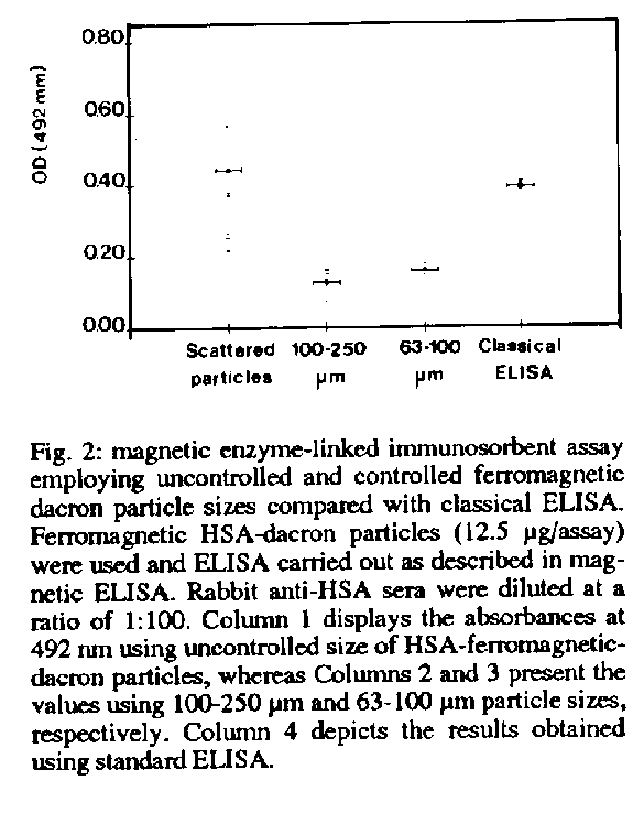
The critical influence of the ferromagnetic-dacron particle size is demonstrated in Fig. 2. More scattered values are observed when ferromagnetic-dacron with uncontrolled particle size was used compared with those from controlled particle size and classical ELISA. These data can be better analyzed by the average and standard deviation of the absorbances at 492 nm obtained by using these materials for the magnetic ELISA: uncontrolled particles size - 0.441 +/- 0.216; 100-250 um size - 0.128 +/- 0.031; 63-100 um - 0.158 +/- 0.012. The mean OD value obtained from classical ELISA was 0.391 +/- 0.013. Considering these results, small particles (63-100 um) were selected to be used thoughout this work.
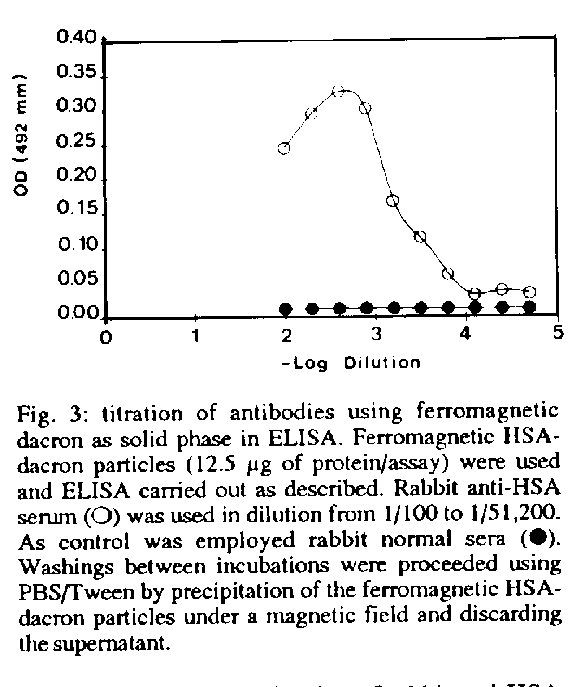
Figure 3 shows a typical antibody titration by using ferromagnetic dacron (63 - 100 um) as solid phase in ELISA. A ratio of 2.5 was still estimated between absorbances at 492 nm of the immunized rabbit sera at the dilution of 1/51,200 (4,82 ug/100 ml of sample) and of the normal rabbit serum diluted at 1/100.
Table presents the titration of rabbit anti-HSA serum using magnetic enzyme-linked immunosorbent assay employing ferromagnetic dacron and the recuperation of this derivative. Firstly, the method showed to be effective using 12.5 ug of HSA per assay comparing with those values obtained for the normal rabbit sera. Furthermore, the same water insoluble HSA-ferromagnetic-dacron derivative used for a second time, after a previous treatment to remove the complex with the first antibody (rabbit anti-HSA serum) and the second antibody (pig anti-rabbit IgG conjugated to peroxidase), presented reliable values to evaluate the titration of rabbit anti-HSA serum. The data are relatively higher than those described for the first assay because of some remnant antibody not removed by the treatment with glycin/HCl buffer although the correlation factor (F) between absorbances of immunized and normal sera remained similar. Finally, a third use of the same derivative also showed significant values to estimate the titration of rabbit anti-HSA serum.
The summary of these findings indicates that antigen-ferromagnetic- dacron derivative can be used for enzyme-linked immunosorbent assay with some advantages: magnetic field replaces centrifugation or filtration and the antigenic complex can be used more than once. However it is important to observe that relatively higher amounts of antigen were required in this technique with some loss of sensivity if compared with polystyrene matrix, probably due to steric hindrance of antigenic sites. Probably the big amount of protein required for immobilization modify the presentation of epitopes (Kricka et al. 1980, Venkatesan & Wakelin 1993). This phenomenon was also observed when complex antigenic extract (parasite crude extract) were used (not shown). Optimization of this method will certainly offer best results. So, it is possible to conclude that this method should be economic relevant for immunological purposes, specially considering its recuperation. In our laboratory, the diagnosis of some tropical diseases by using this method is under investigation.
ACKNOWLEDGMENTS
To Dr Paulo Paes de Andrade for providing encouragement and support.
REFERENCES
Camargo ZP, Guesdon JL, Drouhet E, Improvisi L 1984. Magnetic enzyme-linked immunosorbent assay (MELISA) for determination of specificIgG in paracoccidioidomycosis. Sabouraudia 22: 291-299.
Camargo ZP, Unterkircher C, Drouhet E 1986. Comparison between magnetic enzyme-linked immunosorbent assay (MELISA) and complement fixation test (CF) in the diagnosis of paracoccidioidomycosis. J Med Vet Mycology 24: 77-79.
Carneiro Leao AMA, Oliveira EA, Carvalho Jr LB 1991. Immobilization of protein on ferromagnetic dacron. Appl Biochem Biotechnol 33: 3-58.
Carvalho Jr LB , Melo EHM, Vasconcelos ARA, Lira RR 1986. Glucose oxidase immobilised on gel beads polyacrylamide and polyethylenetherephtalate. Arq Biol Tecnol 29: 525-531.
Carvalho Jr LB, Silva MPC, Melo EHM 1987. Activity of immobilized alpha-amylase. Braz J Med Biol Res 20: 521- 526.
Coughlan MP, Kierstan MP, Border PM, Turner APF 1988. Analytical applications of immobilized proteins and cells. J Microbiol Meth 8: 1-50.
Goldstein L, Freeman A, Blassberger D, Granot R, Sokolovsky M 1977. Chemically modified polymers containing isocyanide functional groups as support for enzyme immobilization, p.153-167. In Z Bohak, N Sharon (eds) Biotechnological Applications of Proteins and Enzymes, Academic Press, New York.
Guesdon JL, Avrameas S 1977. Magnetic solid phase enzyme- immunoassay. Immunochem 14: 443-447.
Guesdon JL, Avrameas S 1981a. Solid phase enzyme immunoassays. Appl Biochem Bioeng 2: 207-232.
Guesdon JL, Avrameas S 1981b. Magnetic solid-phase enzyme immunoassay for the quantitation of antigens and antibodies: application to human immunoglobulin E. Meth Enzymol 73: 471-482.
Hersh LB, Yaverbaum S 1975. Magnetic solid-phase radioimmunoassay. Clin Chem Acta 63: 69-72.
Hudson L, Hay FC 1989. Antibody interaction with antigen, p. 207- 263. In L Hudson, FC Hay (eds) Practical Immunology, 3rd ed., Blackwell Scientific Publications, Oxford.
Kemeny DM, Challacombe SJ 1988. Microtitre plates and other solid phase supports, p. 31-55. In DM Kemeny, SJ Challacombe (eds) ELISA and other solid phase immunoassays, John Wiley & Sons, New York.
Kennedy JF, Melo EHM, Jumel K 1989. Immobilized biosystems in research and industry, p. 297-313. In GE Russell Biotechnology and Genetic Engineering Reviews, Intercept, Newcastle.
Kricka LJ, Carter TJN, Burt SM, Kennedy JH, Holder RL, Halli Day MI, Telford ME, Wisdom GB 1980. Variability in the adsorption properties of microtitre plates used as solid supports in enzyme immunoassay. Clin Chem 26: 741- 744.
Lowry OH, Rosebrough NJ, Farr AL, Randall RJ 1951. Protein measurement with the folin phenol reagent. J Biol Chem 193: 265-275.
Montenegro SML, Almeida AMP, Carvalho AB, Carvalho Jr LB 1991. The use of dacron plates for dot-enzyme linked immunosorbent assay (dot-ELISA). Mem Inst Oswaldo Cruz 84: 461-465.
Montenegro SML, Almeida AMP, Carvalho AB, Carvalho Jr LB 1993. Standartization of the dot enzyme-linked immunosorbent assay (Dot- ELISA) for experimental plague. Mem Inst Oswaldo Cruz 88: 119-123.
Mosbach K, Andersson L 1977. Magnetic ferrofluids for preparation of magnetic polymers and their application in affinity chomatography. Nature 270: 259-261.
Oliveira EA, Silva MPC, Figueiredo ZMB, Carvalho Jr LB 1989. Immobilization of proteins on plates of dacron. Appl Biochem Biotechnol 22: 109-114.
Paus E, Nustad K 1989. Immunoradiometric assay for alpha, gamma- and gamma,gamma-enolase (neuron-specific enolase) with use of monoclonal antibodies and magnetizable polymer particles. Clin Chem 35: 2034-2038.
Powell LW 1990. Immobilized biocatalyst technology, p. 369- 394. In WM Fogarty, CT Kelly (eds) Microbial Enzymes and Biotechnology, 2nd ed., Elsevier, London.
Robinson GA, Hill AO, Philo RD, Gear JM, Rattle SJ, Forrest CG 1985. Bioelectrochemical enzyme immunoassay of human choriogonadotrophin with magnetic electrodes. Clin Chem 31: 1449-1452.
Venkatesan P, Wakelin D 1993. ELISAs for parasitologists: or lies, damned lies and ELISAs. Para-sitol Today 9: 228-232.
Voller A, Bidwell DE, Bartlett A, Edwards R 1977. A comparison of isotopic and enzyme-immunoassays for tropical parasitic diseases. Trans R Soc Trop Med Hyg 71: 431-437.
Weetall HH 1970. The insolubilized L-asparaginase implant: a preliminary report. J Biomed Mat Res 4: 597-599.
Copyright 1994 Fundacao Oswaldo Cruz - FIOCRUZ The following images related to this document are available:Line drawing images[oc94039c.gif] [oc94039b.gif] [oc94039a.gif] |
| |||||||||
