 |
| About Bioline | All Journals | Testimonials | Membership | News |
|
||||||
|
||||||
Reversibility of Muscle and Heart Lesions in Chronic, Trypanosoma cruzi Infected Mice after Late Trypanomicidal TreatmentMA Segura, E Molina de Raspi *, MA Basombrio *
Code Number: OC94044
Size of Files:
    Text: 17K
    Graphics: Line Drawings (Gif) - 23K
Facultad de Ciencias Naturales * Laboratorio de Patologia Experimental, Facultad de Ciencias de la Salud, Universidad Nacional de Salta, Calle Buenos Aires l77, 4400, Salta, Argentina
The effect of trypanomicidal treatment upon established histopathological Trypanosoma cruzi induced lesions was studied in Swiss mice. The animals were inoculated with 50 trypomastigotes and infection was allowed to progress without treatment for 99 days. After this period, the animals were divided in three groups, treated for 30 days with either placebo, benznidazole (200 mg/kg/day) or nifurtimox (100 mg/kg/day). These treatments induced 94 and 100% cure rates respectively as detected by xenodiagnosis and reduction of antibody levels. Autopsies and histopathological studies of heart, urinary bladder and skeletal muscle performed on day 312 after infection showed almost complete healing without residual lesions. As long periods were allowed between infection, treatment and autopsy, the results indicate that tissue lesions depend, up to advances stages, on the continuous presence of the parasite.
Key words: Trypanosoma cruzi - chronic infection - chemotherapy - histopathology
Drug treatment of Chagas' disease is mainly undertaken in acute patients, using two drugs against the ethiologic agent Trypanosoma cruzi: Benznidazole (Bz) and Nifurtimox (Nx) (WHO 1991). Besides the toxicity of these drugs, a serious therapeutic problem is that most patients first present long after the acute stage. At this time, parasitological follow up of infection is difficult and antiparasitic therapy is generally considered as overdue (MSP report 1983).
Experimental studies in mice are useful to test terms and schedules for effective treatment. Moreover, they allow some analysis of pathogenic mechanisms: the search for histological cure, several months after parasitological cure, seems to be a good test for the putative immunopathologic component of tissue lesions (Teixera et al. 1975, Cossio et al. 1980). The effects of late antiparasitic treatment in mice with T. cruzi induced lesions, established after 100 days of infection, were studied by Laguens et al.(1983) and Andrade et al. (1989). In the first study, lesions were recorded shortly after treatment, finding a remarkable healing effect in skeletal and gut muscle but little, if any, improvement in heart condition. Andrade et al. (1988) studied tissue lesions 90-100 days after treatment. They also found a clear healing effect, but complete tissue recovery was only found in 21-57% of the animals. In both studies, it was difficult to rule out whether autoreactive cells or antibodies could play a role in maintaining residual lesions. In this work we used Bz and Nx to test in mice the effect of late treatment upon advanced, chronic lesions induced by T. cruzi. The results showed a remarkable healing effect, reaching 100% with Nx, and a good correlation, both in mice with and without treatment, between residual parasitemia and lesion. This finding bears on generally accepted immunopathogenic mechanisms.
MATERIALS AND METHODS
Mice and parasites - Seventy female, outbred Swiss mice were used. They were 40 days old when the experiment started. The animals were infected intraperitoneally with 50 blood trypomastigotes of the COB isolate of T.cruzi, a parasite line obtained from Triatoma infestans in the field and passaged five times in mice.
Serological reactions - Blood was obtained by sectioning the tail tip of anesthesized mice. Direct agglutination of formalin-killed epimastigotes was tested with twofold serum dilutions on microtiter plates.
Histology - The complete heart, urinary bladder and quadriceps muscle of each mouse were fixed in 10% formalin, embedded in paraffin and histologic sections were stained with hematoxylin-eosin. Quantitation of lesions was based on a double blind analysis, performed by three observers, of three frontal sections of each organ, each taken at approximately l mm of each other.
The degree of mononuclear infiltration was classified as follows: 0 = no lesions; 1 = tissues presenting a total of one to four foci of infiltration in all sections; 2 = more than four foci and 3 = extensive or confluent areas of infiltration, often associated with degenerative or fibrotic lesions.
Detection of parasitemia - To test the presence of T. cruzi in blood, two methods were sequentially used: microhematocrit (MH) and xenodiagnosis (X). For MH (Freilij et al. 1983) 50ul of blood were collected from the tail tip of mice and centrifuged in a capillary pipette. T. cruzi was concentrated and detected microscopically in the leukocyte interphase. When parasites were not found, the animals were anesthesized and subjected to X. Ten, third instar, laboratory reared T. infestans were allowed to feed on each mouse for 15 min in the dark. Thirty and 60 days later the presence of parasites was studied in fresh mounts of the pooled bug's feces.
Drug treatments - Mice received 0.2 ml suspensions by an intragastric cannula containing either 100 mg/kg of Nx or 200 mg/kg of Bz in 2% carboxymethyl cellulose. The drugs were given daily for 30 days.
Experimental schedule - After infection (day 0) all animals were kept for 99 days without treatment and then divided into three experimental groups. The first (A, 22 mice) was tested with the drug vehicle alone, the second (B, 13 mice) received Bz and the third (C, 13 mice) received Nx. MH and/or X were applied on days 30 and 196. Serological determinations were performed on day 306 and autopsies on day 312. Since a few mice died during the experiments and a few serum or tissue specimens were lost, the numbers under "results" may be slightly less than the total of mice in the group.
Statistical analysis - The significance of differences was calculated with the Fisher's exact test for rates of infection and with the Mann-Whitney's "U" test for serological and histopathological data.
RESULTS
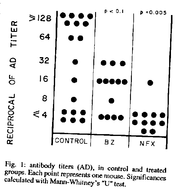
Parasitological and serological monitoring of infection - T. cruzi was detected in all mice on day 30 post infection. Parasites were still detected in 65 % (17/26) of non treated mice on day 130. At this time none of the 18 Bz treated mice revealed parasitemia by either MH or X and only one of the 16 Nx treated mice had positive X. Serological data of day 306 revealed a high antibody level (X+/-SE of titer: 24+/-10) in the control group and significant reductions in treated groups, which were stronger for Nx (4 +/- 1, p = 0.005) than Bz (12 +/- 5, p < 0.1) (Fig. 1). The detection of parasitemia on day 196 was predictive as to whether a mouse would present histologic alterations at autopsy (Table).
TABLE
Correlation between detection of parasitemia and histopathological alterations in infected, non treated mice
====================================================
             Histopathological alterations (+/total)
             ---------------------------------------
                            Urinary
                Heart       bladder      Muscle
----------------------------------------------------
Parasitemia   7/10 (70%)   8/11 (73%)   10/11 (91%)
Non           1/6  (17%)   2/8  (25%)    2/9  (22%)
parasitemia
              p = 0.056    p = 0.050    p = 0.031
====================================================
Histopathological evaluations of infected, non-treated mice - No macroscopic abnormalities were observed in any mouse at the time of autopsy. The microscopic lesions consisted essentially of lymphomonocytic infiltrates, centered around perivascular areas, each organ presenting particular features of distribution and damage. Heart: out of 19 mice, lesions were found in seven. These were discrete in extention and predominated in atria and perivalvular tissues. Hypertrophy of muscle fibers was found in four mice and pericardial fibrotic thickening in two. Urinary bladder: out of 19 mice, lesions were found in nine. These consisted of inflammatory infiltrates affecting the muscular layer and sometimes reaching extensive and confluent areas. Submucosal edema was a common finding. Skeletal muscle: this tissue presented the most extensive lesions observed and affected 12/22 mice. An intense chronic myositis with areas of acute inflammation was the main finding. Small infiltrates consisted only of lymphocytes. Moderate infiltrates often included plasmocytes. Severe infiltrates included microabscesses surrounded by polynuclear and lymphoplasmocytic cells or by areas of histiocytic proliferation and fibrosis. Small arteries often presented wall hyalinization and thickening. Amastigote nests were found in four mice.
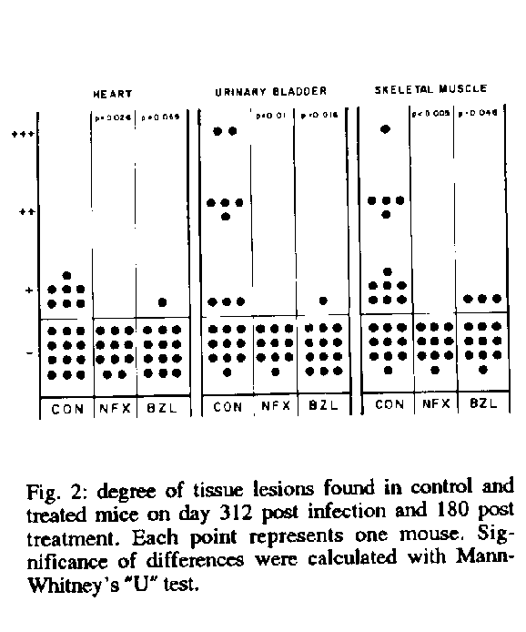
Histopathological findings after drug treatments - No histologic alterations were found in any of the Nx-treated mice. Only four of the Bz-treated animals presented slight tissue alterations. These consisted of isolated foci of lymphocytic infiltration in heart and urinary bladder (one mouse) and similar foci in skeletal muscle (three mice). The comparison of mice with lesions between treated and control groups showed that late treatment with both drugs had significantly cleared the alterations in the three tissues analized (Fig.2).
DISCUSSION
This study shows that established and severe histological alterations found in mice 312 days after T. cruzi infection, can be almost completely eliminated by antiparasitic treatment started as late as 99 days post infection. This finding has implications for both pathogenic mechanisms and therapeutic guidelines.
Much evidence has accumulated showing that tissue lesions in Chagas' disease are mediated by cellular or humoral immunologic effectors (Hudson 1985, Kierszenbaum 1985). While the pathogenic mechanism is open for a variety of analytical aproaches, an issue of major concern for therapy is whether lesions can progress in the absence of infection. Induction of tissue lesions in non infected animals has been obtained experimentally by transference of immune cells (Laguens et al. 198l, Hontebeyrie-Joskowicz et al. 1987) and with T. cruzi antigens (Teixeira et al. 1975, Segura et al. 1980). Although cell transfer experiments succeeded in showings that tissue damage can be immunologically mediated, they did not exclude that residual infection had been the triggering mechanism of self-reactivity in the donor animals, since the transferred "immune" cells were derived directly, or after culture, from animals with ongoing chronic T. cruzi infections. Myosistis and myocarditis were experimentally induced in mice (Segura et al. 1980, Laguens et al. 1981) and rabbits (Teixera et al. 1975) by injection of antigenic extracts of T. cruzi. However the infection schedules were heavy, involving weekly inocula of epimastigote antigens for nine months in rabbits or four doses of over 1 g of epimastigote fractions in mice. Under such regimes even normal tissue antigens (Cossio et al. 1984) or extracts from unrelated parasites (Cabeza Meckert et al. 1984) are pathogenic. In parasitologically cured but still seropositive animals, T. cruzi antigens have been detected by immunoelectron microscopy in the spleen and inflammatory lesions (Andrade et al. 1988, Ben Younes-Chenoufi et al. 1988). Thus, these results provide no ground to believe that tissue lesions found in parasitologically cured animals may be progressive rather than residual in nature.
Treatments initiated during the chronic stage of experimental Chagas' disease have been seldom evaluated histopathologically. Laguens et al. (1983) found an important reduction of tissue lesions in chronic mice after Bz or Nx treatment. In their work, tissues were studied shortly (1 to 60 days) after treatment. In our design, this period was prolonged to 180 days, assuming that autoimmune mechanisms, not triggered by residual parasites, might need such period to develop. However, we found no lesions which might be attributed to autoimmunity. As we had completed these experiments, we learned about a confirmatory study by Andrade et al. (1989), which reinforces the concept that whenever T. cruzi is therapeutically eliminated, the disease does not progress and residual lesions are cleared. The late reversibility of tissue lesions here found indicates that they depend, up to advanced stages, on the continuous presence of the parasite.
ACKNOWLEDGEMENTS
To Alejandro Uncos and Eduardo Isasmendi for their valuable technical assistance.
REFERENCES
Andrade SG, Freitas LAR, Peyrol S, Pimentel AR , Sadigursky M 1988. Trypanosoma cruzi antigens detected by immunoelectron microscopy in the spleen of mice serologically positive but parasitologically cured by chemotherapy. Preliminary report. Rev Soc Bras de Med Trop 21: 41-42.
Andrade SG, Magalhaes JB, Pontes AL 1989. Terapeutica da fase cr& circ;nica da infeccao experimental pelo Trypanosoma cruzi com o Benzonidazol e o Nifurtimox. Rev Soc Bras Med Trop 22: 113-118.
Ben Younes-Chennoufi A, Hontebeyrie-Joskowicz M, Tricottet V, Eisen H, Reynes M , Said G 1988. Persistence of Trypanosoma cruzi antigens in inflammatory lesions of chronically infected mice. Trans R Soc Trop Med Hyg 82: 77-83.
Cabeza Meckert P, Cazzulo JJ, Segura EL, Ruiz AM, Gelpi R , Laguens RP 1984. Induction of heart alterations by immunization with subcellular fractions from Crithidia fasciculata. Experientia 40: 171-173.
Cossio PM, Bustuoabad E, Patemo R, Iorn MB, Casanova MR, Podesta N, Bolomo R, Arana M, Pasqualini CD 1984. Experimental myocarditisn induced in Swiss mice homologous heart immunization resembles chronic experimental Chagas' disease. Clin Exp Immunol 33: 165-175.
Cossio PM, Diez C, Laguens RP, Arana RM 1980. Inmunopatologia de la Enfermedad de Chagas. Hechos y perpectivas. Medicina (Buenos Aires) 40: 222-230.
Freilij H, Muller L, Gonzalez Cappa SM 1983. Direct micromethod for diagnosis of acute and congenital Chagas'disease. J Clin Microbiol 18: 327-330.
Hontebeyrie-Joskowicz M, Said G, Milon G, Marchal G, Eisen H 1987. L3T4+T cells able to mediate parasite-specific delayed-type hypersensitivity play a role in the pathology of experimental Chagas'disease. European J Immunol 17: 1027-1033.
Hudson L 1985. Autoimmune phenomena in chronic chagasic cardiopathy. Parasitol Today 1: 6-9.
Kierszenbaum F 1985. Autoimmunity in Chagas Disease: cause or symptom? Parasitol Today 1: 4-6.
Laguens RP, Cabeza Meckert P, Chambo G, Gelpi RJ 1981. Enfermedad de Chagas cronica en el raton. II. Transferencia de la enfermedad cardiaca por celulas inmunocompetentes. Medicina (Buenos Aires) 41: 40-44.
Laguens RP, Cabeza Meckert P, Chambo JG, Gelpi R. 1983.Enfermedad de Chagas cronica en el raton. IV. Efecto de drogas trypanomicidas. Medicina (Buenos Aires) 43: 126-131.
MSP report 1983. Normas para la Atencion Medica del Infectado Chagasico. Ministerio de Salud Publica. Argentina 17 pp.
Segura EL, Cabeza Meckert P, Esteva M, Gelpi R, de Campanini AR, Subias E, Laguens RP l980. Accion de las fracciones subcelulares de Trypanosoma cruzi sobre la enfermedad de Chagas cronica en el raton. I. Induccion de cardiopatia en ausencia de infeccion. Medicina (Buenos Aires) 40: 807 (Abstract).
Teixera ARL, Teixera ML, Santos Buch CA 1975. The immunology of experimental Chagas disease. IV. Production of lesions in rabbits similar to those of chronic Chagas'disease in man. Am J of Pathol, 80: 163-178.
WHO 1991. Control of Chagas disease. Report of a WHO Expert Committe. Geneva: World Health Organization, Technical Report Series, No. 811.
Copyright 1994 Fundacao Oswaldo Cruz - FIOCRUZ The following images related to this document are available:Line drawing images[oc94044b.gif] [oc94044a.gif] |
| |||||||||
