 |
| About Bioline | All Journals | Testimonials | Membership | News |
|
||||||
|
||||||
Leishmaniasis Disseminated by Leishmania braziliensis in a Mare (Equus cabalus) Immunotherapy and Chemotherapy AssaysEGO Barbosa-Santos, MCA Marzochi, W Urtado (*), F Queiros (*), J Chicarino (***), RS Pacheco (**)
Code Number: OC94045
Size of Files:
    Text: 17K
    Graphics: Photographs (Jpg) - 67.6K / Halftones (Gif) - 112K
Departamento de Ciencias Biologicas, Escola Nacional de Saude Publica - FIOCRUZ, Rua Leopoldo Bulhoes 1480, 21041-210 Rio de Janeiro, RJ, Brasil * Clinica de Tratamento para Equinos "Horse Master", Niteroi, RJ, Brasil ** Departamento de Bioquimica e Biologia Molecular *** Hospital Evandro Chagas, Instituto Oswaldo Cruz, Rio de Janeiro, RJ, Brasil
Cutaneous disseminated lesions caused by Leishmania sp. were found in a pregnant mare (Equus cabalus) from a rural city in the State of Rio de Janeiro, Brazil. Before delivering, treatment was undertaken by immunotherapy followed by chemotherapy. Histopatology and serology were performed during treatment, as well as the biochemical characterization of the parasite (L. braziliensis) that was isolated from one of the lesions.
Key words: mucocutaneous leishmaniasis - disseminated lesions - horse - therapeutic
CASE REPORT
A 13-years-old, "Quarto de Milha", pregnant mare, from Sapucaia, State of Rio de Janeiro, presented several nodular and ulcerate cutaneous lesions along many parts of the body. The diagnosis was carried out by the identification of Leishmania amastigotes in a tissue biopsy of a cutaneous nodular lesion. Afterwards the animal was transferred to a specialized clinic for treatment.
The leishmaniasis skin test with P10000 antigen (Marzochi & Barbosa-Santos 1988) was applied in the upper part of the muzzle. Blood samples to detect IgG anti-leishmania antibody using an indirect immunofluorescence technique (IFT) were collected during the treatment. Nodular and ulcerate lesions were biopsied for histopathological exams prior to delivery and during immunotherapy and chemotherapy.
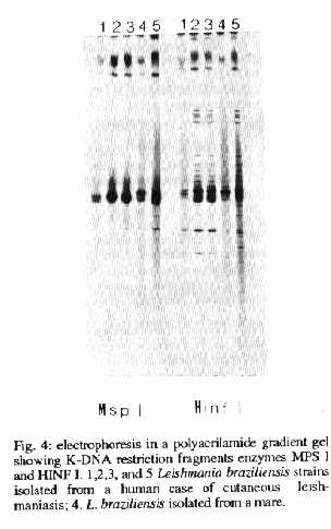
Biochemical characterization was performed by the analysis of restriction K-DNA profile, as previously shown using restriction enzymes MspI and HinfI (Pacheco et al. 1984). A standard sample of a laboratory strain of Leishmania braziliensis was used, which had originated from a human case from Jacarepagua, in the city of Rio de Janeiro, Brazil and the WHO (1990) reference strain of L. braziliensis.
Maintenance and in vivo retrieval of the strain was carried out by subcutaneous inoculation of the isolated parasite in the foot pad of young hamsters (Cricetus auratus). The animals were observed for eight months and subsequently killed. Liver and spleen fragments were then collected and introduced in a culture medium.
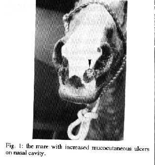
The delayed-type hypersensitivity reaction to the P10000 skin test was positive after 48 hr (diameter of induration area: 9.0mm). During three months before delivery, the clinical aspect of the mare and antibody levels remained unchanged, with lesions on genital organs, right hind thigh and nasal cavity (Fig. 1), IFT antibody titer was 1:160. Histological cuts showed an inflamatory infiltrate by lymphocytes, histiocytes, plasmocytes, polymorphonuclear cells and Leishmania amastigotes.
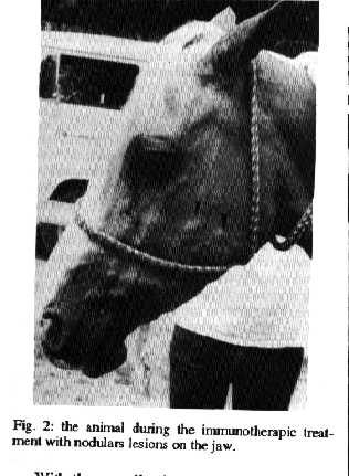
Immunotherapy was undertaken after delivery and during nursing of the foal. After the first series of proposed applications no change was observed either in the lesions or in the seric antibody levels. After the second application an aggravation of the clinical condition of the mare was observed. With the onset of the nodular and ulcerate lesions, apparently distributed along the superficial lymphatic draining system, affecting the four limbs, the teats and lateral parts of the neck and the jaw (Fig. 2); antibody titers increased to 1:320.
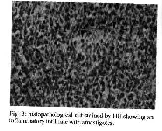
Histopathological analysis of the nodular lesions demonstrated connective and muscular tissues with a chronic inflamatory infiltrate, mainly composed of lymphocytes, plasmocytes and epithelioid cells, which formed granulomas, as well as necrosis, cellular debris and Langhans-type giant cells, several amastigotes could also be seen (Fig. 3).
With the complication of the clinical condition of the mare, conventional chemotherapy was adopted. After the first series of antimony, a stabilization in the progress of the lesions was observed. At the beginning of the second treatment series a regression in the nodular and ulcerate lesions was observed and seric antibody titers remained stable at 1:80 until the end of the 11-months observation period.
Histopathological analysis of the cutaneous nodules showed presence of a minor inflammatory infiltrate around vessels, mainly composed of monocytes, lymphocytes and plasmocytes. Vascular proliferation was also observed, with some vessels exhibiting a proeminent endothelium.
At the end of the second chemotherapy series the nodules had disappeared and the cutaneous lesions had also healed. A histopathological exam showed a sparse inflammatory infiltrate, composed of monocytes, especially around superficial and deep blood vessels. Fragments of nasal scars that were inoculated in culture medium and in hamsters were negative in respect to parasite isolation.
The IFT on blood samples taken from the foal upon birth and one month later presented titers of 1:40 and 1:80, respectively, remained unchanged during the months following suckling. The foal was subsequently separated from its mother and taken to a rural property in Sapucaia. During the following year, when the foal was observed, no lesions which could resemble leishmaniasis were detected.
The K-DNA eletrophoretical profile of the isolated sample from an ulcerate lesion was compatible with the L. braziliensis pattern with the two restrition enzymes used (Fig. 4).
The P10000 skin test and serological avaliation were carried out on other domestic animals where the studied mare lived. The results revealed that two equines (50%) and three dogs (100%) were positive for both tests, although no one had presented scars or cutaneous lesions suggestive of an infection of cutaneous leishmaniasis.
DISCUSSION
In southeast Brazil American tegumentary leishmaniasis (ATL) has been endemic in early modified rural and periurban environments, which are subject to occasional epidemics (D'Utra e Silva 1915, Menezes et al. 1974, Marzochi 1992, Passos et al. 1993). The epidemiology of ATL has suffered profound changes, probably due to the attempts at establishing new biocenoses between the parasite and its hosts.
One of the changes is the participation of domestic animals, like dogs (Coutinho et al. 1985, Falqueto et al. 1986, Aguilar et al. 1989) and equines (Vexenat et al. 1986, Aguilar et al. 1989, Yoshida et al. 1990, Barbosa-Santos et al. 1991), in the ATL cycle by L. braziliensis in areas where the transmission is in intra and peridomiciliar environments (Grimaldi Jr et al. 1989, Marzochi 1992, Souza et al. 1992).
The first observation of cutaneous lesions in horses (Equus caballus) was made by Mazza (1927) in Argentina. In 1959 Alencar described infection in E. asinus in the State of Ceara, Brazil. New cases have been reported in endemic areas of ATL in Venezuela, where equines are considered domestic reservoirs of the parasite (Pons & Londres 1968, Bonfante-Garrido et al. 1981, Aguillar et al. 1984).
The histopathological examination of the lesion fragments stained by HE, obtained before and immediatly after delivery, showed a characteristic granulomatous, exudative, necrotic reaction. According to Magalhaes et al. (1986) this histopathological reaction pattern represents a step in the natural evolution of infection by L. braziliensis.
The negative results obtained with immunotherapy do not agree with those reported by Convit et al. (1982). This difference in results might be due to the preparation of the immunotherapic suspension, which must include an antigen capable of displaying a mosaic of epitopes with immunogenic characteristics. Another factor which may have affected the results is the BCG strain, which should not have its adjuvant characteristics in the immune response affected by cultivation and maintenance procedures (Lagrange et al. 1978). The BCG suspension would otherwise induce an adverse reaction, with the supression of the immune response (Barbosa-Santos 1985). Thus, the biological association of BCG and L. braziliensis should, modulate cellular and humoral responses in this parasite infection, as was demonstrated by Goncalves da Costa et al. (1988).
As soon as chemotherapy was initiated the number of lesions decreased. Towards the end of the second treatment series complete healing was observed.
Histopathological examination of ulcerate material from the nasal cavity revealed an inflammatory infiltrate by monocytes around small blood vessels, which is in accordance to the histological pattern of cellular exudative reactions. These reactions are typically found in cases of disease regression under treatment (Magalhaes et al. 1986).
Seric antibody titers, although reduced to 1:80, remained at similar levels during the observation period, that resemble observed levels in humans after treatment (Souza et al. 1982). Serology of the foal, which was positive on birth (1:40) could be traced back to passive transfer of antibody from the mother. The absence of clinical signs of leishmaniasis in the foal during the following year of observation does not eliminate the possibility of an unapparent infection, that could have been mechanically induced by the presence of Leishmania in the teats and genital mucosa of the foal's mother.
The K-DNA eletrophoretical profile of the isolated sample showed no equivalence with the expected pattern of bands of L. braziliensis, although the former is compatible with the latter pattern. It is possible that the mare had been infected by a wild strain of L. braziliensis, which supports the possibility of occurrence of an enzootic cycle of leishmaniasis in that region. As regards the results of other animals which were negative for skin test and antibody, research may indicate that ATL can exist in subclinical forms.
After 12 months of observation no cutaneous lesions reappeared in the treated mare, even during the next pregnancy, when altered metabolism could cause multiplication of eventual remnant parasites.
The authors believe this is the first record of clinical cure by treatment with the pentavalent antimony derivate of an equine with naturally acquired mucocutaneous leishmaniasis in the form of multiple lesions.
ACKNOWLEDGMENT
To Dr Hooman Momen for English revision of the manuscript.
REFERENCES
Aguilar CM, Fernandez R, Fernandez E, Deane LM 1984. Study of an outbreak of cutaneous leishmaniasis in Venezuela. The role of domestic animals. Mem Inst Oswaldo Cruz 79:181-195.
Aguilar CM, Rangel EF, Garcia L, Momen H, Grimaldi Jr, Vargas Z 1989. Zoonotic cutaneous leishmaniasis due to Leishmania (Viannia) braziliensis associated with domestic animals in Venezuela and Brazil. Mem Inst Oswaldo Cruz 84: 19-28.
Alencar JE 1959. Um caso de leishmaniose tegumentar em Equus asinus. XIV Congresso Brasileiro de Higiene, Niteroi, RJ, Brasil.
Barbosa-Santos EGO 1985. Modulacao da resposta imune na leishmaniose tegumentar do Novo Mundo. Thesis MSc. Instituto Oswaldo Cruz, Rio de Janeiro, RJ, Brasil, 82pp.
Barbosa-Santos EGO, Marzochi MCA, Urtado W, Queiros F, Chicarino J 1991. Immunotherapy and chemotherapy of cutaneous and disseminated cutaneous leishmaniasis in a horse in Brazil. Am J Trop Med Hyg 45 (Suppl.): 119.
Bonfante-Garrido R, Melendez E, Torre R, Morillo N, Arredondo C, Urdaneta I 1981. Enzootic equine cutaneous leishmaniasis in Venezuela. Trans R Soc Trop Med Hyg 75: 471-474.
Convit J, Rondon A , Ulrich M , Bloom B, Castellanos PL, Pinardi ME , Castes M, Garcia L 1982. Immunotherapy versus chemotherapy in localized cutaneous leishmaniasis. The Lancet 1: 401-404.
Coutinho SG, Nunes MP, Marzochi MCA, Tramontino N 1985. A survey for american cutaneous and visceral leishmaniasis among 1342 dogs from areas in Rio de Janeiro (Brazil) where the human disease occurs. Mem Inst Oswaldo Cruz 80: 17-22.
D'Utra e Silva O 1915. Sobre a leishmaniose tegumentar americana e seu tratamento. Mem Inst Oswaldo Cruz 7: 213-243.
Falqueto A, Coura JR, Coutinho GB, Grimaldi Jr, Sessa PA, Carias VRD, Claudino JA, Aires ATJ 1986. Participacao do cao no ciclo da transmissao da leishmaniose tegumentar no municipio de Viana, Estado do Espirito Santo, Brasil. Mem Inst Oswaldo Cruz 81: 155- 163.
Goncalves da Costa SC, Barbosa-Santos EGO, La-grange PH 1988. Vaccination of mice against Leishmania mexicana amazonensis with microssomal fraction associated with BCG. Ann Inst Pasteur 139: 143-156.
Grimaldi Jr, Tesh RB, McMahon-Pratt D 1989. A review of the geographic distribution and epidemiology in the New World. Am J Trop Med Hyg 4: 687-725.
Lagrange PH, Hutrel B, Ravisse P 1978. La reaction locale granulomatense apr& grave;s vaccination par le BCG chez la souris. I- Description. Ann Immunol 129C: 529-546.
Magalhaes AV, Moraes MAP, Raic NA, Llanpos-Cuentas EA, Costa JML, Cuba-Cuba CA, Marsden PD 1986. Histopatologia da leishmaniose tegumentar por Leishmania braziliensis braziliensis. I - Padroes histologicos e estudo evolutivo das lesoes Rev Inst Med Trop Sao Paulo 28: 253-262.
Marzochi MCA 1992. Leishmanioses no Brasil. As leishmanioses tegumentares. JBM 63: 82-104.
Marzochi MCA, Barbosa-Santos EGO 1988. Evaluation of a skin test on the canine mucocutaneous leishmaniasis diagnosis. Mem Inst Oswaldo Cruz 82: 391-392.
Mazza S 1927. Leishmaniasis cutanea en el caballo observacion de la misma en el perro. Bol Inst Clin Quirurgico (Buenos Aires) 3: 462-464.
Menezes JA, Reis VL, Vasconcelos JA 1974. Pequeno surto de leishmaniose tegumentar americana em Macau (Cordeiro). Rev Soc Bras Med Trop 8: 143-153.
Pacheco RS, Lopes UG, Grimaldi Jr, Momen H 1984. Schizodeme analysis of Leishmania isolates and comparison with some techniques, p. 57-95. In JA Rioux, Leishmania taxonomie et phylogenese. Application Eco-Epidemiologique, International CNRS/ISERM, Montepellier.
Passos VMA, Falcao AL, Marzochi MCA, Gontijo CF, Dias EC, Barbosa- Santos EGO, Guerra HL, Natz N 1993. Epidemiological aspects of American cutaneous leishmaniasis in a periurban area of the metropolitan region of Belo Horizonte, Minas Gerais, Brazil. Mem Inst Oswaldo Cruz 88: 103-110.
Pons F, Londres H 1968. Leishmaniasis tegumentaria en el asentamiento campesino de Zipayare. Aspectos epidemiologicos, clinicos y imunologicos. Su importancia en el reforma agraria. Kasmera 3: 5-59.
Souza WJS, Coutinho SG, Marzochi MCA, Toledo LM, Gottelieb MV 1982. Utilizacao da reacao de imunofluorescencia indireta no acompanhamento da terapeutica da leishmaniose americana. Mem Inst Oswaldo Cruz 77: 247-253.
Souza WJ, Sabroza PC, Santos CS, Souza E, Henrique MF, Coutinho SG 1992. Montenegro skin test for American cutaneous leishmaniasis carried out on school children in Rio de Janeiro, Brazil: an indicator of transmission risk. Acta Tropica 52: 111-119.
Vexenat TA, Barreto AC, Rosa AC, Salles CC, Magalhaes AV 1986. Infeccao natural de Equus asinus por Leishmania b. braziliensis, Bahia. Mem Inst Oswaldo Cruz 81: 237- 238.
WHO 1990. Control of leishmaniasis 793: 54-55.
Yoshida EL, Correa FM, Marques SA, Stolf HO, Dillon NL, Momen H, Grimaldi Jr 1990. Human, canine and equine (Equus caballus) leishmaniasis due to Leishmania braziliensis (Leishmania braziliensis braziliensis) in the south-west region of Sao Paulo, Brazil. Mem Inst Oswaldo Cruz 81: 237-238.
Copyright 1994 Fundacao Oswaldo Cruz - FIOCRUZ The following images related to this document are available:Halftone images[oc94045d.gif] [oc94045b.gif] [oc94045c.gif] [oc94045a.gif]Photo images[oc94045c.jpg] [oc94045a.jpg] [oc94045d.jpg] [oc94045b.jpg] |
| |||||||||
