 |
| About Bioline | All Journals | Testimonials | Membership | News |
|
||||||
|
||||||
RESEARCH NOTE
The Cave Organ of Triatominae (Hemiptera, Reduviidae) under Scanning Electron Microscopy
Silvia Catala
Code Number: OC94057
Size of Files:
    Text: 7.5K
    Graphics: Photographs (jpg) - 118K / Halftones (Gif) - 190K
Centro de Investigaciones Entomologicas y Catedra de Biologia Celular, FCEFN, Universidad Nacional de Cordoba, Casilla de Correo 395, Cordoba, 5000, Argentina
Key words: Triatominae - antennal sensilla - Chagas disease
Triatominae bugs, vectors of Chagas disease, have a four segmented antennae with several hundreds of sensilla on the surface. Chemoreceptors as well as thermo-hygrore-ceptors and mechanoreceptors have been identified on Triatoma infestans and Rhodnius prolixus antennae (VB Wiglesworth &: JD Gillet 1934 J Exp Biol 11: 120-139, S Mc Iver &: R Siemicki 1984 J Morphol 180: 19-28, 1985 J Morphol 183: 15-23, C Lazzari 1990 PhD Thesis, S Catala &: C Schofield 1994 J Morphol 219: 193-204).
The cave organ, a sensorial receptor placed on the pedicel of Triatominae antennae, was first observed by R Barth (1952 Bol Inst Oswaldo Cruz 5: 1-4) who described it as a cuticular invagination with a ellipsoid cavity and a slender channel opening to the surface. The original name "cova das cerdas" (cave of hairs) was given because of the numerous setae covering the cavity walls. A layer of bipolar cell cover the inner surface of the cave and their axons converge together to form a single nerve.
There is no evidence about the function of this enigmatic organ. Looking at its particular structure and its occurrence in haemato- phagous Heteroptera, Barth (loc. cit.) supposed "cova das cerdas" to be a thermoreceptor. Lazzari (loc. cit.) hypothesized a possible role in perception of infrared radiation. A similar organ apparently detecting infrared radiation (from forest fires) has been shown for the Buprestid beetle Melanophila acuminata (W Evans 1966 Ann Ent Soc Am 5: 873-877).
The objective of this paper was to describe the cave organ morphology as seen under scanning electron microscopy (SEM) and to compare some details of its structure on different genera.
Adult antennae from nine Rhodnius species (R. pictipes, R. nasutus, R. prolixus, R. robustus, R. ecuadoriensis, R. paraensis, R. neglectus, R. pallescens and R. neivai) and four Triatoma species (T. infestans, T. sordida, T. guasayana and T. patagonica), were analyzed under SEM. The Rhodnius specimens came from collections of the Natural History Museum (London), Fundacao Oswaldo Cruz (Rio de Janeiro, Brazil), and Instituto Evandro Chagas (Belem, Brazil). The Triatoma specimens were supplied by Servicio Nacional de Chagas (Argentina).
Antennae of fifth stage nymphs were also studied for R. pictipes, T. infestans and T. sordida. Each antennae was sputter coated with three layers of gold-palladium and observed with SEM (Hitachi S 800 and Hitachi S 2500).
Two live R. nasutus were also used to cryofracture the antennal pedicel. Each pedicel was fixed in 0.5% glutaraldehyde/0.5% para formaldehyde in 15M phosphate buffer pH 7.4, treated with 1% osmium tetroxide and cryo-protected with 15, 30 and 50% DMSO (di-methyl- sulphoxide). Then the pedicel was frozen using liquid Nitrogen (-40 oC) and fractured to expose the cavity of the cave organ. Fractured material was thaw in 50% DMSO, washed with buffer and kept for several days in 0.1% osmium tetroxyde at 4 oC. After two more hours in 1% osmium tetroxide the material was dehydrated by increasing acetone concentration. After critical point drying, freeze-fractured material was sputter coated with three layers of gold-palladium and observed.
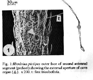
The cave organ aperture was observed on the outer surface of the second antennal segment (pedicel), very close to the first trichobothria, on all Rhodnius and Triatoma species (Fig. 1 ).
Fig. 1: Rhodnius pictipes outer face of second antennal segment (pedicel) showing the external aperture of cave organ. x 200. t: first tricobothria.
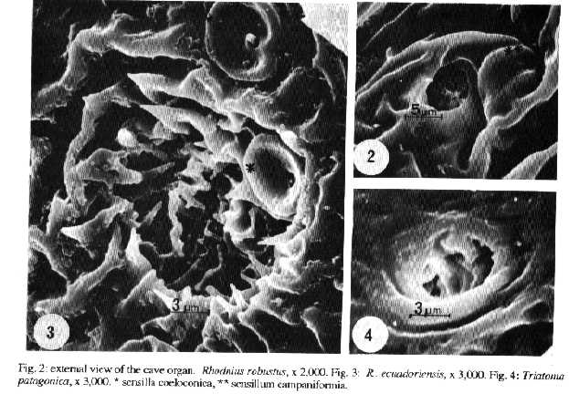
Within the genus Rhodnius the pedicel cuticle is highly folded and gives an ornate appearance to the cave aperture (Figs 2, 3) with diameter between 6 um and 20 um. The edge shows spines or small tubercles, and is sometimes closely associated with campaniformia (Fig. 2) or coeloconic sensilla (Fig. 3). On Triatoma species the aperture looks simpler and smaller. The aperture diameters range from 3 to 20 um for the Rhodnius and Triatoma species studied (Fig. 4).
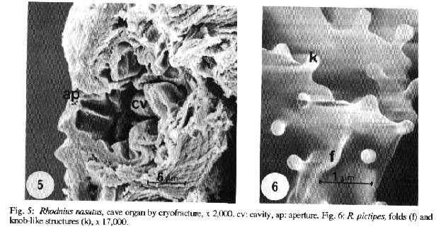
As evidenced by cryofracture of R. nasutus pedicel, the cave organ has a spacious chamber 14-20 um diameter under the antennae surface (Fig. 5).
All the inner cuticular surface appears deeply folded and shows knob-like structures with a rounded head (0.3 um diameter) supported by a brief stalk (up to 0.17 um) (Fig.6). Some of these structures are only domes, less than 0.2 um height. We do not see pores on the wail, even at high magnification (data not shown). This description of cave organ as seen under SEM differs from that of Barth (loc. cit.) using light microscopy (LM). Probably, different genus have different cave organ morphology.
Fig. 2: external view of the cave organ. Rhodnius robustus x 2,000.
Fig. 3: R. ecuadoriensis, x 3,000.
Fig. 4: Triatoma patagonica, x 3,000. * sensilla coeloconica, ** sensillum campaniformia.
Fig. 5: Rhodnius nasutus, cave organ by cryofracture, x 2,000. cv: cavity, ap: aperture.
Fig. 6: R. pictipes, folds (f) and knob-like structures (k), x 17,000.
Barth (loc. cit.) found the cave organ in adults as well as in nymphs. We were not able to find the cave organ aperture on nymphs and the same results were pointed out by Reparaz and Hack (1979 FACENA 3: 153-156) and Lazzari (loc. cit.) suggesting that the organ is closed and non-functional in nymphs.
Electrophysiological studies carried out by Lazzari (loc. cit.) and Lazzari and Mwicklein (unpublished data) showed that the sense cells of the cave organ were not modified by chemical stimulation with carbon dioxide, lactic or burytic acid but thermal stimulation evidenced a clear modification of the electrical activity.
New morphological studies, at the ultrastructural level could help to understand the real function of this enigmatic receptor of Triatominae bugs.
Acknowledgements - To Electron Microscopy Unit and Medical and Veterinary Entomology Division (Natural History Museum, London, GB, where I developed my work) for advice and support. To Natural History Museum, Fundacao Oswaldo Cruz, Instituto Evandro Chagas, and Servicio Nacional de Chagas for providing the specimens. To Drs Chris Schofield and David Gorla who encouraged my work.
Copyright 1994 Fundacao Oswaldo Cruz - FIOCRUZ The following images related to this document are available:Halftone images[oc94057c.gif] [oc94057b.gif] [oc94057a.gif]Photo images[oc94057a.jpg] [oc94057c.jpg] [oc94057b.jpg] |
| |||||||||
