 |
| About Bioline | All Journals | Testimonials | Membership | News |
|
||||||
|
||||||
Comparative Study of Four Species of Trichuris Roederer, 1761 (Nematoda, Trichurinae) by Scanning Electron Microscopy Reinalda Marisa Lanfredi, Wanderley De Souza, Delir CorrEa Gomes* Programa de Biologia Celular e Parasitologia, Instituto de Biofisica Carlos Chagas Filho, Universidade Federal do Rio de Janeiro, Ilha do Fundao, 21941-900 Rio de Janeiro, RJ, Brasil *Laboratorio de Helmintos Parasitos de Vertebrados, Departamento de Helmintologia, Instituto Oswaldo Cruz, Av. Brasil 4365, 21045-900 Rio de Janeiro, RJ, Brasil
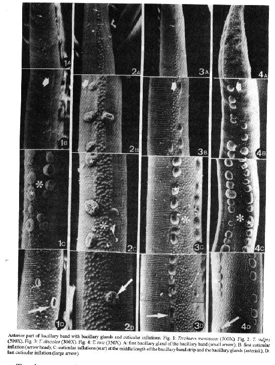
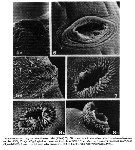
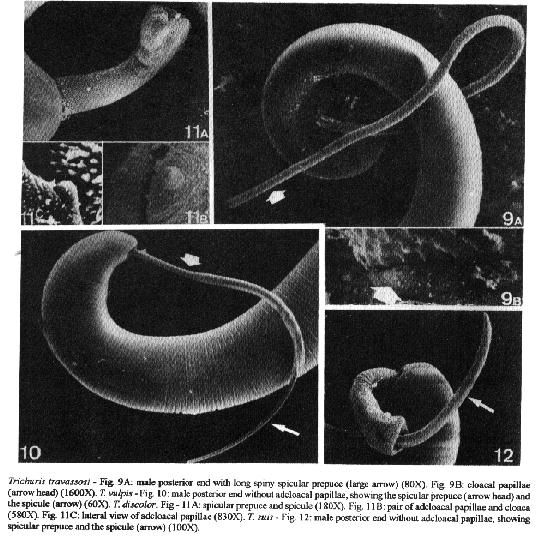
A comparative morphological study of Trichuris travassosi, T. vulpis, T. discolor and T. suis was performed using scanning electron microscopy. Cuticular inflation associated with the bacillary band, vulva and male external genital appendages were analyzed. Qualitative and quantitative analyses of these structures were made for each species; they are of taxonomic value. Key words: Trichuris - morphology - bacillary band - vulva - male genital apparatus Whipworm identification is still difficult. Chandler (1930) discussed the problem and presented characters for a reliable determination of trichurids. Skrjabin et al. (1970) reviewed the species and pointed out that various characters are used for the description of the species. Scanning electron microscopy (SEM) has been used by several groups to study whipworm external structures such as the bacillary band, vulva and male genital apparatus [Sheffield (1963), Jenkins (1969), Batte et al. (1977), Wright (1978), Tenora et al. (1992)] but only Barus et al. (1977,1978), Zaman (1984), Pfaffenberger and Best (1989), Lanfredi (1990) and Gomes et al. (1992) demonstrate the taxonomic importance of these structures. In the present study the SEM was employed to analyze and compare the surface of four species of Trichuris and the studied structures demonstrated to be valuable for their identification. MATERIALS AND METHODS The four species of whipworms Trichuris travassosi Gomes, Lanfredi, Pinto & De Souza, 1992 from Oryzomys nigripes (Olfers, 1818), Trichuris vulpis (Froelich, 1789) from Canis familiaris L., 1758, Trichuris discolor (Linstow, 1906) from half blood cattle Bos taurus L. 1758 x Bos indicus L., 1758 and Trichuris suis (Schrank, 1788) from Sus scrofa L., 1758 were collected by necropsy of naturally infected hosts, and fixed in AFA solution (2% acetic acid, 3% formaldeyde and 95% of 70% alcohol). Part of the samples was used for worm identification by light microscopy according to Skrjabin et al. (1970) and Knight (1971). Specimens were deposited at Instituto Oswaldo Cruz Helminthological Collection (CHIOC). For SEM studies, samples were dehydrated in an ethanol series (50 - 100 GL), critical point dried in CO2, coated with gold and examined in a Jeol JSM25SII scanning electron microscope operating at an accelerating voltage of 15KV. For each character analyzed, at least five specimens of each species were studied. All measurements are in micrometers (um). In this work we adopted the name spicular prepuce to designate the cuticular structure with or without spines that everts from the cloacal lining, previously referred as spicular sheath. This designation actually reports to the cuticle that involves the spicule, according to Gomes et al. (1992). RESULTS The four species studied presented typical characters for the genus Trichuris. The body is divided into two parts; an anterior long and thin portion where the lateral bacillary band is located and a posterior part containing reprodutive organs. It is formed by rows of bacillary glands that appear at a variable distance from the anterior end, with one bacillary gland per cuticular striation, increasing in number posteriorly. Another cuticular structure, the cuticular inflations, are present bordering either side of the anterior bacillary band (Figs 1A-4D). The posterior part is thicker. At the transition region between the anterior and posterior parts of the body, the bacillary band ends with sparce bacillary glands. The vulva opens near the junction of the anterior and posterior parts. At this point a small dilatation of the body is observed, and the cuticle becomes smooth, losing its peculiar striation (Figs 5-8). The anus is subterminal. The male cloacal aperture is located at the posterior end of the parasite. In some specimens the spicule emerges from the spicular aperture and may be covered in different extents by the cloacal prepuce (Figs 9A-12). Trichuris travassosi Gomes, Lanfredi, Pinto & De Souza, 1992 Bacillary band - The bacillary band begins at a distance of 70-98 from the anterior extremity of the body, which at this point is 28-37 wide (Fig. 1A). The first cuticular inflations appear at the point where the bacillary band has two bacillary glands per cuticular striation. Its width is 10-13; body width 40-47 (Fig.1B). Cuticular inflations are present on beside and within region occupied by bacillary band sides (Fig. 1C). They are 104-120 in number. At the last cuticular inflation the bacillary band is 30-31 wide; there are 4-6 bacillary glands and the body width is 51-53 (Fig. 1D). The cuticular inflations look like round or elongated bulbs, sometimes collapsed (Fig. 1C). They are 6-31 long and 7-17 wide. Cuticular inflations may be contiguous or up to 120 (Fig. 1C). Vulva - The vulval opening is a transversal slit, formed by cuticular projections resembling a lip. It is covered by a spineless wrinkled radiated cuticule, resembling a crater. It is 31-39 long and 17-25 wide (Fig. 5). The studied specimens do not present everted vagina. Male genital apparatus - The cloacal aperture presents conspicuous adcloacal papillae (Figs 9A-B). The cloacal prepuce, when everted, can be very long, partially covering the spicule (Fig. 9A). It measures 36-38 in diameter at the proximal end and 34-47 at the distal end. The cloacal prepuce is covered by sparsely distributed spines. At the proximal end (Fig. 13A) spines are thicker, becoming thinner and sparser towards the posterior region (Fig. 13A-D). In most specimens examined the spicule was totally covered by the cloacal prepuce. Trichuris vulpis (Froelich, 1789) Smith, 1908 Bacillary band - The bacillary band begins at a distance of 79-85 from the anterior end body, which at this point is 26-32 wide (Fig. 2A) and consists of a row of large bacillary glands that protrude from the cuticle. The first cuticular inflation is 11-12 wide and appear where the bacillary band has three bacillary glands per cuticular striatiation; the body is 50-52 wide at this point (Fig. 2B). Cuticular inflations are 26-31 in number; they are 7-21 wide and 7-26 long, and sparsely distributed on each side of the anterior bacillary band (Figs 2B-2D). The last cuticular inflations occur where the bacillary band has six to eight bacillary glands and its width is 30-31 and the body is 65-67 wide (Fig. 2D). The cuticular inflations generally are rounded in shape and most of them were colapsed (Figs 2A-D); they are 7 wide and 10-26 long. The distance between the cuticular inflations range up to 198. Vulva - The vulval opening is a transversal opening. Its anterior edge is semicircular and the posterior edge shows a central elevation, covered by a spineless cuticle circularly wrinkled. It is 60-99 long and 46-50 wide (Fig. 6). The studied specimens do not present everted vulva. Male genital apparatus - Adcloacal papillae absent (Fig. 10). When everted the spicular prepuce can be very long, with a diameter of 30-40 at the proximal end and 52-64 at the distal end. At the proximal part the cloaca, the spicular prepuce is covered by small triangular spines (Fig. 14A) which become smaller (Fig. 14B), less frequent, and even disappear (Fig. 14C) at the distal end (Fig. 14D). The spicule is 20-38 wide at the proximal part and 14-23 at the distal end. Trichuris discolor (Linstow, 1906) Ransom, 1911 Bacillary band - The bacillary band begins at a distance of 110-150 from the anterior extremity of the body, which at this place is 50-56 wide (Fig. 3A). The first cuticular inflation is 15-18 wide and occurs where the bacillary band has three to four bacillary glands per striation; the body width is 79-88 at this point (Fig. 3B). The last cuticular inflation appears where the bacillary band presents 18-20 bacillary glands per striation; it is 67-76 wide and the body width is 122-129 at this point (Fig. 3D). Cuticular inflations are round 131-137 in number. Some of them are elongated (Fig. 3C); they are 6-18 wide and 6-26 long. They are located on both sides of the bacillary band (Figs 3B-D). The largest distance between each other is 122. Vulva - The vulval opening is transversally ellipsoid, 80-83 long and 37-40 wide. The surface of the cuticle presents a large number of spines 1.5-4.0 wide and 2.0-7.5 long. The cuticle around the vulva is smooth, with very thin and small spines located in the proximal portion (Fig. 7). The studied specimens do not present everted vagina. Male genital apparatus - The cloacal aperture presents a pair of adcloacal papillae easily seen when the spicular prepuce is inside (Fig. 11B). The spicular prepuce is sleeve-like, 57-92 in diameter at the proximal end and 91-148 at the distal end. It is totally and densely covered by thick spines (Fig. 11A) which are uniform in shape and size (Figs 15A-B). Most of our specimens did not present spicular prepuce distended or fully distended, allowing observation of the cloacal aperture and adcloacal papillae, but making impossible the observation of the prepuce in its total length. Trichuris suis (Schrank, 1788) Smith, 1908 Bacillary band - The bacillary band begins at a distance of 77-80 from the extremity of the body which at this point is 51-53 wide (Fig. 4A). The first cuticular inflation occurs where the bacillary band is 18-19 wide and has 4-5 bacillary glands and the body is 83-85 (Fig. 4B). At the last cuticular inflation the bacillary band presents 21-28 bacillary glands, is 81-84 wide and the body is 115-128 (Fig. 4C). The cuticular inflations are 145-151 in number. Generally they are round, 14-21 wide and 14-30 long, and located on both sides of the bacillary band (Figs 4B-D). The largest distance between each other is 91. Vulva - The vulval opening is oval, 58-63 long and 34-35 wide. It is covered by a spiny cuticle. The spines are 1.0-2.5 wide at the base and 4.0-6.5 long (Fig. 8). The cuticle around the vulva is smooth with evenly distributed spines. During oviposition the vagina is everted and even in this situation spines are observed in the cuticle (Fig. 8B). Male genital apparatus - Adcloacal papillae absent (Fig. 12). The spicular prepuce is campanulate, with a diameter of 49-59 at the proximal end and 79-114 at the distal end (Fig. 12). It is totally and densely covered by uniformly pointed spines (Fig. 16B). The spicule is 33-35 thick at the proximal part and is generally distended (Fig. 12). DISCUSSION The four species examined can be easily differentiated by SEM observations of cuticular structures initially proposed in the comparison with data on the analyzed structures, according to other authors (Table). Trichuris travassosi and T. vulpis present similar dimensions at the anterior end but, can be easily distinguished by the number and disposition of cuticular inflations and number of bacillary glands (Figs 2A-D, 4A-D). Similarly T. discolor and T. suis are larger than the preceding species but also can be differentiated by the number, shape and disposition of cuticular inflations and bacillary glands (Figs 1A-D, 3A-D). Wright (1975) described part of the T. myocastoris bacillary band using SEM where crater-like cuticular inflations are sparsely distributed on the bacillary band margins. His analyses of the number of inflations, based on light microscope observations, may be inaccurate, because, depending on the worm position, some inflations may be overlooked. Based on SEM we agree with him, that the anterior part of the nematode must be straight and carefully mounted on the stub under stereoscopic microscope with the bacillary band upside or it becomes impossible to count the cuticular inflations. Zaman (1984) reported 40 to 90 cuticular inflations in T. trichiura, with diameters ranging from 10 to 20, distinguishing it from closely related morphotype, T. suis. The vulva in T. travassosi and T. vulpis is spineless, but in the former, the cuticle bears radially arranged wrinkles (Figs 5A-B) while in the latter it bears concentric wrinkles (Fig. 6). In addition it is longer and wider than that of T. travassosi. The vulva in T. discolor and T. suis is armed with spines, but in the former, it is longer and the spines are more developed (Figs 7, 8A-B). According to Knight (1971), Knight and Uhazy (1973) and Tenora et al. (1992) the vagina in T. discolor does not evert and there is no comment about spines, but our specimens showed spines on its margins, easily seen by SEM (Fig. 7).
------------------------------------------------------------
Species Bacillary Vulva Adcloacal Spicular SpiculeReference
             band       papillae  prepuce
-------------------------------------------------------------
T. travassosi   X    X     X       X     X Gomes et al.
1992
T. vulpis       X    X     X       X     X Present data
T. discolor     X    X     X       X     X Present data
                X^b        X       X      Tenora et al. 1992
T. suis         X    X     X       X     X Present data
                                       X^a Barus et al. 1977
                X^c                        Batte et al. 1977
T. myocastoris  X                 X^a      Gomes et al.
1992
                X^c                        Wright 1975
T. elatoris                        X     X
Pfaffenberger &
                                           Best 1989
T. dipodomis                       X     X
Pfaffenberger &
                                           Best 1989
T. lani         X                          Barus et al.
1978
T. globulosus   X                          Barus et al.
1978
                                   X^a     Barus et al. 1977
T. skrjabini        X                      Barus et al.
1978
                                   X^a     Barus et al. 1977
T. ovis             X                      Barus et al.
1978
T. cervicaprae                     X^a     Barus et al.
1977
T. muris                    X      X     X Wright 1978
T. trichiura    X^c         X      X     X Zaman 1984
------------------------------------------------------------
^a: detail of spicular prepuce spines^b: detail of posterior bacillary band ^c: detail of anterior cuticular inflations Barus et al. (1978) reported that, T. skrjabini, T. ovis and T. lani present a characteristic vulval appendage that projects above the body surface, and in T. globulosus as a small internal hemispherical vulval appendage that protrudes with a cuticular internal vulval aperture at tip. These characters distinguish the above species from the four species presented in this study. Zaman (1984) also described the vulval aperture of T. trichiura as being surrounded by an elevated, deeply indented rim-like structure. It can be differentiated from that of T. suis by its shape and size. Trichuris travassosi and T. discolor present adcloacal papillae but can be distinguished by the shape of the spicular prepuce (Figs 9A-B, 11A-C) and the shape and disposition spines on the spicular prepuce (Figs 13A-D, 15A-B).
Undoubtedly, SEM morphological studies may be regarded as very important to the differentiation of Trichuris species and the significance of each character will be better evaluated, when all the described species have been analyzed by this method. ACKOWLEDGMENTS To Drs Maria Conceicao Zocoller, Wanda Coutinho and Carlos Graeff-Teixeira, for the supply of trichurid specimens; to Adriana L Rangel for the help in preparing specimens for light and scanning electron microscopy; to Dr Roberto Magalhaes Pinto for the English review. REFERENCES Batte EG, Lamb MC, Mise RE, Talim SD 1977. Parasitology of swine Trichuriasis. Am J Vet Res 38: 1075-1079. Barus V, Kotrla B, Tenora F 1977. A scanning electron microscopy study of spicular sheath of some trichurids (NEMATODA). Folia Parasitol 20: 107-110. Barus V, Kotrla B, Tenora F 1978. Scanning electron microscopic study of vulva of some trichurids (Nematoda). Folia Parasitol 25: 31-34. Chandler AC 1930. Specific characters in the genus Trichuris with a description of a new species Trichuris tenuis from a camel. J Parasitol 16: 198-208. Gomes DC, Lanfredi RM, Pinto RM, De Souza W 1992. Description of Trichuris n.sp. (Nematoda Trichuridae) from a Brazilian rodent by light and scanning electron microscopy. Mem Inst Oswaldo Cruz 87: 1-10. Jenkins T 1969. Electron microscope observations of the body wall of Trichuris suis Schrank 1788. (Nematoda: Trichuroidea). The cuticle and Bacillary band. Z Parasitenk 32: 374-387. Lanfredi RM 1990. Estudo morfologico de quatro especies do gEnero Trichuris Roederer, 1761 (Nematoda: Trichuridae) por microscopia eletronica de varredura. PhD Thesis. Universidade Federal Rural do Rio de Janeiro, Itaguai, RJ, Brasil, 75pp. Knight RA 1971. Redescription of Trichuris discolor (Von Linstow, 1906) and T. skrjabini (Baskakov, 1924) from domestic ruminants in the United States and comparisons with T. ovis (Abidgaard, 1975). J Parasitol 57: 302-310. Knight RA, Uhazy LS 1973. Redescription of Trichuris (= Trichocephalus) schumakovitschi (Savinkova, 1967) from Canadian Rocky Mountain Bighorn Sheep (Ovis canadensis canadensis). J Parasitol 59: 136-140. Pfaffenberger GS, Best T 1989. Trichuris elatoris sp. n. (Nematoda: Trichuridae) from Texas kangoroo rat (Dipodomys elator). Proc Helminthol Soc Wash 56: 76-81. Sheffield HD 1963. Electron microscopy of bacillary band and stichosome of T. muris, T. vulpis. J Parasitol 49: 998-1009. Skrjabin KI, Shikhobalova NP, Orlov IV 1970. Trichocephalidae and Capillariidae of animals and man and diseases caused by them. Essentials of Nematodology, Vol VI. Translated by Dr A Birron. Edited by D Greenberg Israel Program for Scientific Translations Ltd. Keter Press Wiener Binder Ltd, Jerusalem, 257pp. Tenora F, Ooi H, Stanek M, Kamiya M 1992. Some novel features of male posterior end of Trichuris discolor as revealed by scanning electron microscopy. Jap J Parasitol 41: 487-491. Wright KA 1975. Cuticular inflations in whipworms Trichuris sp. Int J Parasit 54: 461-463. Wrigth KA 1978. Structure and function of male copulatory apparatus of the nematodes Capillaria hepatica and Trichuris muris. Can J Zool 56: 651-662. Zaman V 1984. Scanning electron microscopy of Trichuris trichiura (Nematoda). Acta Tropica 41: 287-292.
Copyright 1995 Fundacao Oswaldo Cruz
The following images related to this document are available:Halftone images[oc95096c.gif] [oc95096b.gif] [oc95096d.gif] [oc95096a.gif]Photo images[oc95096b.jpg] [oc95096a.jpg] [oc95096c.jpg] [oc95096d.jpg] |
| |||||||||
