 |
| About Bioline | All Journals | Testimonials | Membership | News |
|
||||||
|
||||||
Lymnaea cousini Jousseaume, 1887, from Ecuador (Gastropoda: Lymnaeidae) W Lobato Paraense Departamento de Malacologia, Instituto Oswaldo Cruz, Av. Brasil 4365, 21045-900 Rio de Janeiro, RJ, Brasil
Code Number: OC95121
Size of Files:
    Text: 16K
    Graphics: Line Drawings (gif) 63K
A description is given of the shell, renal organ, reproductive
system and radula of topotypic specimens of Lymnaea
cousini Jousseaume, 1887. A diagnosis between it and four
other lymnaeids which also occur in South America and were
previously studied by the author (L. columella, L.
diaphana, L. viatrix and L. rupestris) is presented.Key words: Lymnaea cousini - Lymnaea columella - Lymnaea diaphana - Lymnaea viatrix - Lymnaea rupestris - morphology - taxonomy - Ecuador
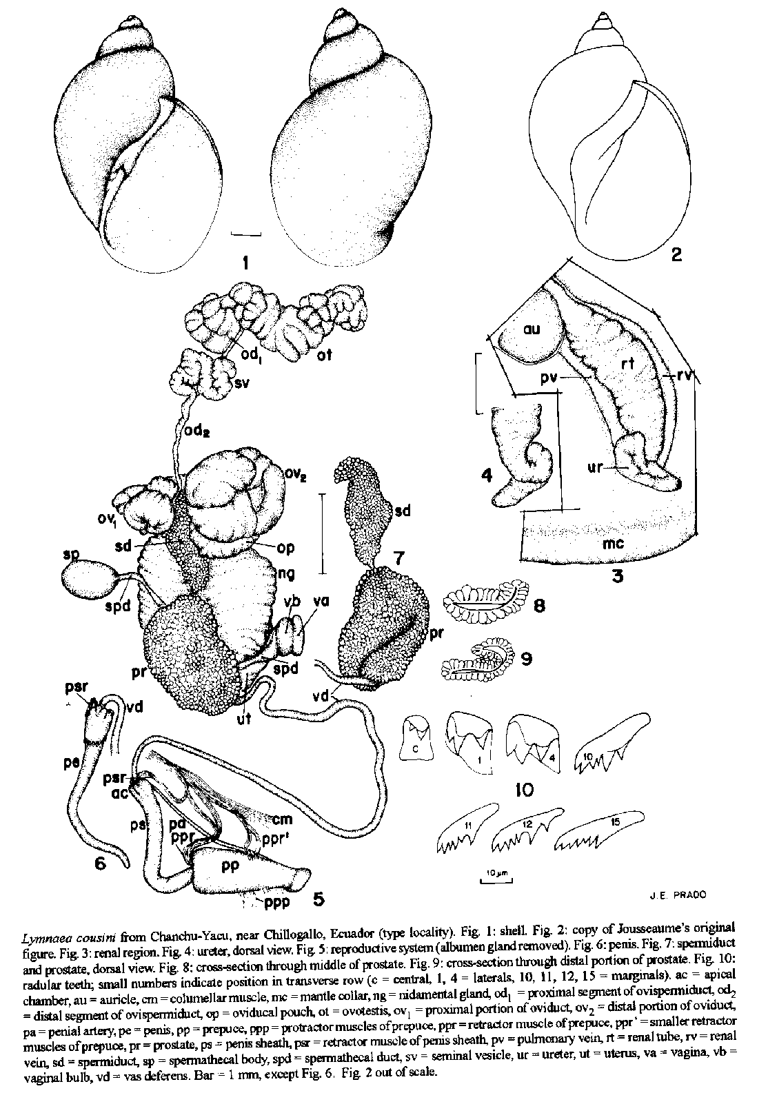
Among the molluscs collected by Auguste Cousin in Ecuador there is a species described and figured by Jousseaume (1887) under the name of Lymnaea cousini: "Testa ovato-conica, tenuiscula, malleata, subtilissime striata, interdum lineis elevatis circumdata, corneo-fusca; spira conica, acuta; sutura impressa, subexcavata; anfracti 4 convexi; ultimus ventricosus, apertura ovalis, columella alba, margo dexter tenuis, acutus, vix reflexus. Dimensions: long., 10 alpha 14 mm; gr.diam., 6 alpha 10 mm; p.diam., 5 alpha 6 mm; ouverture, long., 7 alpha 10 mm; larg., 4 alpha 6 mm. Coquille ovale alpha spire conique; son test mince, fragile, luisant et subtransparent, est ornee de stries longitudinales fines, serrees, et de petites cotes circulaires, peu reguliores, tres espacees, alpha peine saillantes et lisses. Sa couleur est d'un corne fauve qui prend pres du peristome et au sommet une legere teinte rougeatre. La spire est formee de quatre tours convexes et arrondis, separes par une suture profonde; leur developpement sa'effectue avec rapidite surtout le dernier tour qui forme alpha lui seul plus des 7/8 de la coquille. Les tours embryonnaires lisses forment alpha l'extremite de la coquille un petit sommet aigu. Le tour suivant est legerement strie et le dernier est orne de stries et de cordons qui, par leur entrecroisement et leur disposition, produisent de petites facettes alpha la surface de la coquille. 'ouverture de forme ovale, est recouverte, chez les individus tres adultes, d'une legere croute cretacee, luisante et blanche; le peristome presente un bord externe, mince, tranchant et legerement dejete en dehors, surtout alpha la partie anterieure, alors que posterieurement il est presque droit. Le bord columellaire assez epais et blanchatre, decrit une courbe et forme en penetrant dans l'ouverture un leger bourrelet; il est relie alpha l'extremite posterieure du bord externe par une couche d'enduit blanchatre assez large surtout au niveau de l'ombilic qu'elle obture presque completement; alpha ce niveau existe un large sillon que limite en dedans le bord columellaire. Habitat. Cette espece, peu variable quant alpha sa forme, la coloration et les ornements, presente au point de vue de la taille de tres grandes differences. Elle m'a ete envoyee par M Cousin, qui l'a recueillie alpha Chanchu-Yacu, pres de Chillogallo, canton de Quito." On April 23, 1965, I visited the above-mentioned type locality, about 10 km southwest of Quito, collecting from a pond a sample of this lymnaeid which was abundant among watercresses. MATERIALS AND METHODS This study is based on 24 shells and 10 dissected specimens. Each of the latter was previously killed by gradual immersion in water heated to 70 C, with the aperture upward, so carefully as to minimize its retracting back to the shell. After 15 sec the snail was completely plunged for 20 additional sec and then transferred to water at room temperature. The animal (under water) was drawn from the shell with a forceps applied to the cephalopedal mass and fixed in slightly modified Railliet-Henry's fluid (distilled water 930 ml, sodium chloride 6 g, formalin 50 ml, glacial acetic acid 20 ml). The radulae were separated from the buccal mass by digestion for 12 hr in a solution of NaOH at 56 C. They were then rinsed in tap water and mounted in a drop of glycerin on a microscopic slide, with the dorsal (toothed) surface upwards as in the living animal. Measurements were made on camera lucida drawings. Voucher specimens were deposited at the malacological collections of Instituto Oswaldo Cruz (no. 1112), Museum of Zoology-University of Michigan, and Natural History Museum- London. DESCRIPTION The largest shell (Fig. 1) is 8.5 mm long and 6.0 mm wide, and has five whorls; spire length 3 mm, aperture length 6 mm, aperture width 4 mm. The following ratios were calculated from 20 specimens, 6.6-8.5 mm long (means +/- SD): shell width/shell length = 0.54-0.65 (0.59 +/- 0.03); spire length/shell length = 0.31-0.38 (0.35 +/- 0.02); aperture length/shell length = 0.61-0.69 (0.65 +/- 0.02); aperture length/spire length = 1.59-2.23 (1.88 +/- 0.18). Thus the shell tends to be one and a half times as long as it is wide, and its aperture tends to be two thirds as long as the shell or twice as long as the spire. A description of the qualitative shell characters would be superfluous, since the specimens of the present sample agree perfectly with Jousseaume's, whose original figure is reproduced in this paper (Fig. 2). The cephalopedal mass is yellowish gray. The melanic pigmentation of the mantle is distributed into discrete flecks over the roof of the hypopeplar cavity and tends to diffuse on the rest of the mantle roof. The renal tube (Fig. 3, rt) extends straightly from the right side of the pericardial region toward the mantle collar (mc), bordered by the renal vein (rv) on the right and the pulmonary vein (pv) on the left. On reaching the septum between the pulmonary and hypopeplar cavities, just behind the osphradium, it comes back upon itself and, after a short course, bends sharply cephalad and rightward between the first loop and the pulmonary-hypopeplar septum, forming a ureter (ur) which tapers to a subterminal meatus behind the pneumostome. Fig. 4 shows a dorsal view of the terminal portion of the renal tube. The reproductive system is shown in Figs 5-9. The ovotestis (ot) has a ginger-like appearance, composed of acini pressed against each other around a collecting canal which continues into the ovispermiduct. The latter has a very short smooth-walled proximal segment (od1) followed by a bosselated swelling, the seminal vesicle (sv), and then narrows cephalad into a distal segment (od2) which empties into the carrefour. The albumen gland (not figured) has no special characteristics, and covers the carrefour and the origins of the oviduct and ovispermiduct. The oviduct arises ventrally from the carrefour as a tube of bosselated wall. It follows a convolute course (ov1) around the region of the carrefour, between the albumen and nidamental glands, so that its distal portion (ov2 ) gets in touch with the proximal portion. Near its distal end, at a point hidden by its terminal folds, the oviduct is connected with a wrinkle-walled sac, the oviducal pouch (op), which projects from its right side and with which it communicates through a narrow orifice. Then the oviduct proceeds cephalad, continuing into the nidamental gland (ng). As usual with lymnaeids, the nidamental gland is convex dorsally and flattened ventrally, and its outer surface is crossed by numerous parallel grooves that give it a striated appearance. Its ventral surface has a shallow longitudinal depression, coincident with the raphe, over which the distal portion of the spermiduct and the proximal portion of the prostate run. The nidamental gland narrows suddenly into a smooth-walled uterus (ut), which bends to the right and continues into a short vagina (va). The vagina has a bulbous appearance due to a local thickening of the wall musculature forming a sphincter or sphincter-like structure. The spermathecal body (sp) varies in shape from more or less elongated to globoid (egg-shaped in Fig. 5), depending on the amount of its contents and its degree of contraction on fixation. The spermathecal duct (spd) is uniformly thin and about thrice as long as the body. The spermiduct (sd) emerges from the carrefour, beside the oviducal origin, showing no diverticulum or lateral pouch at its beginning (Fig. 7, sd). It runs distalward as a ribbon of granular surface appressed to the ventral side of the nidamental gland, and suddenly diminishes in caliber to merge into the prostate (pr). The prostate is about half as bulky as the nidamental gland, and has the same granular appearance as the spermiduct. Its dorsal surface, appressed to the ventral side of the distal half of the nidamental gland and to the uterus, is flattened on its proximal half, and then shows a fissure formed by the folding of its left margin. As a result, the prostate lumen, initially slit-like, gradually takes on a J-shaped appearance in cross-section (Figs 8, 9). Some specimens, however, show a lengthwise fissure. The fissured distal end of the prostate shows ventrally two rounded protuberances, from whose convergence the vas deferens (vd) arises. The penis sheath (ps) is somewhat swollen at the proximal end owing to the presence of a circlet of minute knobs corresponding to inner apical chambers (ac) communicating with the sheath lumen; it is regularly cylindric and about one and a half times as long as the prepuce (pp). As the penis sheath is frequently more or less deeply intussuscepted into the prepuce, an accurate measurement of the lengths of the two organs, in such cases, is only possible in longitudinal section. The penis (Fig. 6) is about as long as the sheath, tapering to a slender point where the penis duct opens terminally. The prepuce is at least twice as wide as the penis sheath in the examined specimens; this proportion is artificially increased if the prepuce is fixed in a contracted state or is dilated by an intromittent penis sheath. The inner surface of the prepuce shows a series of circular folds, the caudalmost of which surrounds the penis sheath opening as a thickened ridge, the sarcobellum. The extrinsic muscles of the penial complex are usually two main retractors, two smaller retractors and two smaller protractors. The main retractors arise side by side from the columellar muscle (cm). One of them, the penis sheath retractor (psr), is inserted into the head of the penis sheath, and the other, the prepuce retractor (ppr), into the junction of the penis sheath with the prepuce. Not infrequently does the former give off a slip that merges in the latter. These two main retractors may be fused at their origin, splitting at a variable distance from their insertion. The smaller extrinsic muscles are inserted into the preputial wall. A group of retractors (ppr') arise from a branch of the columellar muscle, and a group of protractors (ppp) originate on the right wall of the head. A branch of the cephalic artery, the penial artery (pa), runs along the prepuce and penis sheath to reach the head of the latter. The radula of the largest specimen has 71 transverse rows of teeth, with the formula 24-1-24 (6 laterals, 2 intermediates, 16 marginals). The central tooth has a small cusp and a minute accessory cusp high on its left. The laterals are tricuspid. Radular teeth are shown in Fig. 10. REMARKS So far the only anatomical observation on Lymnaea cousini was made by Hubendick (1951), who showed in his Fig. 183 the penial complex and the vaginal region of the single specimen he had the opportunity to examine. The vagina is slim and tapering, without the bulbous swelling described above (Fig. 5, vb), and the penial complex closely resembles the one represented in Fig. 5 of this paper. The prepuce is said to have “dark spotso, which quite probably resulted from accumulation of pigment in the tissues of the infected snail (Hubendick's specimen had its internal organs destroyed by trematode larvae), as observed by Agersborg (1924). L. cousini is considered by Hubendick (1951) identical with L. ubaquensis Piaget, 1914 (from lake Ubaque, Cundinamarca province, Colombia), and with L. bogotensis Pilsbry, 1935 (from Bogotbeta). His specimen of Fig. 183 (L. ubaquensis) proceeds from Valdivia, Chile, and a shell from “Ecuadorois shown in his Fig. 327b. There follows a comparison between L. cousini and the four South American lymnaeid species I have studied so far: viatrix, rupestris, columella and diaphana (see Paraense, 1976, 1982, 1983, 1984, respectively). The shell of cousini is more broadly conic than in the other species. Its aperture is wider than in viatrix, columella and diaphana, and comparable to that of rupestris; it is about twice as long as the spire, as well as in viatrix, columella and rupestris, the same length to a little longer in diaphana. The body whorl is more convex than in viatrix, columella and diaphana, similar to that of rupestris. The suture is well-marked, as in viatrix, columella and diaphana, deeply channelled in rupestris, giving the latter's spire a loosely wound appearance. It must be emphasized that shell polymorphism is a common occurrence among gastropods, so that significant variation may occur in the mentioned characters. The following anatomic differences are noteworthy. Kidney: similar to that of columella, with two distinct flexures in the ureter (Figs 3, ur, 4); in the other three species the ureter is straight. - Ovotestis: the projection of its image (Fig. 5, ot) occupies about half the area of the nidamental gland; the proportion is about 1:1 in viatrix, diaphana and rupestris, and 1/3 to 1/7 in columella. - Vagina: with a bulbous swelling, absent in the other four. - Spermiduct: narrower than the prostate, as in viatrix, diaphana and rupestris; about the same width as the prostate in columella. - Prostate: proximal half flattened,distal half with an oblique dorsal fissure (some specimens show a lengthwise fissure); a longitudinal fissure is present in viatrix and diaphana, and none at all in columella and rupestris. - Penis sheath: longer than the prepuce; shorter in viatrix, about as long in rupestris, as long to shorter in diaphana, much shorter in columella. - Radula: lateral teeth tricuspid, as in columella and diaphana; predominantly bicuspid in viatrix and rupestris. ACKNOWLEDGEMENTS This paper is based on specimens collected during a trip supported by the Pan American Health Organization. To Dr Antonio Menna, then Representative of the PAHO in Ecuador, and to Prof. Luis A Leon, University of Quito, for their inestimable help during my work in that country. REFERENCES Agersborg HPK 1924. Studies on the effect of parasitism upon the tissues. I. With special reference to certain gastropod molluscs. Quart J Microscop Sci 68 (new series): 361-401. Hubendick B 1951. Recent Lymnaeidae: their variation, morphology, taxonomy, nomenclature, and distribution. Kungl Svenska Vetenskapsakad Handl (4th ser) 3: 1-223. Jousseaume F 1887. Mollusques nouveaux de la Republique de l'Equateur. Bull Soc Zool France 12: 165-186. Paraense WL 1976. Lymnaea viatrix: a study of topotypic specimens (Mollusca: Lymnaeidae). Rev Brasil Biol 36: 419-428. Paraense WL 1982. Lymnaea rupestris sp. n. from southern Brazil (Pulmonata: Lymnaeidae). Mem Inst Oswaldo Cruz 77: 437-443. Paraense WL 1983. Lymnaea columella in northern Brazil. Mem Inst Oswaldo Cruz 78: 477-482. Paraense WL 1984. Lymnaea diaphana: a study of topotypic specimens (Pulmonata: Lymnaeidae). Mem Inst Oswaldo Cruz 79: 75-81. Research Fellow Received 6 March 1995 Accepted 5 May 1995 Copyright 1995 Fundacao Oswaldo Cruz
The following images related to this document are available:Line drawing images[oc95121a.gif] |
| |||||||||
