 |
| About Bioline | All Journals | Testimonials | Membership | News |
|
||||||
|
||||||
Low Occurrence of Arthritic Manifestations in Patients with Pulmonary Tuberculosis. T Cell Subsets and Humoral Studies Diana Dlugovitzky, Ariel Torres, Maria C Hourquescos*, Maria J Svetaz*, Norberto Quagliato**, Eduardo Valentini***, Beatriz Amigot***, Osvaldo Molteni, Oscar Bottasso**** Catedra de Microbiologia ****Instituto de Inmunologia, Facultad de Ciencias Medicas, Universidad Nacional de Rosario, Santa Fe 3100, Rosario (2000), Argentina *Seccion Inmunologia, Laboratorio Central, Hospital Centenario **Departamento de Reumatologia, Hospital Provincial ***Servicio de Tuberculosis, Policlinico Carrasco, Rosario, Argentina
Code Number: OC95126
Size of Files:
    Text: 26K
    Graphics: Line Drawings (gif) 22K
Given the suspected role of mycobacteria in the establishment
of disorders with an autoimmune background and joint damage, a
study was conducted to analize whether rheumatic symptoms were
likely to be present in tuberculosis (TB) patients. To this
end, 330 patients with a bacteriologic confirmation of
tuberculosis were investigated for the presence of arthritic
complaints. The latter were recorded in five of them with
rheumatic symptoms mostly involving interphalangeal and
metacarpophalangeal joints, and preceding the clinical
manifestations of the TB illness. Three out of these five
patients remained arthritic by the time of the bacteriologic
conversion and fulfilled the criteria for the diagnosis of
rheumatoid arthritis. In the two remaining patients sputum
negativization was accompanied by a disappearance of rheumatic
manifestations. These patients were also assessed for their
peripheral levels of major T cell subsets as well as for the
presence of autoantibodies. Comparisons with a series of
non-arthritic TB cases, rheumatoid arthritis patients, and
controls revealed that presence of rheumatic manifestations
was associated with a different profile of autoantibody
formation and T cell subset changes. Evidence recorded in the
present study indicates that joint affectation in TB is a rare
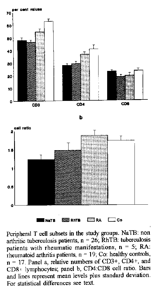
event, being rather the exception than the rule.Key words: tuberculosis - rheumatoid arthritis - rheumatic manifestations - autoantibody - T lymphocytes Several lines of investigations suggest that the immune response against mycobacteria may play a role in the pathogenesis of disorders with an autoimmune background like rheumatoid arthritis (RA) (Shoenfeld & Isenberg 1988). Indeed, autoantibody phenomena are often detected in sera of tuberculosis patients (Singer et al. 1962, Lindqvist et al. 1970, Sela et al. 1987), whereas raised levels of anti-mycobacterial antibodies were reported in sera of patients with RA as was the presence of mycobacteria-reactive T cells in the synovium (Bahr et al. 1989, Tsoulfa et al. 1989, Holoshitz et al. 1989). Supporting this view, studies on adjuvant-induced arthritis have concurred in demonstrating an etiologic role of mycobacteria in the elicitation of the autoimmune arthritis that results in rats following the induction with an oil suspension of dead mycobacteria (Pearson & Wood 1964, Van Eden et al. 1985, Van Eden 1990). From a clinical standpoint, arthritic episodes were recorded in cancer patients undergoing immunotherapy with BCG (Torisu et al. 1978) and in some tuberculosis (TB) patients but the actual existence of such inflammatory arthritis in TB is still a subject of debate (Poncet 1897, Isaacs & Sturrock 1974). From the foregoing, a study was conducted to get a clearer picture on whether RA-like symptoms are likely to be present in our series of TB patients. Attempts were also made to analyze whether such condition, if present, correlated with a different profile of major T cell subsets and autoantibody formation. MATERIALS AND METHODS Patients - Careful examination for the presence of any arthritic sign or symptom, in all TB patients admitted to Carrasco Hospital during a 2-year period, enabled us to detect five patients (3 men and 2 women) who complained of some RA-like manifestations. Patients presented with morning stiffness and swelling of proximal interphalangeal and/or metacarpophalangeal joints. Concomitant arthritic disorders such as psoriatic arthritis, polymyalgia rheumatica, SjögrenÆs syndrome and systemic lupus erythematosus were excluded. Furthermore, none of these patients had a familial history of autoimmune diseases. This first study group was referred to as rheumatic TB patients (RhTB). To compare immunological data, three additional groups were simultaneously included. Group 2 comprised 26 TB inpatients (13 men and 13 women) presenting no arthritic manifestations (NaTB). Both groups were composed by patients with active pulmonary TB who had sputum positive for acid-fast bacilli and chest X-rays clearly consistent with tuberculosis. Pulmonary TB is usually classified on the basis of the extent and type of X-ray findings into four types; mild, patients with a single lobe involved, and without visible cavities; moderate, patients presenting unilateral involvement of two or more lobes with cavities, if present, reaching a total diameter no greater than 4 cm; moderate plus, patients as above or also with affectation of one or two contralateral lobes but cavities' diameter totalizing a value higher than 4 cm; severe, bilateral disease with massive affectation and multiple cavities. Types of TB in these two patient groups were as follows: RhTB, two moderate plus and three severe; NaTB, 14 moderate plus and 12 severe. Patients were on their first week of antituberculous treatment when blood samples were collected. The third group included 19 patients (4 men and 15 women) with RA, according to the American Rheumatism Association criteria (Arnett et al. 1988). Duration of disease ranged from 6 months to 14 years with a mean of 5 years. Seven out of them were newly diagnosed patients, whereas most of the remaining ones were receiving therapy with prednisone (maximum dose 10mg/day) and non steroid antiinflammatory drugs. The control group consisted of 17 healthy individuals (7 men and 10 women) living in the same area with a similar socioeconomic background. Age distribution in years (means +/- sd) for the four groups showed the following: RhTB 46 +/- 8, NaTB 41 +/- 13, RA 46 +/- 8, controls 38 +/- 12 (statistically non significant). Enumeration of T-cell subsets - Leukocytes isolated from whole fresh heparinized venous blood were separated by centrifugation on 'Ficoll-Trysoyon'. The buffy coat was removed and after washing three times in buffered-saline phosphate (BSP) cells were adjusted to a concentration of 7 x 10³ cells/ml in BSP. Two hundred and fifty ml of this cellular suspension were incubated for 30 min at 4oC with 10 ml of monoclonal mouse antibodies reacting against CD3, CD4 and CD8 structures (Dakopatts, Denmark). Upon that, lymphocytes were washed three times in azide-PBS and then incubated under similar conditions with 5 ml of fluorescein-labelled goat anti-mouse-immunoglobulin. After three washing the cells were mounted in slides and counted under immunofluorescence light. The number of positive cells was expressed as the percentage of 200 counted cells. Autoantibody measurements - Antinuclear antibodies (ANA) were demonstrated by indirect immunofluorescence using rat liver frozen sections as substrate. Bound immunoglobulin was detected with goat anti-human immunoglobulin conjugated to FITC (Pasteur, France). Rheumatoid factor (RF) was measured by an agglutination test that utilizes IgG-coated polystyrene beads (Artritest, Wiener Lab. Argentina). Double-stranded anti DNA antibodies (dsDNA) were tested using an indirect immunofluorescence test with Crithidia luciliae as substrate. Counter immunoelectrophoresis was performed for the detection of antibodies to extractable nuclear antigen (ENA) with calf thymus powder as substrate. Statistical analysis - Data regarding the levels of T lymphocyte subpopulations were analyzed by the Kruskall-Wallis and Mann-Whitney U tests. Chi square, and Fisher test were used to compare the prevalence of autoantibody production among groups. A probability value of 0.05 was set as the limit of statistical significance. RESULTS According to the purpose of the study, an approach was established by which patients assisted for TB in Carrasco Hospital Rosario were subjected to a systematic survey for the presence of any arthritic complaint. A total of 330 patients with clinical, radiological, and bacteriological confirmation of pulmonary TB were registered during a 2-year period. As stated in Methods, five of these patients reported to be experiencing rheumatic complaints, which preceded for 1 to 8 months (four patients) or coincided (one patient) with the clinical manifestations of TB. The latter mostly consisted of cough, fever, weight loss, or lack of appetite. Polyarthritis had a subacute onset with involvement of proximal interphalangeal and metacarpo-phalangeal joints, wrists, and knees in one case. Some joints appeared warm, in turn, and painful with palpation or motion. Information available at the time of bacteriologic negativization, that is two to three months following initiation of antibacillary treatment, revealed that three of the five RHTB patients persisted with joint affectation and a positive RF test. Such patients fulfilled the criteria of the American Rheumatism Association for the diagnosis of RA (Arnett et al. 1988). The two remaining patients, whose RF tests were initially negative or weakly positive, appeared recovered from the rheumatic manifestations. In both cases arthritic complaints manifested by the time that TB became symptomatic. The whole series of TB patients showed a striking response to antituberculous treatment with the three conventional drugs (isoniazid, streptomycin, and rifampicin), which reflected in bacteriologic conversion, disappearance of infectious symptoms and radiological improvement. Data regarding the levels of peripheral T cell populations in the four study groups are depicted in Fig. Results are presented in per cent values as no statistical differences in the absolute numbers of lymphocytes/ml were registered among groups. Both TB groups and patients with RA had a significant reduction in the level of CD3+ cells compared with healthy controls, with differences more accentuated in the RhTB and NaTB groups (p < 0.001, panel a). The same was true for the levels of CD4+ lymphocytes (p < 0.001, same panel). In turn, numbers of CD8+ cells appeared significantly decreased in RhTB and RA patients when compared to the values recorded in NaTB and controls (p < 0.001, panel a). Such changes reflected, in turn, on substantial differences among groups as to the balance of CD4+ and CD8+ cell populations. As shown in Fig. panel b, patients from the NaTB group had a lowered CD4:CD8 cell ratio, which was statistically different not only from RA patients and controls but also from the RhTB group (p < 0.01). Further analysis of the two latter groups revealed no significant differences between them. CD4:CD8 cell ratio of RA patients was above the other groups and appeared statistically significant even in relation to controls (p < 0.05). Within the RhTB, levels of CD3+, CD4+, and CD8+ cells in patients who continued to be arthritic were comparable to those recorded in patients whose rheumatic manifestations resolved upon specific chemotherapy. The results of serological tests are summarized in Table I. As expected many RA patients yielded positive results when searched for the presence of RF and ANA. Few patients from the NaTB group showed autoantibody production that reacted against, ENA, dsDNA, and IgG. Such pattern contrasted with the findings recorded in RhTB patients who showed a significantly higher prevalence of RF, and ANA positive sera (p < 0.02 and p < 0.01, respectively). With the exception of one patient showing a weakly positive RF test, RF and ANA were detected in the three RhTB patients who remained arthritic. Titers of RF in the whole sample ranged from 1:20 to 1:160 with median values being a little higher in the RA group. A similar trend was observed for ANA whose titers oscillated between 1:20 and 1:320. With regard to this, a homogeneous staining pattern was the one most frequently seen within groups. We next proceeded to select subjects with at least one positive test for comparison of the overall prevalence of positive autoantibody sera in the study groups. As shown in the same Table, autoantibody formation largely prevailed in the RhTB and RA groups, which accounted for a significant statistical difference when compared to NaTB patients and controls (p < 0.01). In the RA group, duration of disease, activity, or corticosteroid therapy made no differences as to the laboratory results. TABLE I. Distribution of autoantibodies in the study population
-----------------------------------------------------
Groups   ANA     RF     ENA    dsDNA    Overall
                                        prevalence
-----------------------------------------------------
NaTB    0(0)     5(19)    2(7.7)  4(15)   10(38.5)
RhTB    3(60)a   4(80)b   0(0)    0(0)    4(80)c
RA      9(47)a   15(79)b  0(0)    0(0)    16(84)c
Co      1(6)     1(6)     0(0)    0(0)    2(17.6)
-----------------------------------------------------
Results are presented as a number of positive sera (%). ANA:
antinuclear antibody; RF: rheumatoid factor; ENA: extractable
nuclear antigen; dsDNA: double-stranded DNA; NaTB: non
arthritic tuberculosis patients; RhTB: patients with
tuberculosis and rheumatic manifestations; RA: rheumatoid
arthritis patients; Co: healthy controlsa: p<0.01 in comparison to NaTB and Co b: p<0.02 and p<0.01 in comparison to NaTB and Co, respectively c: p<0.01 in comparison to NaTB and Co TABLE II. Relative numbers of T cell subsets in the study population when classified according to the presence of autoantibody (AuAb), ANA, and RF positive sera
------------------------------------------------------------
Status   n        CD3      CD4     CD8      CD4:CD8
------------------------------------------------------------
AuAb-    35      53+/-12 33+/-9  22.4+/-4.6  1.46+/-0.42
AuAb+    32      52+/-6  34+/-5  20.8+/-2.3a 1.63+/-0.34
ANA-     54      54+/-7  34+/-6  22.4+/-2.5  1.52+/-0.33
ANA+     13      54+/-6  36+/-4  19.5+/-1.8a 1.80+/-0.29b
RF-      42      54+/-8  34+/-7  22.8+/-2.5  1.50+/-0.33
RF+      25      53+/-5  34+/-4  20.6+/-2.3a 1.67+/-0.33c
-------------------------------------------------------------
Data are presented as mean +/-sd a, b, and c: significantly different from their respective seronegative counterparts, p<0.001, p<0.02, and p<0.04, respectively (Mann-Whitney U test)
Lastly, we attempted to learn whether autoantibody production correlated with a different profile of T cell populations. To this end, patients were separated according to their serological results into positives or negatives and then analyzed for the levels of T cell subsets. Table II shows that patients showing at least one positive test and those ANA or RF positive had fewer CD8+ cells than the seronegative ones (p < 0.001). In the two latter cases differences also extended to the CD4:CD8 ratios (p < 0.02 and p < 0.04, respectively). DISCUSSION Evidence recorded in the present study indicates that joint affectation in TB is rather the exception than the rule. To this fact, it should be added that three of these five patients presented a RA-like syndrome whose onset seemed to be merely coincidental with the manifestation of the TB illness. Such statement appears likely when considering that 95% confidence limits for the rate of RA-like manifestations (3/330 = 0.9%) indicate that the true prevalence of such arthritis in the population, from which the sample of 330 was taken, was between 0% to 1.9%. Only in two patients, could we detect a certain form of reactive arthritis that resolved concomitantly with the mycobacterial disease. Also, it should be stressed that this kind of joint affectation did not appear to fit with the clinical features of tuberculous rheumatism in which polyarthritis often developed in absence of a clinically overt TB (Isaacs & Sturrock 1974). Questions as to how exactly TB infection can result in polyarthritis remain unanswered. However, it seems clear that some TB-associated immunologic events may act as a precipitating factor capable enough to put into play the cascade of events leading to joint inflammation. Assessment of lymphocyte subsets by using monoclonal antibodies made evident that RhTB patients displayed an intermediate profile of T cell changes when compared with the ones recorded in NaTB and RA patients. While CD3+ and CD4+ lymphocytes were decreased in both NaTB and RhTB patients, CD8+ cells, that remained within normal levels in the NaTB group, appeared lowered in RhTB as did in RA patients. CD4+ cells are thought to be the dominant lymphocytes in the cellular immune response to Mycobacterium tuberculosis given the reciprocal relationship between T cell responsiveness and extent of disease in patients with tuberculosis (Toosi et al. 1986). Therefore, the reduced CD4 cell counts in our series of TB patients may be in line with the former demonstration since most of these patients had an advanced disease. Beck et al. (1985) also observed a reduction of CD4/CD8 ratios in TB patients with changes predominantly due to a CD4 lymphopenia rather than any affectation of CD8+ cells. These authors reported that lowered levels of CD4+ cells could not be related to the radiological severity of the disease while information on the actual extent of lung involvement was not provided in their study. Data regarding CD8+ cells and their lowered levels in RhTB and RA may be interpreted, at least, in two different and not mutually exclusive ways. In general terms, T cells bearing the CD8 surface marker have been shown to express cytolitic as well as 'suppressor' function. Since reduced CD8+ cell numbers were found associated with autoantibody formation, the former phenomenon might be reflecting a particular decrease of CD8+ cells with downregulatory activity on autoantibody secreting cells. An additional hypothesis, which is currently under evaluation, raises the possibility that lowered CD8+ cell values were linked to the production of lymphocytotoxic antibodies specific for such subset. Presence of lymphotoxins have been demonstrated in patients with mycobacterial diseases other than TB, i.e., lepromatous leprosy and also with RA (DeHoratius 1980). Abnormal humoral immune response in patients with TB has long been recognized (Lindqvist et al. 1970, Sela et al. 1987). As suggested for leprosy (Masala et al. 1979), induction of autoantibody synthesis is believed to result from the chronic presence of mycobacterial antigens in host cells that leads to polyclonal B-cell activation, though molecular mimicry between self-antigens and M. tuberculosis structures may also take part in this process (Shoenfeld et al. 1987). In keeping with other studies (Lindqvist et al. 1970, Sela et al. 1987), sera from our TB patients were found to react against dsDNA and IgG, and ENA-binding antibodies were also detected in one case. Contrastingly, none of these serum samples revealed the presence of antinuclear antibodies. Reports exist in the literature indicating that ANA are more likely to prevail in patients under treatment with anti-tuberculous drugs, isoniazid in particular (Cannat & Seligmann 1966, Rothfield et al. 1978). Such demonstration may help to explain the absence of ANA in our series of TB patients since blood was collected at the start of treatment. Results obtained in RhTB patients showed not only a greater prevalence of seropositivity but also a shift in the pattern of autoantibody specificities. RhTB patients did show ANA positivity and RF was present in most of them. Whether such facts are simply epiphenomenic or play a contributory role in the generation of polyarthritis remains unkown. Despite that, it is worth commenting that occurrence of RF in TB and RA has been associated with complement activation (Reyes et al. 1983). A process that is broadly known for its proinflammatory effects. Despite the limitations of the present study, evidence reported here does not appear to indicate a relationship between TB and joint affectation. Also, we cannot discard the presence of an unknown factor as partly responsible for the reactive arthritis. With regard to this, in a recent study we have demonstrated that some humoral abnormalities of lepromatous leprosy are more likely to occur when patients are concomitantly infected with the hepatitis B virus (Cabrini et al. 1991). ACKNOWLEDGEMENTS To Mrs Silvia Luchesi for her secretarial assistance. REFERENCES Arnett FC, Edworthy SM, Bloch DA, McShane DJ, Fries JF, Cooper NS, Healey LA, Kaplan SR, Liang MH, Lutura HS, Medsger TA, Mitchell M, Neustadt DH, Pinals RS, Schaller JG, Sharp RL,Wilder RL, Hunder GG 1988. The American Rheumatism Association 1987 revised criteria for the classification of rheumatoid arthritis. Arthritis Rheum 31: 315-324. Bahr GM, Rook GAW, Al Saffar M, van Embden J, Stanford JL, Behbehani K 1989. An analysis of antibody levels to mycobacteria in relation to HLA type: evidence for non-HLA-linked high levels of antibody to the 65 kDa heat shock protein of M.tuberculosis in rheumatoid arthritis. Clin Exp Immunol 74: 211-215. Beck JS, Potts RC, Kardjito T, Grange JM 1985. T lymphopenia in patients with active pulmonary tuberculosis. Clin Exp Immunol 60: 49-54. Cabrini JM, Bottasso OA, Margasin S, Schujman L, Mangiaterra L, Morini JC 1991. Biological false positive test for syphilis in lepromatous leprosy with concomitant Hepatitis B virus infection. J Invest Allergol Clin Immunol 1: 45-48. Cannat A, Seligmann M 1966. Possible induction of antinuclear antibodies by isoniazid. Lancet 1: 185-187. DeHoratius R 1980. Lymphocytotoxic antibodies, p. 151-174. In RS Schwartz, Progress in Clinical Immunology Vol 4. Grune & Stratton, New York. Holoshitz J, Koning F, Coligan JE, DeBruyn J, Strober S 1989. Isolation of CD4- CD8- mycobacteria-reactive T lymphocyte clones from rheumatoid arthritis synovial fluid. Nature (London) 339: 226-229. Isaacs AJ, Sturrock RD 1974. Poncet's disease-fact or fiction? A reappraisal of tuberculous rheumatism. Tubercle 55: 135-142. Lindqvist KJ, Coleman RE, Osterland KC 1970. Autoantibodies in chronic pulmonary tuberculosis. J Chron Dis 22: 717-725. Masala C, Amendolea MA, Nuti M, Riccarducci R, Tarabini CGL, Tarabini CG 1979. Autoantibodies in leprosy. Int J Lepr 47: 171-175. Pearson CM, Wood FD 1964. Studies of polyarthritis and other lesions induced in rats by injection of mycobacterial adjuvant: I. General clinical and pathologic characteristics and some modifying factors. Arthritis Rheum 2: 440-459. Poncet A 1897. De la polyarthrite tuberculeuse deformante ou pseudorheumatisme chronique tuberculeux. Congr Fr Chir 1: 732-739. Reyes PA, Maluf JG, Curd JG, Vaughan JH 1983. Association of rheumatoid factor with complement activation in rheumatoid arthritis and other diseases. Clin Exp Immunol 53: 391-396. Rothfield NF, Bierer WF, Garfield JW 1978. Isoniazid induction of antinuclear antibodies; a prospective study. Ann Intern Med 88: 650-652. Sela O, El-Roeiy O, Isenberg DA, Kennedy RC, Colaco CB, Pinkhas J, Shoenfeld Y 1987. A common anti-dna idiotype in sera with active pulmonary tuberculosis. Arthritis Rheum 30: 50-56. Shoenfeld Y, Isenberg DA 1988. Mycobacteria and autoimmunity. Immunol Today 9: 178-182. Shoenfeld Y, Vilner Y, Coates ARM, Rauch J, Lavie G, Shaul D, Pinkhas J 1987. Monoclonal anti-tuberculosis antibodies react with DNA, and monoclonal anti-DNA autoantibodies react with M. tuberculosis. Clin Exp Immunol 66: 255-263. Singer JM, Plotz CM, Peralta FM, Lyons HC 1962. The presence of anti-gamma globulin factors in sera patients with active pulmonary tuberculosis. Ann Intern Med 56: 545-551. Toossi Z, Kleinherz ME, Ellner JJ 1986. Defective IL-2 production and responsiveness in human tuberculosis. J Exp Med 163: 1162-1172. Torisu M, Miyahara T, Shinohara N, Ohsato K, Sonozaki H 1978. A new side-effect of BCG immunotherapy: BCG-induced arthritis in man. Cancer Immunol Immunother 5: 77-83. Tsoulfa G, Rook GAW, Bahr GM, Sattar MA, Behbehani K, Young DB, Mehlert A, van Embden JDA, Hay FC, Isenberg DA, Lydyard PM 1989. Elevated IgG antibody levels to the mycobacterial 65-kDa heat shock protein are characteristic of patients with rheumatoid arthritis. Scand J Immunol 30: 519-528. Van Eden W 1990. Heat-shock proteins in autoimmune arthritis: A critical contribution based on the adjuvant arthritis model. APMIS 98: 383-394. Van Eden W, Holoshitz J, Nevo A, Frenkel A, Klajman A, Cohen I 1985. Arthritis induced by an anti-mycobacterial T cell clone that responds to cartilage proteoglycan. Proc Natl Acad Sci USA 82: 5064-5067. Copyright 1995 Fundacao Oswaldo Cruz
The following images related to this document are available:Line drawing images[oc95126a.gif] |
| |||||||||
