 |
| About Bioline | All Journals | Testimonials | Membership | News |
|
||||||
|
||||||
RESEARCH NOTE Toxigenic and Invasive Capacities: Possible Pathogenic Mechanisms in Arcobacter cryaerophilus H Fernandez, G Eller*, J Paillacar, T Gajardo, A Riquelme Institute of Clinical Microbiology and *Institute of Immunology, Universidad Austral de Chile, Casilla Postal 567, Valdivia, Chile
Code Number: OC95128
Size of Files:
    Text: 6K
    Graphics: Photos (jpg) 38K / Halftones (gif) 94K
Key words: Arcobacter cryaerophilus - pathogenic
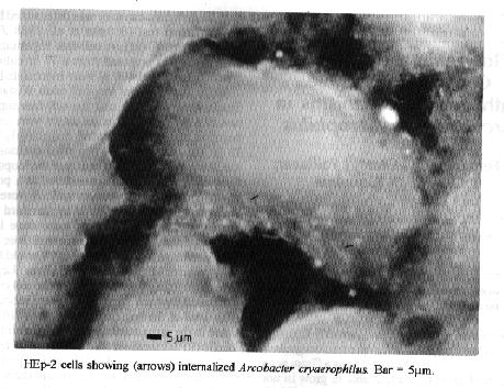
mechanisms - invasiveness - toxigenicityArcobacter cryaerophilus, formerly known as aerotolerant Campylobacter-like microorganisms and Campylobacter cryaerophila, is a Gram negative curved or S shaped rod, able to grow in normal O2 tension and optimal temperature of 30 C (P Vandamme, H Goosens 1992 Zbl Bakt 276: 447-452). It has been isolated from aborted animal fetuses, from tissues and feces from cattle, swine and poultry and also from human stool, abdominal content and blood as well as from surface water and sewage (S Neill et al. 1985 Int J Syst Bacteriol 35: 342-356, M Lambert et al. 1987 J Clin Microbiol 25: 706-713, W Tee et al. 1988 J Clin Microbiol 26: 2469-2473, S Stampi et al. 1993 Epidemiol Infect 110: 633-639). This isolation spectrum suggests a wide natural distribution and reflects the zoonotic character of A. cryaerophilus. However, the factors of pathogenicity and the clinical significance of this bacteria remains unknown. The aim of this study was to determine the existance of toxigenic and invasive capacities in two strains of A. cryaerophilus isolated from an aborted bovine fetus (strain 7625) and from swine feces (strain 62C). Both strains were isolated on blood agar using a filtration method (T Steele, S McDermontt 1984 Pathology 16: 263-265), phenotypically identified (Vandamme, Goosens loc. cit.) and maintained under freezing temperature (-35 C) until use. The toxigenic capacity was determined by the rat ileal loop test (S Saha et al. 1988 J Med Microbiol 26: 87-91). A cell-free supernatant of each strain was prepared from a 72 hr culture in Brucella broth and 400 ul were inocculated individually into two ileal loops of adult Wistar rats. Sterile Brucella broth and the cell-free supernatant of a toxigenic C. jejuni strain were used as negative and positive control respectively. After 18 hr, the rats were killed by ether overdose and the ileum examined. Distention of the loops with fluid accumulation was considered as a positive result and electrolyte concentrations were measured in the intestinal fluids by standard flame spectrophotometry. The tests were done in two animals simultaneously and repeated three times. Invasion assays were done in 18 hr old HEp-2 cells cultures (120,000 cells/ml). Four Leighton tubes containing a coverslip with the cell cultures were used for each strain. They were inocculated with 1 ml of the bacterial suspensions (106 c.f.u) made in the same medium used for growing HEp-2 cells (RPMI medium with 10% fetal calf serum) and incubated during 3 hr (infection period), at 37 C in a CO2 incubator. Following the washing (10X) of the tubes, 1 ml of the cell growing medium was added and the cell monolayers were reincubated for further 4 hr (multiplication period) under the same conditions mentioned above. Then, the tubes were washed three times and the coverslips with the cell monolayers were stained with 0.01% acrydine orange in GeyÆs solution for 45 sec, rinsed with Hanks balanced salts solution, counterstained for 45 sec with 0.05% crystal violet in 0.15 N NaCl, rinsed again, mounted on slides and examined under epifluorescence microscopy. Internalized bacteria were seen as green-fluorescing curved rods (Fig.). Both strains produced distention of the ileal loops with fluid accumulation and enhanced electrolytes concentrations. The concentrations of Na+ and Cl- (mEq/l) in the intestinal fluids produced by strains 7625 and 62C were 135 and 114, and 134 and 117 respectively whereas, in the negative control loop, they were 26 and 40. Toxin production in C. jejuni was previously demonstrated using this biological model (Saha et al. loc. cit.). Fluids and electrolyte excretion to the intestinal lumen of the rat was also demonstrated in C. jejuni (H Fernandez et al. 1983 Infect Immun 40: 429-431), suggesting the presence of a toxigenic substance as described for other toxigenic bacteria (R Sullivan, T Asano 1971 Am J Phisiol 220: 1793-1797, F Klipstein et al. 1976 Infect Immun 14: 1004-1010).
This results suggest that toxigenicity and invasion could be pathogenic mechanisms by which A. cryaerophilus may produce disease. However, further studies are necessary to elucidate the pathobiology of this bacteria as well as their clinical and epidemiological significance. Moreover, specific isolation media need to be developed, since most strains are sensitive to some of the antibiotics used in Campylobacter selective media (J Kiehlbauch et al. 1992 Antimicrob Agents Chemother 36: 717-722) and are unable to grow at 42 C. These facts delay the isolation of A. cryaerophilus, that may pass undetected (J Penner 1988 Clin Microbiol Rev 3: 157-172) in man and animals biological products in which more attention is given to other gram negative curved rods such as the well known Campylobacter species. Currently, studies focusing these aspects are in course in our laboratory.
Copyright 1995 Fundacao Oswaldo Cruz
The following images related to this document are available:Halftone images[oc95128a.gif]Photo images[oc95128a.jpg] |
| |||||||||
