 |
| About Bioline | All Journals | Testimonials | Membership | News |
|
||||||
|
||||||
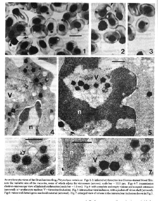
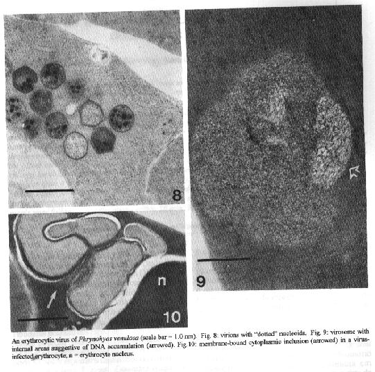
An Erythrocytic Virus of the Brazilian Tree-frog, Phrynohyas venulosa AP Alves de Matos, I Paperna*, R Lainson**/+ Electron Microscopy Unit, Pathologic Anatomy Department, Curry Cabral Infectious Diseases Hospital, R da Beneficencia, P-1000 Lisboa, Portugal *Department of Animal Sciences, Faculty of Agriculture, Hebrew University of Jerusalem, Rehovot 76-100, Israel **Secao de Parasitologia, Instituto Evandro Chagas, Caixa Postal 691, 66017-970 Belem, PA, Brasil
Code Number: OC95133
Size of Files:
    Text: 7K
    Graphics: Photos (jpg) 205K / Halftones (gif) 247K
Blood erythrocytes of Brazilian tree-frogs, Phrynohyas
venulosa were found to frequently contain single, small,
densely staining inclusions. Electron microscopy showed these
to be icosahedral viral particles which measured from 250-280
nm in diameter: they were devoid of an envelope, and thus
differed from previously described viruses of frog
erythrocytes. The infected erythrocytes lacked a crystalline
body.Key words: viral particles - erythrocytes - Phrynohyas venulosa - tree-frogs - Brazil Erythrocytic icosahedral viruses have been found in anurans from North America (Gruia-Gray et al. 1989), Africa (Alves de Matos & Paperna 1993) and neotropical America. In the latter geographic region they have been recorded in the frog Leptodactylus ocellatus from the State of Rio de Janeiro, Brazil (Sousa & Weigel 1976) and the toad Bufo marinus from Costa Rica (Speare et al. 1991). In this communication we report the finding of erythrocytic icosahedral virus in the tree-frog, Phrynohyas venulosa from the State of Para, North Brazil. MATERIALS AND METHODS Blood was collected from a clipped toe, and thin films were air-dried, fixed in absolute methyl alcohol, and stained with Giemsa (30 drops of stain to 15.0 ml of distilled water buffered to pH 7.4) for 1 hr. Blood collected into a glass capillary tube was allowed to coagulate before extraction and fixation in 2.5% glutaraldehyde in 0.1m cacodylate buffer (pH 7.4) for 24 hr at 4oC. After post-fixation in 1.0% osmium tetroxide in the same buffer, and consecutive rinsing in the buffer and distilled water, the material was stained, en-bloc, in uranyl acetate. The clot was then rinsed in distilled water, dehydrated in graded ethanol, and embedded in Agar 812 medium (Agar Company, U.K.). Thin sections, cut on a LKBIII microtome with a diamond knife, were stained on grids with uranyl acetate and lead citrate and examined with a JEOL 100S transmission electron microscope. RESULTS Densely stained, purple-red inclusions were found in the erythrocytes of 7 out of 33 tree-frogs collected in Capanema, 50 km East of Belem, Pará, between October and December 1992. Among the positive frogs, three developed heavy parasitae-mias. The inclusions were usually single, and uniformly small (about 1.5-2.5 mm in diameter). The erythrocytic vacuoles associated with the inclusions were also small (Figs 1-3) and unlike the large ones described for erythrocytic viral infections in other anurans. Crytalline bodies were absent. Electron-microscopic examination revealed virions 250-280 nm in diameter, of hexagonal outline and compatible with an icosahedral shape (Figs 4-7). The virion nucleoid was either uniformly dense (Fig. 7), or made up of concentric dense and lucent layers (Fig. 6). Some others possessed a dotted pattern (Fig. 8). The polygonal shell (capsid) was closely applied, but a number of empty or even peeled-off capsids were seen (Figs 5-7). Virions occurred in an intranuclear inclusion (Fig. 5) or in the cytoplasm (Figs 4,6). One cytoplasmic inclusion assemblage contained empty and defective particles, and also one with a long extension derived from the capsid (Fig. 4). Virosomes without virus had an internal fibrilar area resembling accumulated DNA (Fig. 9). Membranous inclusions occurred in the cytoplasm of some cells, but the virion-associated envelopes demonstrated in other anuran erythrocytic viruses were lacking (Fig. 10). DISCUSSION Comparing the presently described material with that recorded from other anurans, the virus of P. venulosa approximates most closely to the size range (200-300 nm) of erythrocytic viruses from Rana pipiens (Bernard et al. 1968). These, and all other anuran erythrocytic viruses (with the exception of those from B. marinus, Speare et al. 1991), however, possess a virion-associated envelope, which is lacking in the organism from P. venulosa. Again, the infected erythrocytes harbouring the presently described virus have no crystalline body - commonly seen associated with the erythrocytic viruses of L. ocellatus, Rana catesbeiana and Ptychadena anchietae (Sousa & Weigel 1976, Gruia-Gray et al. 1989, Alves de Matos & Paperna 1993). The absence of crystalline bodies, and the small size of the vacuoles in the infected erythrocytes, does not seem to reflect a particular stage of differentiation of the virus in P. venulosa, because these same characteristics were present in all the material collected from seven different, infected frogs, taken at various intervals during the months of October-December. We conclude that frog erythrocytic viruses are probably host-specific, and that the crystalline body is not, as earlier implied (Alves de Matos & Paperna 1993), characteristic of all anuran infections, but is lacking in some. REFERENCES Alves de Matos AP, Paperna I 1993. Ultrastructure of erythrocytic virus of the South African anuran Ptychadena ancietae. Dis Aquat Org 16: 105-109. Bernard GW, Cooper EL, Mandell ML 1968. Lamellar membrane encircled viruses in the erythrocytes of Rana pipiens. J Ultrastr Res 26: 8-16. Gruia-Gray J, Petric M, Desser SS 1989. Ultrastructural, biochemical and biophysical properties of an erythrocytic virus of frogs from Ontario, Canada. J Wildlife Dis 25: 497-506. Sousa MA, Weigel DR 1976. The viral nature of Toddia Franca, 1912. Mem Inst Oswaldo Cruz 74: 213-230. Speare R, Freeland WJ, Bolton SJ 1991. A possible iridovirus in erythrocytes of Bufo marinus in Costa Rica. J Wildlife Dis 27: 457-462.
Copyright 1995 Fundacao Oswaldo Cruz
The following images related to this document are available:Halftone images[oc95133b.gif] [oc95133a.gif]Photo images[oc95133a.jpg] [oc95133b.jpg] |
| |||||||||
