 |
| About Bioline | All Journals | Testimonials | Membership | News |
|
||||||
|
||||||
Memorias do Instituto Oswaldo Cruz
Vol. 90(6), Nov./Dec. 1995
Research Note: Scanning Electron Microscopic Observations on Goezia spinulosa (Diesing, 1839) (Nematoda: Anisakidae) from Arapaima gigas (Cuvier, 1817) Helio Martins de Araujo Costa+, Marcos Pezzi Guimaraes+, Dagmar Diniz Cabral*, Maria Jose Santos Mundim* Departamento de Parasitologia, Instituto de Ciencias Bioligicas, Universidade Federal de Minas Gerais, Caixa Postal 486, 30161-970 Belo Horizonte, MG, Brasil *Departamento de Ciencias Fundamentais para a Saude, UFU, Uberlandia, MG, Brasil
Code Number: OC95143
Size of Files:
Text:
Graphics: Line Drawing (gif) - 13K
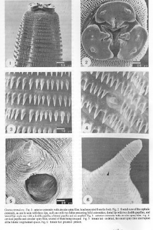
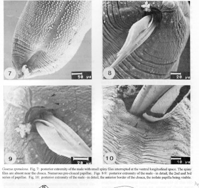
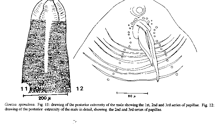
Photographs (jpg) - 83KKey words: Goezia spinulosa - Arapaima gigas - Nematoda - Anisakidae - morphology This nematode was first described by KM Diesing (1839 Ann Wien Mus Naturg 2: 219-242), who named it Lecanocephalus spinulosus. R Drasche (1884 Verh K K Zool bot ges 33: 107-118) re-examined Diesing's findings and redescribed them. A Railliet and A Henry (1915 Bull Soc Path Exot 8: 270-274) placed this parasite in the genus Goezia (Zeder, 1800). L Travassos and JFT Freitas (1964 Publicaoes Avulsas do Museu Emilio Goeldi 2: 3-16) examined four specimens of Arapaima gigas, all of them parasitized by Goezia spinulosa. A detailed account of this nematode was given by HA Baylis (1927 Parasitol 19: 35-47), who reported the existence of 12-15 pairs of pre-anal papillae in the male, laterally located, as well as an isolate papilla on the anterior anal labium. L Travassos et al. (1928 Arch Inst Biol 1: 5-68) carrying out a study on the helminthologic fauna of fresh water fish in Brazil were able to corroborate the data obtained by Baylis ( loc. cit.). JFT Freitas and H Lent (1946 Rev bras Biol 6: 215-222) studied specimens of G. spinulosa from Astronotus ocellatus (Agassiz) and found 21 pairs of caudal papillae in the males (six pos anal and two adanal); the spicules length were 0.41-0.52 mm and they were alate. E Santos et al. (1979 Atas Soc Biol 20: 11-19) found this same number of papillae and the same measurements of the spicules that were alate. MI Haman (1984 Neotropica 30: 55-61) refers to the findings of this species in Pseudoplatystoma coruscans (Agassiz) in Argentina and his description gives little new contribution to the knowledge of the species; the spicules had the same size and measured 0.25 -0.51 mm in length. The examined specimens were collected from pirarucu (A. gigas) coming from the Araguaia River, and presented the following characters: about 13.8 mm and 16.0 mm in length by 0.69 and 0.73 in maxima width for the males and females, respectively; a well defined cephalic extremity (Fig. 1), with three large, distinct and depressed lips, each lip showing internally two lobes with bifid extremities; dorsal lip with two double papillae and sub-ventral lips, each one presenting a double sub-ventral papilla, a single lateral papilla and one amphid (Fig. 2); the dentigerous borders recorded by Baylis (loc. cit.) could not be observed; triangular oral opening; body covered with circular spiny files (Figs 1-3), which increase in size gradually in direction to the middle portion of the body, and next decrease, always gradually, in direction to the posterior extremity; the spines are unlike each other, even when placed in the same file. Distinct cervical papilla situated nearly the 18th file of spines (Fig. 4). In the female, the spiny files are extended throughout the tail, being interrupted right through the lateral longitudinal spaces (Fig. 5); distinct phasmids (Fig. 6). At the posterior extremity of the male, the spiny files are interrupted throughout the ventral longitudinal space and are absent near the cloaca (Fig. 7); tail short-sized; two strong and alate spicules, in disagreement with J Kaur and S Khera (1991 Acta Parasitol Polonica 36: 51-54); they measure 0.51 - 0.58 mm in lenght, with bifid distal extremities. For the purpose of observation, the caudal papillae could be divided into three series: the first series with at least eight pairs distributed laterally on the pre-cloacal region covered with spines (Fig. 7); the second one with about 12 pairs situated on the unspiny region, and appearing to be a continuation of those located on the spiny region (Figs 8, 11). These last papillae reach nearly the half of the lateral border of the cloaca, with a little interruption up to the beginning of the third series, composed of six pairs which surround the posterior half of the cloaca (Figs 9 and 12). The isolate papilla described by Baylis (loc. cit.) appears clearly on the anterior border of the cloaca (Fig. 10). Goezia spinulosa. Fig. 11: drawing of the posterior extremity of the male showing the 1st, 2nd and 3rd series of papillae. Fig. 12: drawing of the posterior extremity of the male in detail, showing the 2nd and 3rd series of papillae.
+CNPq fellowship
Received 2 January 1995
Accepted 1 August 1995
Copyright 1995 Fundacao Oswaldo Cruz, FIOCRUZ
The following images related to this document are available:Photo images[oc95143b.jpg] [oc95143a.jpg]Line drawing images[oc95143c.gif] |
| |||||||||
