 |
| About Bioline | All Journals | Testimonials | Membership | News |
|
||||||
|
||||||
Four New Species of Oswaldocruzia (Nematoda: Trichostrongylina, Molineoidea) Parasitizing Amphibians and Lizards from Ecuador
Badreddine Ben Slimane, Marie-Claude Durette-Desset^+
Laboratoire de Protozoologie et Parasitologie coniparee de l'E.P.H.E. & Laboratoire de Biologie Parasitaire, Protistologie, Helminthologie, associe au C.N.R.S., Museum National d'Histoire Naturelle, 61 Rue Buffon, 75231 Paris Cedex 05, France
Code Number: OC96063
Size of Files:
    Text: 30.7K
    Graphics: Line Drawings (gif) 183K
[FIGURES AT END OF TEXT] Description of four new species of Oswaldocruzia parasitizing Iguanidae and Leptodactylidae from Ecuador, demonstrate that they are morphologically close to each other. Like most of the other neotropical and holarctic Oswaldocruzia, they are characterized by spicules with three main branches: blade, shoe and fork; the division of the fork within the distal third of the spicule length appears to be characteristic of the neotropical species. - Oswaldocruzia bainae n. sp. parasitizing Anolis chrysolepis and Anolis fuscoauratus possesses a synlophe visible only on transversal sections of the body. It is composed of rounded and not pointed ridges. - Oswaldocruzia tcheprakovae n. sp. parasitizing Eleutherodactylus altamazonicus is closely related to O. bainae, but the synlophe is present only in the anterior and posterior extremities of the body. - Oswaldocruzia cassonei n. sp. parasitizing Eleutherodactylus lanthanites is closely related to O. taranchoni, Ben Slimane and Durette-Desset, 1995, a parasite of Bufo marinus from Brazil. It is differentiated by the synlophe and the measurements. - Oswaldocruzia petterae n. sp. parasitizing Leptodactylus pentadactylus is closely related to O. chambrieri, Ben Slimane and Durette-Desset, 1993, parasitizing Bufo and Eleutherodactylus in the same region. It is differentiated since, for an equivalent length of the body, the ridges are almost two times fewer and the spicules smaller.
Key words: Oswaldocruzia n. spp.- Nematoda - Trichostrongylina - lguanidae - Leptodactylidae - Ecuador
In this study, we continue the review of the genus Oswaldocruzia Travassos, 1917, a cosmopolitan parasite of Amphibians and Reptiles. The diagnosis of the species relies on new morphological criteria, particularly the synlophe characteristics in oesophageal region, the relative arrangement of rays 6 and 8 of the caudal bursa and the acute spicular morphology. Six species of Oswaldocruzia were described in Ecuador (cf Ben Slimane & Durette-Desset, 1993, 1995), but only from Amphibians. We describe below three new species in Amphibians and one in Reptiles.
MATERIALS AND METHODS The Nematodes were collected in the small intestine of Ecuadorian Leptodactylidae and Iguanidae (neotropical fauna, guyano-brazilian sub-zona). The study of the synlophe is based on the Durette-Desset (1985) method; the nomenclature of the synlophe in oesophageal region follows Ben Slimane et al. (1993). More particularly, the cervical alae are defined as one or more latero-ventral ridges, more developed than the other ridges. The nomenclature of the caudal bursa follows Durette-Desset and Chabaud (1981), concerning the relative arrangement of rays 6 and 8 follows that of Durette-Desset et al. (1992). The spicules were studied after dissection and the nomenclature is that of Ben Slimane et al. (1993). The material was stored in 70^o ethanol and deposited in the Helminthological Collections of the Museum National d'Histoire Naturelle of Paris (MNHN) and in those of the Museum d'Histoire Naturelle of Geneve (MHNG).
DESCRIPTION
The species are closely related to each other and to the other species previously studied in the same region. Some characters do not provide specific differences and can be defined similarly for all the species. Head: cephalic vesicle present without anterior swelling. En face view: buccal aperture triangular, with 6 externo- labial papillae, 4 cephalic papillae and 2 amphids. Small dorsal oesophageal tooth present. Anterior extremity: excretory pore situated within distal third of oesophagus. Triangular-shaped deirids, posterior to excretory pore. Well developed excretory glands. Musculo- glandular separation of oesophagus acutely visible at nerve ring level. Caudal bursa: with 2-3 pattern which tends towards 2-1-2 i.e extremities of rays 4 directed towards anterior of body, nearer those of rays 3 than rays 5. Rays 2 and 3 joined along, rays 5 and 6 joined along. Rays 8 arising on root of dorsal ray and overlapped by rays 6 except in their distal extremity (type III). Rays 9 arising on dorsal ray before division of the latter into two branches of which internal ones are longest. Thick dorsal ray. Gubernaculum absent. Genital cone: 15 um high by 15 um wide at its proximal part, bearing on anterior lip a large papilla zero and 2 min papillae 7 on posterior lip. Spicules: divided proximally into three main branches: extemo- lateral branch or blade, interno-dorsal branch or shoe, interno- ventrat branch or fork. Fork divided within distal third of spicule. Female: didelphic with very short infundibula.
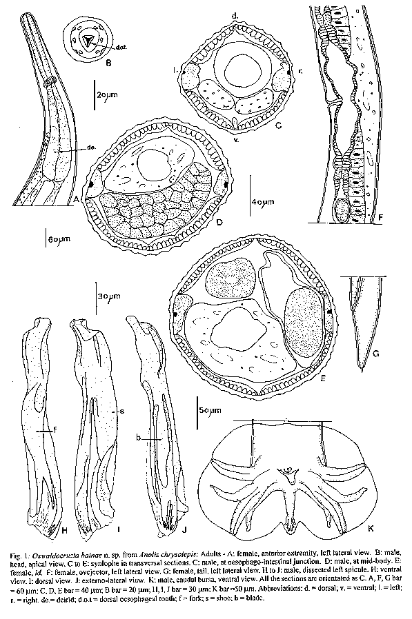
Oswaldocruzia bainae n. sp.
Type-material: holotype male, allotype female MHNG-INVE 19484; 1 male, 3 females paratypes MNHN 212 MD Host: Anolis chrysolepis (Iguanidae) Site: small intestine Locality: San Pablo, Ecuador Voucher specimens: from the same site and the same locality as the types In 9 Anolis chrysolepis: 11 males , 5 L4 males, 16 females, 10 L4 females, MHNG-INVE 19485 to 19491. 7 male, 1 L4 male, 9 females, 1 entsheathed LA female, 2 L4 females MNHN 206 MD-207 MD. In 6 Anolis fuscoauratus: 12 males, 3 females: MHNG-INVE 19492-19497; 2 males , 4 females: MNHN 219 MD.
ADULTS
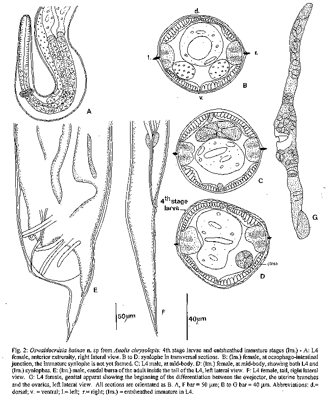
Small nematodes with anterior part of body coiled or not. Cervical alae absent. Synlophe: (studied in 1 male and 1 female paratypes, 1 male and 1 female from A. chrysolepis 2 males from A. fuscoauratus. Numbers in brackets correspond to voucher specimens). In both sexes, cuticle bears longitudinal 'pseudoridges' or undulations, uninterrupted whole length of body but obvious only in transversal sections (Fig. 1C). In male 65% (74 to 77%) of ridges appear in oesophageal region, within 67% (74 to 78%) of dorsal ridges and 63% (71 to 81%) of ventral ridges. In female 81% (71%) of ridges appear in oesophageal region within same dorsal and ventral ratio. Ridges disappear just anterior to caudal bursa in male and at phasmids level in female. In male 27 (30, 34, 26) ridges at oesophago-intestinal junction (Fig. 1C) and 40 (40, 44, 35) at mid-body (Fig. 1D). In female, 44 (34) ridges at oesophago-intestinal junction and 54 (48) at mid-body (Fig. 1E). In transversal sections, rounded ridges are more or less regularly spaced. Holotype-male: 7100 um long and 130 um wide at mid-body. Cephalic vesicle 75 um long and 40 um wide. Nerve ring, excretory pore and deirids 190 um, 380 um and 400 um from apex, respectively. Oesophagus 530 um long. Caudal bursa illustrated in Fig. 1K. Spicules 190 um long, blade divided at its distal part into two small branches which are subdivided into numerous processes; fork distally divided at 17% of whole length of spicule (Fig. 1H-J). Allotype-female: 12350 um long and 190 um wide at mid- body. Cephalic vesicle 90 um long and 40 um wide. Nerve ring, excretory pore and deirids 190 um, 370 um and 390 um from apex, respectively. Oesophagus 540 um long (Fig. 1A). Vulva 4150 um from caudal extremity, vagina vera 50 um dividing vestibule 380 um long into two equivalent parts. Sphincters both 40 um long and infundibula both 30 um long (Fig.1F). Uterine branches both 2450 um long with 70 eggs. All eggs at morula stage, 70 um long and 50 um wide. Tail 160 um long and 70 um wide at level of anus, with caudal spine 16 um long (Fig. 1G). ENTSHEATHED IMMATURE STAGES: (studied in 1 male and 1 female from A. chrysolepis). Male: 3200 um long and 90 um wide at mid-body. Nerve ring, excretory pore and deirids 170 um, 310 um and 330 um from apex, respectively. Oesophagus 390 um long; beginning of formation of bursate rays (Fig. 2E). Female: 4100 um long and 90 um wide at mid-body. Nerve ring, excretory pore and deirids 150 um, 250 um and 250 um from apex, respectively. Oesophagus 370 um long; tail 150 um long and 40 um wide at level of anus with caudal spine 18 um long. Synlophe: same shape as in adult (Fig. 2D). 4TH STAGE LARVA: (studied in 1 male and 1 female from A. chrysolepis) Head: cephalic vesicle absent (Fig. 2A). Synlophe: in both sexes composed of two lateral alae holded by chitinous skeleton whole along the body (Fig. 2C). Male: 3400 um long and 80 um wide at mid- body. Nerve ring, excretory pore and deirids 175 um, 300 um and 320 um from apex, respectively. Oesophagus 390 um long. Genital apparat 1450 um long. Female: 3250 um long and 70 um wide at mid-body. Nerve ring, excretory pore and deirids150 um, 240 um and 240 um from apex, respectively. Oesophagus 370 um long (Fig. 2A). Genital apparat 610 um long (Fig. 2G). Tail 110 um long by 30 um wide at anus level (Fig. 2F). DISCUSSION The specimens from Ecuadorian A. chrysolepis and A. fuscoauratus have no specific differentiation each other and belong to the genus Oswaldocruzia Travassos, 1917. Among the numerous known species in the genus, those which present, as Ecuadorian specimens, spicules divided proximally into three main branches with a fork distally divided (within the distal third) seem characteristic of the neotropical zone. The specimens described above possess: (1) a relative arrangement of the rays 6 and 8 of type III, i.e the rays 8 arise on root of the dorsal ray and are overlapped by the rays 6 except at their distal extremity; (2) a spicular blade distally divided into numerous processes; (3) a poor developed synlophe with undulations and not sharp ridges. Only one other neotropical species has the same synlophe: 0. peruensis Ben Slimane et al. (1995), a parasite of Peruvian Stenocercus roseiventris. It differs from the Ecuadorian specimens by the relative arrangement of the rays 6 and 8 (type II) and by the presence of the cervical alae. We consider the specimens from Anolis spp. as belonging to a new species 0swaldocruzia bainae n.sp., named after our colleague, Dr Odile Bain.
Oswaldocruzia tcheprakovae n.sp.
Type-material: holotype male, allotype female MHNG-INVE
19506, 1 L4 male, 5 female paratypes MNHN, 178 MD ADULTS Small nematodes, with anterior part of body coiled. Cervical alae absent. Synlophe: (studied in the holotype and 1 paratype female). In both sexes, absence of synlophe except in anterior and posterior extremities of body where ridges longitudinal. According to sex and level of body, ridges are rounded (undulations) or pointed. In anterior extremity, ridges appear behind cephalic vesicle. In male, dorsal ridges disappear at about 640 um from apex and ventral ones at about 700 um. In female (7000 um long), dorsal ridges disappear at about 1200 um from apex (i.e three times length of oesophagus) and ventral ones at about 2400 um (i.e a third of whole length). In posterior extremity, dorsal ridges of male (3650 um long) visible at about 1400 um from caudal bursa in transversal section of body but in toto, only at approximatively 450 um; ventral ridges visible at about 250 um. In female, ventral ridges appear at ovejector level and dorsal ones at anus level. In male, 16 dorsal ridges at 1400 um to caudal bursa (Fig. 3I). In female, 26 ridges (13 dorsal, 13 ventral) at oesophago-intestinal junction (Fig. 3B), 13 ventral ridges at ovejector level (Fig. 3G) and 21 ridges at anus level (Fig. 3H). In transversal section, pointed ridges are orientated perpendicularly to body surface. Undulations and ridges are both with same size and irregularly spaced. Holotype-male: 3650 um long and 70 um wide at mid- body. Cephalic vesicle 50 um long and 30 um wide. Nerve ring, excretory pore and deirids 140 um, 230 um and 250 um from apex, respectively. Oesophagus 370 um long. Caudal bursa illustrated in Fig. 4H. Spicules not dissected, 130 um long. Blade divided at its distal part into two small branches. Subdivision of small branches not seen. Fork distally divided at 28% of whole length of spicule. Allotype-female: 7350 um long and 100 um wide at mid-body. Cephalic vesicle 50 um long and 30 um wide. Nerve ring, excretory pore and deirids 150 um, 280 um and 300 um from apex, respectively. Oesophagus 400 um long (Fig. 4A). Vulva 2600 um from caudal extremity. Vagina vera 30 um long dividing vestibule 180 um long into two parts, posterior one being slightly shorter. Sphincters both 30 um long and infundibula both 25 um long (Fig. 4D). Anterior uterine branch 1800 um long with 31 eggs, posterior uterine branch 1700 um long with 28 eggs. All eggs at morula stage 70 um long and 40 um wide. Tail 140 um long and 50 um wide at level of anus, with caudal spine 15 um long (Fig. 4C). 4TH STAGE LARVA MALE: 2000 um long and 50 um wide at mid- body. Head without cephalic vesicle. Nerve ring, excretory pore and deirids 110 um, 180 um and 190 um from apex, respectively. Oesophagus 360 um long. Synlophe composed of two lateral ridges orientated perpendicularly to body surface and supported by a chitinous skeleton (Fig. 4K).
DISCUSSION With a synlophe mainly composed of rounded and not pointed ridges, the species can be related to O. peruensis Ben Slimane et al. (1995) parasite of Sternocercus roseiventris in Peru and O. bainae n.sp. parasite of Anolis spp. It is closely related to the latter since the specimens described above, have an arrangement of rays 6 and 8 of type III and no cervical alae as O. bainae. But the specimens of Eleutherodactylus altamazonicus can be distinguished from all the neotropical species by its very particular synlophe lacking ridges in the median part of the body. We therefore consider the specimens from Eleutherodactilus altamazonicus as belonging to a new species Oswaldocruzia tcheprakovae n.sp. named after our colleague Roselyne Tcheprakoff.
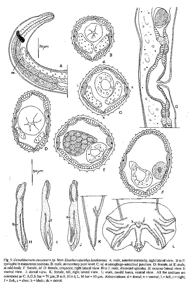
Oswaldocruzia cassonei n.sp.
Type-material: holotype male, allotype female MHNG-INVE
19477, 4 males, 1 female MNHN, 169 MD ADULTS Small nematodes, curved on ventral line. Cervical alae absent. Synlophe: (studied in 2 male and 1 female paratypes, 2 males, parasites of E. lanthanites and 1 male, parasite of E. conspicillatus. Numbers in brackets correspond to voucher specimens). In both sexes, cuticle bears uninterrupted longitudinal ridges. In male, 85-93% of ridges appear in oesophageal region within 79- 100% of dorsal ridges and 87-100% of ventral ridges. In female 86% of rides appear in oesophageal region within same dorsal and ventral ratio. Ridges disappear just anterior to caudal bursa in male and at phamids level in female. In male, 29, 31 (28, 28, 26) ridges at oesophago-intestinal junction (Fig. 5C) and 32, 35 (30, 33, 34) ridges at mid- body (Fig. 5E). In female, 36 ridges oesophago-intestinal junction (Fig. 5D) and 42 ridges at mid-body (Fig. 5F). In transversal section, ridges same size, orientated perpendicularly to body surface with regular spacing. Holotype male: 4200 um long and 100 um wide at mid- body. Cephalic vesicle 50 um long and 30 um wide. Nerve ring, excretory pore and deirids 130 um, 210 um and 230 um from apex, respectively. Oesophagus 380 um long (Fig. 5A). Caudal bursa illustrated in Fig. 6M. Spicules 125 um long. Blade with spatulate extremity, fork distally divided at 23% of whole length of spicule (Fig. 5 H, I, J). Allotype-female: 9200 um long and 130 um wide at mid-body. Cephalic vesicle 65 um long and 35 um wide. Nerve ring, excretory pore and deirids 160 um, 270 um and 290 um from apex, respectively. Oesophagus 480 um long. Vulva 2850 um from caudal extermity. Vagina vera 30 um long dividing vestibule 220 um long into two equivalent parts. Sphincters both 30 um and infundibula both 20 um long (Fig. 5G). Anterior uterine branch 1900 um long with 38 eggs, posterior uterine branch 1800 um long with 26 eggs. All eggs at morula stage, 70 um long and 50 um wide. Tail 120 um long and 50 um wide at level of anus with caudal spine 15 um long (Fig. 5K).
DISCUSSION
In the neotropical region, the sole species closely related to the specimens described above is Oswaldocruzia taranchoni Ben Slimane and Durette-Desset (1995) a parasite of Bufo marinus from Pernambuco, Brazil which has both the caudal bursa of type III and the spicular blade with spatulate extremity. Unlike the specimens described above, in O. taranchoni, the sexual dimorphism concerning the size is slighter (male 6.4-5 mm, female 7.35 mm), the ridges are more numerous (53-75 at mid-body in females) and not pronounced, and the division of the spicular fork is deeper. We consider the specimens from Eleutherodactylus as belonging to a new species Oswaldocruzia cassonei n. sp named after our colleague Jimmy Cassone.
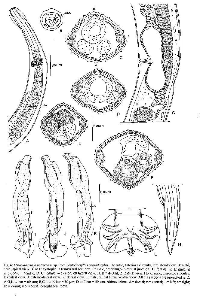
Oswaldocruzia petterae n. sp.
Type material: holotype male, allotype female MHNG-INVE
19500, 1 male, 1 female paratypes MNHN 153 MD ADULTS
Small nematodes, with anterior part of body coiled. Cervical alae absent. Synlophe: (studied in the male and the female paratypes; in 2 males and 2 females from voucher material. Numbers in brackets correspond to voucher specimens). In both sexes, cuticle bears longitudinal ridges over whole length of body. In male 82-100% of ridges appear in oesophageal region within 87-100% of dorsal ridges and 77-100% of ventral ridges. In female 90% of ridges appear in oesophageal region within same dorsal and ventral ratio. Ridges disappear just anterior to caudal bursa in male and at phasmids level in female. In male, 23 (26, 27) ridges at oesophago-intestinal junction (Fig. 6C) and 28 (26, 30) at mid-body (Fig. 6E). In female, 34 (33, 31) ridges at oesophago-intestinal junction (Fig. 6D), and 38 (41, 38) at mid-body (Fig. 6F). In transversal section, ridges are orientated perpendicularly to body surface, same size and regularly spaced except in oesophageal region where the ridges in front of lateral fields are more spaced. Holotype-male: 5000 um long and 90 um wide at mid-body; cephalic vesicle 65 um long and 40 um wide. Nerve ring, excretory pore and deirids 190 um, 330 um and 350 um from apex, respectively. Oesophagus 470 um long (Fig. 6A). Caudal bursa illustrated in Fig. 6 L. Spicules 140 um long; blade distally divided into 6 processes, fork distally divided at 21% of whole length of spicule (Fig. 6 I, J, K). Allotype-female: 9000 um long and 120 um wide at mid-body; cephalic vesicle 80 um long and 35 um wide. Nerve ring, excretory pore and deirids 200 um, 350 um and 370 um from apex, respectively. Oesophagus 510 um long Vulva 3100 um from caudal extremity. Vagina vera 35 um long dividing vestibule 240 um long into two equivalent parts. Sphincters both 25 um long and infundibula both 25 um long (Fig. 6G). Anterior uterine branch 2200 um long with 55 eggs, posterior uterine branch 2200 um long with 52 eggs. All eggs at morula stage 70 um long and 50 um wide. Tail 180 um long and 65 um wide at level of anus, with caudal spine 15 um long (Fig. 6H). DISCUSSION
The specimens from Leptodactylus are mainly characterized by (1) a cephalic vesicle without proximal swelling; (2) a synlophe with numerous ridges regularly spaced; (3) the absence of the cervical alae; (4) a caudal bursa of type III and (5) the spicular blades divided into numerous processes at their distal extremities. The only species sharing the same characters is Oswaldocruzia chambrieri Ben Slimane and Durette-Desset (1993), a parasite of Bufo and Eleutherodactylus from the same region as the specimens studied above. The extra-branches present on the spicular shoes of O. chambrieri are not observed in any of the Leptodactylus specimens. Therefore, this character is not constant within the same species and cannot be used as a specific character. Only two differences indicate a speciation: the number of ridges at mid-body level and the length of the spicules both in relation to the size of the specimen. In a female of O. chambrieri, 9 mm long, the number of ridges is 38 and the length of the spicules is 140 um. In a 6.4 mm long female parasite of Leptodactylus, the number of ridges is 54 and the length of the spicules is 190 um. We therefore consider the specimens from Leptodactylus, belonging to a new species Oswaldocruzia petterae n. sp. named after our colleague Dr Annie Petter.
REFERENCES
Ben Slimane B, Durette-Desset MC 1993. Quatre nouvelles especes du genre Oswaldocruzia Travassos, 1917 (Nematoda: Trichostrongyloidea) parasites d'Amphibiens d'Equateur. Rev Suisse Zool 100: 113-136. Ben Slimane B, Durette-Desset MC 1995. Identification d'Oswaldocruzia subauricularis (Rudolphi, 1819) et O. mazzai Travassos, 1935 et description de deux nouveaux Oswaldocruzia (Nematoda, Trichostrongylina, Molincoidea) parasites de Bufonidae neotropicaux. Rev Suisse Zool 102: 635-653. Ben Slimane B, Durette-Desset MC, Chabaud AG 1993. Oswaldocruzia (Trichostrongyloidea) parasites d'Amphibiens des Collections du Museum de Paris. Ann Parasit Hum Comp 68: 88-100. Ben Slimane B, Verhaagh M, Durette-Desset MC 1995. 0swaldocruzia peruensis n. sp. (Nematoda: Trichostrongylina) parasite d'un Iguanidae de Perou. Bull Mus Natl Hist Nat Paris 4eme ser 17: 77-82. Durette-Desset MC 1985. Trichostrongyloid Nematodes and their Vertebrate hosts: Reconstruction of the phylogeny of a parasitic group. Adv Parasitol 24: 239-306. Durette-Desset MC, Chabaud AG 1981. Nouvel essai de classification des Nematodes Trichostrongy-loidea. Ann Paras hum comp 56: 297-312. Durette-Desset MC, Nasher AK, Ben Slimane B 1992. 0swaldocruzia arabica n.sp.(Nematoda, Trichostrongyloidea) parasite d'un Bufonidae de la peninsule arabique et remarques sur des especes proches. Bull Mus Natl Hist Nat Paris 4e ser 14: 693-703. ^+Corresponding author. Fax: 33-1-40 7934 99.
E-mail: mcdd@cimrs1.mnhn.fr Fig. 2: Oswaldocruzia bainae n. sp from Anolis chrysolepis: 4th stage larvae and entsheathed immature stages (Im.) - A: L4 female, anterior extremity, right lateral view. B to D: synlophe in transversal sections. B: (Im.) female, at oesophago-intestinal junction, the immature synlophe is not yet formed. C: L4 male, at mid-body. D: (Im.) female, at mid-body, showing both L4 and (Im.) synlophes. E: (Im.) male, caudal bursa of the adult inside the tail of the L4, left lateral view. F: L4 female, tail, right lateral view. G: L4 female, genital apparat showing the beginning of the differentiation between the ovejector, the uterine branches and the ovaries, left lateral view. All sections are orientated as B. A, F bar = 50 um; B to G bar = 40 um. Abbreviations: d.= dorsal; v. = ventral; l.= left; r.= right; (Im.) = entsheathed immature in L4. Fig.3: Oswaldocruzia tcheprakovae n.sp. from Eleutherodactylus altamazonicus. A-I: synlophe in transversal sections. A-H: female (7000 mm long). A: at excretory pore level. B: at oesophago-intestinal junction. C: at 830 um from apex (twice the length of oesophagus). D: at 1180 um from apex. E: at 2360 um from apex, at about level of the first third of body). F: at mid-body. G: at ovejector level. H: at anus level. I: male, at 1400 um from the caudal bursa. J-L: L4 male. J: head, right lateral view. K: synlophe at mid-body. L: tail, right lateral view. All the sections are orientated as B. Abbreviations: d. = dorsal; v. = ventral; 1.= left; r.= right. Fig. 4: Oswaldocruzia tcheprakovae n.sp. from Eleutherodactylus altamazonicus. A-D: female - A: anterior extremity, left lateral view. B: head, apical view. C: tail, left lateral view. D: ovejector, left lateral view. E-H: male. E: caudal bursa, showing the disappearance of the ventral ridges. F: anterior extremity, showing the appearance and the disappearance of the ridges, right lateral view. G: posterior extremity, showing the appearance and the disappearance of the ridges, right lateral view. H: caudal bursa, ventral view. A, C, D bar = 40 um; B bar= 20 um; E bar = 50 um; F, G bar = 60 um; H bar = 30 um. Abbreviations: de.= deirid; d.o.t = dorsal oesophagal tooth. Fig. 5: Oswaldocruzia cassonei n.sp. from Eleutherodactilus lanthanites. A: male, anterior extremity, right lateral view. B to F: synlophe in transversal sections. B: male, at excretory pore level. C: id, at oesophago-intestina1 junction. D: female, id. E: male, at mid-body. F: female, id. G: female, ovejector, right lateral view. H to J: male, dissected spicules. H: externo- lateral view. I: ventral view. J: dorsal view. K: female, tail, right lateral view. L: male, caudal bursa, ventral view. All the sections are orientated as C. A,G,K bar = 70 um; B to F, H to J, L, M bar = 50 um. Abbreviations: d.= dorsal; v.= ventral; 1.= left; r.= right; f = fork; s = shoe; b = blade; de.= deirid. Fig. 6: Oswaldocruzia petterae n. sp. from Leptodactyllus pentadacylus. A: male, anterior extremity, left lateral view. B: male, head, apical view. C to F: synlophe in transversal sections. C: male, oesophago- intestinal junction. D: female, id. E: male, at mid- body. F: female, id. G: female, ovejector, left lateral view. H: female, tail, left lateral view. I to K: male, dissected spicules. I: ventral view. J: externo-lateral view. K: dorsal view. L: male, caudal bursa, ventral view. All the sections are orientated as C. A,G,H,L bar = 60 um; B,C, I to K bar = 30 um; D to F bar = 50 um. Abbreviations: d.= dorsal; v.= ventral; 1.= left; r.= right; de.= deirid; d.o.t=dorsal oesophageal tooth. Copyright 1996 Fundacao Oswaldo Cruz
The following images related to this document are available:Line drawing images[oc96063e.gif] [oc96063d.gif] [oc96063a.gif] [oc96063f.gif] [oc96063c.gif] [oc96063b.gif] |
| |||||||||
