 |
| About Bioline | All Journals | Testimonials | Membership | News |
|
||||||
|
||||||
A Low Stringency Polymerase Chain Reaction Approach to the Identification of Biomphalaria glabrata and B. tenagophila, Intermediate Snail Hosts of Schistosoma mansoni in Brazil Teofania HDA Vidigal/+, Emmanuel Dias Neto */**, Andrew JG Simpson***, Omar S Carvalho Laboratorio de Helmintoses Intestinais *Laboratorio de Parasitologia Celular e Molecular, Centro de Pesquisas Rene Rachou-FIOCRUZ, Caixa Postal 1743, 30190-002 Belo Horizonte, MG, Brasil **Departamento de Bioquimica e Imunologia, ICB, UFMG, Belo Horizonte, MG, Brasil *** Instituto Ludwig de Pesquisas do Cancer, Laboratorio de Genetica do Cancer, Sao Paulo, SP, Brasil
This study was supported in part by CNPq and FAPEMIG.
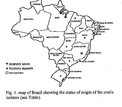
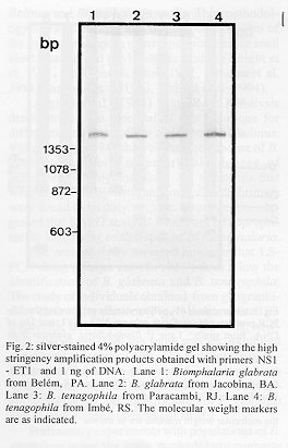
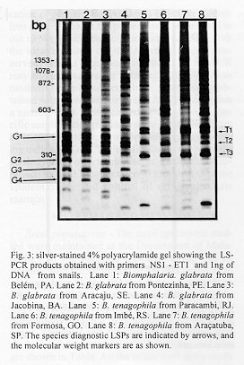
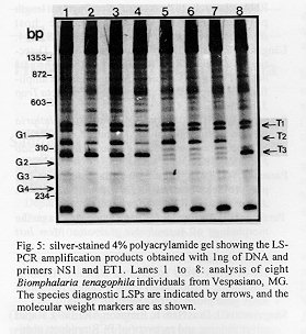
The low stringency-polymerase chain reaction (LS-PCR) with a pair of specific primers for the amplification of the 18S rRNA gene was evaluated as a means of differentiating between the two Schistosoma mansoni intermediate host species in Brazil: Biomphalaria glabrata and B. tenagophila. Individual snails obtained from different states of Brazil were used and the amplification patterns obtained showed a high degree of genetic variability in these species. Nevertheless, 4 and 3 clearly defined specific diagnostic bands was observed in individuals from B. glabrata and B. tenagophila respectively. The detection of snail specific diagnostic bands suggests the possibility of reliable species differentiation at the DNA level using LS-PCR. Key words: polymerase chain reaction - low stringency PCR - Biomphalaria - identification - Brazil Biomphalaria glabrata and B. tenagophila are important intermediate snail hosts of Schistosoma mansoni in Brazil although B. straminea also acts as a host in same areas and B. amazonica and B. peregrina can be infected under artificial conditions (Correa & Paraense 1971, Paraense 1973). These snails exhibit extensive intraspecific variation at the morphological (Paraense & Deslandes 1955, Paraense 1975) and genetic level (Knight et al. 1991, Vidigal et al. 1994). Intraspecific morphological heterogeneity, in particular, complicates snail identification especially of small specimens. An efficient and reliable method of identifying species, would be extremely valuable in the study of the distribution host species both in areas of active transmission of schistosomiasis and non endemic areas, were the presence of susceptible species should be notified to alert the health services. Because of its simplicity and sensitivity, a snail identification test based on the polymerase chain reaction (PCR) would be ideal. As a step in this direction two groups (Langand et al. 1993, Vidigal et al. 1994) have independently undertaken the amplification of host snail DNA using the arbitrarily primed PCR (AP-PCR) (Welsh & McClelland 1990), in order to produce randomly amplified polymorphic DNAs, RAPDs (Williams et al. 1990). However, the results obtained by Vidigal et al. (1994) working within B. glabrata, showed a remarkable degree of intraspecific polymorphism, with the result that the RAPD patterns observed were so variable, that there were no bands common to all the specimens analyzed. This suggested that an AP-PCR based test for species identification would be extremely difficult to develop. As an alternative to AP-PCR we have here explored the related methodology of low stringency-PCR (LS-PCR) where instead of arbitrarily selected primers, primers that specifically amplify defined regions of the genome, are used under the amplification conditions used for AP-PCR (Dias Neto et al. 1993). The result of LS-PCR amplification is a specific fragment defined by the primer used, together with a complex set of other fragments (known as low stringency products or LSPs) derived from low stringency interactions of the primers with other sequences in the target genome. This method has been of value in S. mansoni sex determination (Dias Neto et al. 1993), diagnosis of leptospirosis (Caballero et al. 1994) and the quantification of human papilomavirus infection (Caballero et al. 1995). In the present work we elected to use rRNA gene specific primers for LS-PCR with the idea that since the rRNA gene is highly conserved, related sequences amplified by LS-PCR may also exibit a greater intraspecific stability than randomly amplified sequences. The results obtained were consistent with this hypothesis in that a number of B. glabrata and B. tenagophila specific amplification products were identifiable from all specimens of these two species studied providing the basis of a PCR identification test. The methodology thus offers promise as a molecular approach to snail identification in the context of the epidemiology and control of schistosomiasis mansoni. MATERIALS AND METHODS Snail populations - The snail specimens studied were maintained at the Department of Malacology of the Institute Oswaldo Cruz, Rio de Janeiro, with the exception of the specimens of B. glabrata from Belem, PA and B. tenagophila from Vespasino, MG, which were collected directly from the field. The studies were undertaken using populations obtained from localities shown in the Fig. 1. The dates and the sites of collection as well as the number of specimens in the original samples are shown in Table. All the snails used in the study were reared and maintained at room temperature under identical conditions in aquarium with running water and calcium carbonate as substrate. The snails were exposed to artificial diurnal lighting of 10 hr and fed with lettuce. In all cases, the snails were identified by means of comparative morphology based on the reproductive organs and shells (Paraense l975) and examined for the presence of infection by S. mansoni. None of the snails were found to be infected. DNA extraction - Total DNA was extracted from the foot of the snails basically as described previously (Vidigal et al. 1994). Briefly, the foot of each snail was mechanically disrupted in 50mM Tris HCl pH 8.0, 100 mM NaCl, 50 mM EDTA, 0.5% SDS and incubated with 50 ug/ml proteinase K overnight at 37 C. Following phenol/chloroform extraction and ethanol precipitation, DNA was resuspended in 10 mM Tris-HCl, 1 mM EDTA pH 8.0 and the DNA concentration estimated by comparison with known standards on 2% ethidium bromide stained agarose gels. The LS-PCR technique - The primers used were based on the sequence of the 18s rRNA gene from the bivalve mollusk Placopecten available in GenBank (access number X53899). The primers correspond to a conserved region of the gene and show a high degree of homology with human (access number X03205), S. mansoni (access number X53467), fungi (Neurospora crassa, access number X04971) and other 18s rRNA genes. The protocol used was that previously applied to the sexual determination of human DNA and larval stages of S. mansoni (Dias Neto et al. 1993). DNA samples from each individual were amplified in duplicate using 100 pg and 1 ng of template DNA. Each reaction was undertaken in final volume of 10 ul containing 0.8 units of Taq DNA polymerase (Cenbiot RS, Brazil), 200 uM of each dNTP, 1.5 mM MgCl2, 50mM KCl, 10mM Tris-HCl pH 8.5, together with 6.4 pmoles of the primers NS1 (5'- GTAGTCATATGCTTGTCTCAG - 3') and ET1 (5'- GTCCAGACACTACGGGAAT - 3'). The reaction mix was overlaid with 20 ul of mineral oil and, following an initial denaturation at 95 C for 5 min, was subjected to two cycles through the following temperature profile: 30 C for 2 min for annealing, 72 C for 1 min for extension and 30 sec at 95 C for denaturation followed by 33 cycles where the annealing step was altered to 40 C. In the final cycle, the extension step was for 5 min. Following amplification, 3 ul of each reaction was mixed with 1 ul of 4x sample buffer (0.125% bromophenol blue, 0.125% xylene cyanol, 15% glycerol) and subjected to electrophoresis in 4% polyacrylamide gel (acrylamide/bisacrylamide 29:1) in TBE buffer (2 mM EDTA,10 mM Tris borate pH 8.0). The gels were silver stained by fixing with 10% ethanol/ 0.5% acetic acid for 3 min, staining with 0.2% silver nitrate in the fixing solution for 5 min and reduction with 0.75 M NaOH/0.1M formaldehyde for 5 min (Sanguinetti et al. 1994). High stringency amplification - For specific amplification of the 18s rRNA region, 3 pmol of each primer (NS1 and ET1) and 0.4 units of Taq DNA polymerase (Cenbiot RS, Brazil) per 10 ul reaction mixture were used. The other components of the reaction mixture were as described above for LS-PCR. The specific amplification consisted of an initial cycle with denaturation step at 95 C for 5 min, annealing of primers at 60 C for 2 min and 72 C for 2 min for extension, this was followed by 29 cycles of amplification at 60 C for 2 min, 72 C for 2 min, and 95 C for 45 sec, with an extended incubation at 72 C for 5 min in the final cycle. RESULTS Specimens of B. glabrata and B. tenagophila, derived from various geographical regions were analyzed (Table). In total six populations of B. glabrata and seven of B. tenagophila were studied. When the primer pair NS1 and ET1 used under high stringency amplification conditions, only the specific band, of approximately 1500 bp corresponding to the 18s rRNA gene, was seen in all individuals of these species, indicating the absence of detectable size polymorphism in this gene in the organisms studied (Fig. 2). When LS-PCR was used, complex patterns were produced composed of low stringency products (LSPs) derived from multiple interactions of the primer pair throughout the genomes studied (Fig. 3). Due to the competitive nature of LS-PCR (Caballero et al. 1995) the specific bands shown in Fig. 2 were of weak and variable intensity. The pattern of LSPs was polymorphic for both species however, a number of bands were consistently amplified from all the specimens of each species. The result shown in Fig. 3 is an experiment that formed part of a screening procedure aimed at identifying primers capable of distinguishing B. glabrata and B. tenagophila. The B. glabrata specimens were characterized by LS-PCR with the primers NS1/ ET1 by the presence of one or two LSP doublets between 370 and 480 bp (G1) and strong LSPs of 290, 260 and 240 bp (G2, G3 and G4). The B. tenagophila specimens were characterized by a clear LSP doublet of 500 bp (T1), strong LSPs of 400 and 310 bp (T2 and T3) as well as the absence of major LSPs between 290 and 240 bp. In order to test the generality of specific bands for the two species the number of specimens was increased (Fig. 4) The major banding characteristics described in Fig. 3 were generally maintained for both species. The pair of LSP doublets (G1) appeared as a variable complex of bands. Although the precise details of the complex were variable it was present in one form or another in all B. glabrata specimens and absent from all B. tenagophila specimens. The other B. glabrata bands were consistent in all specimens (although with variable intensity) and absent from all the B. tenagophila specimens. For B. tenagophila the most useful characters were T3 and the absence of detectable LSPs between 290 and 240 bp. T2 was present in all specimens at variable intensity but two of the B. glabrata specimens (lanes 5 and 6) also exhibited a strong LSP of this size. The doublet T1 was not consistent although at least one band of the same size was present in all the specimens tested and in none of the B. glabrata specimens were corresponding LSPs detectable. On the basis of these results, a double blind test was undertaken using of eight specimens collected directly from the field, in the town of Vespasiano, MG, as a test of the LS-PCR identification system (Fig. 5). Individual 1 made the morphological characterization and the DNA extraction. The DNA was then amplified and the diagnostic LSPs evaluated by individual 2. When this profile was compared with the position of the characteristic bands for the two species as marked on the figure the samples were identified as B. tenagophila. The most conclusive features were the presence of T1 and T2, and the absence of bands between 290 and 240 bp. Band T3 is present in all samples although it is considerably weaker in lanes 5 to 7. The DNA based identification was confirmed by the analysis of morphological characters. DISCUSSION The classical methods of identification of freshwater snails of medical importance is difficult because of the variation encountered in the anatomical and morphological characteristics commonly used for separating species (Paraense 1975, Jelnes 1979, 1986, Henricksen & Jelnes 1980). The analysis of anatomy of the reproductive organs and morphological characters of the shell can be time-consuming when several specimens are to be examined. This traditional morphological characters, used by malacologists, for identification of species, have not always been easily utilized by non-specialist. Thus, an easy and reliable method for species identification is important in efforts to control schistosomiasis. Techniques of molecular biology have recently been used to study the variability of basom-matophora mollusks and to identify species of Bulinus and Biomphalaria snails. This methodology allows the direct and detailed examination of the genome and opens up new possibilities for snail identification and phylogenetic studies (Knight et al. 1991, Rollinson & Kane 1991, Strahan et al. 1991, Langand et al. 1993, Vidigal et al. 1994). Langand et al. (1993) using RAPD analysis demonstrated the potential of this technique for differentiating populations and species of Bulinus. Vidigal et al. (1994) showed that the genome of B. glabrata exhibits a remarkable degree of intraespecific polymorphism. All the bands that were amplified by two randomly chosen primers were found to be polymorphic. Those results suggested that RAPD analysis would not be appropriate for identifying related species of Biomphalaria. The present study revealed however that LS-PCR using primers for 18s rRNA does allow the identification of B. glabrata and B. tenagophila. The study of individuals obtained from geographically distinct populations (Fig. 1) demonstrated intraspecific DNA polymorphism by LS-PCR, however specimens from the same species, exhibited some species specific LSPs. The fact that the populations used for this study were obtained from widely separated localities indicate that these primers should allow the identification of B. glabrata and B. tenagophila throughout Brazil. Preliminary experiments with populations of B. straminea using the same methodology suggests that this species is even more variable than B. glabrata and B. tenagophila, no bands are shared between all specimens studied. However at a regional level, shared LSPs were found permitting specific identification in this case (data not shown). Most importantly, B. straminea DNA did not, when used as a template for LS-PCR with the 18s rRNA primers, generate any of the diagnostic LSPs used for identification of B. glabrata and B. tenagophila. Thus, although the methodology cannot be used to conclusively identify B. straminea this snail will not be incorrectly identified as either B. glabrata or B. tenagophila. In summary, the data show that LS-PCR analysis is appropriate for distinguishing B. glabrata from B. tenagophila. The technique requires small amounts of DNA and can be applied to juvenile snails as a means of resolving problems of identification of potential intermediate snail hosts in Brazil, in situations where the classical taxonomy methodology is inconclusive. ACKNOWLEDGMENTS To Dr Wladimir Lobato Paraense and Dr Ligia Correa from the Departamento de Malacologia, Fundacao Oswaldo Cruz, for giving us snails and Dr Izabel de Carvalho Rodrigues from the Instituto Evandro Chagas, Fundacao Nacional de Saude, for giving us snails from Belem, Para. To Dr alvaro Romanha for making available the facilities used in the development of the project and Jose Geraldo Amorim da Silva and Maria angela A dos Santos for their technical assistance. REFERENCES Caballero OLSD, Dias Neto E, Koury MC, Romanha AJ, Simpson AJG 1994. Low-Stringency PCR with diagnostically useful primers for identification of Leptospira serovars. J Clin Microbiol 32: 1369-1372. Caballero OLSD, Villa LL, Simpson AJG 1995. Low stringency PCR (LS-PCR) allows entirely internally standardized DNA quantitation. Nucleic Acids Res 23: 362-363. Correa LR, Paraense WL 1971. Susceptibility of Biomphalaria amazonica to infection with two Brazilian strains of Schistosoma mansoni. Rev Inst Med Trop Sao Paulo 13: 387-390. Dias Neto E, Santos FR, Pena SDJ, Simpson AJG 1993. Sex determination by Low Stringency PCR (LS-PCR). Nucleic Acids Res 21: 763-764. Henricksen UB, Jelnes EJ 1980. Experimental taxonomy of Biomphalaria (Gastropoda: Planorbidae). I. Methods for experimental taxonomic studies on Biomphalaria carried out by horizontal starch gel electrophoresis and staining of twelve enzymes. J Chromatogr 188: 169-176. Jelnes EJ 1979. Experimental taxonomy of Bulinus (Gastropoda: Planorbidae). II. Recipes for horizontal starch gel electrophoresis of ten enzymes in Bulinus and description of internal standard systems and two new species of the Bulinus forskalii complex. J Chromatogr 170: 405-411. Jelnes EJ 1986. Experimental taxonomy of Bulinus (Gastropoda: Planorbidae): the West and North African species reconsidered, based upon an electrophoretic study of several enzymes per individual. Zool J Linn Soc 87: 1-26. Knight M, Brindley PJ, Richards CS, Lewis FA 1991. Schistosoma mansoni: Use of a cloned ribosomal RNA gene probe to detect restriction fragment length polymorphisms in the intermediate host Biomphalaria glabrata. Exp Parasitol 73: 285-294. Langand J, Barral V, Delay B, Jourdane J 1993. Detection of genetic diversity within snail intermediate hosts of the genus Bulinus by using random amplified polymorphic DNA markers (RAPDs). Acta Trop 55: 205-215. Paraense WL 1973. Susceptibility of Biomphalaria peregrina from Brazil and Ecuador to two strains of Schistosoma mansoni. Rev Inst Med Trop Sao Paulo 15: 127-130. Paraense WL 1975. Estado atual da sistematica dos planorbideos brasileiros. Arq Mus Nac Rio de Janeiro 55: 105-128. Paraense WL, Deslandes N 1955. Observations on the morphology of Australorbis glabratus. Mem Inst Oswaldo Cruz 53: 87-103. Rollinson D, Kane RA 1991. Restriction enzyme analysis of DNA from species of Bulinus (Basom-matophora; Planorbidae) using a cloned ribosomal RNA gene probe. J Moll Stud 57: 93-98. Sanguinetti CJ, Dias Neto E, Simpson AJG 1994. Rapid silver staining and recovery of PCR products separated on polyacrylamide gels. BioTechiniques 17: 915-918. Strahan K, Kane RA, Rollinson D 1991. Development of cloned DNA probes for the identification of snail intermediate hosts within the genus Bulinus. Acta Trop 48: 117-126. Vidigal THDA, Dias Neto E, Carvalho OS, Simpson AJG 1994. Biomphalaria glabrata: extensive genetic variation in Brazilian isolates by random amplified polymorphic DNA analysis. Exp Parasitol 79: 187-194. Welsh J, McCleelland M 1990. Fingerprinting genomes using PCR with arbitrary primers. Nucleic Acids Res 18: 7213-7218. Williams JGK, Kubelick AR, Livak KJ, Rafalski JA, Tingey SV 1990. DNA polymorphisms amplified by arbitrary primers are useful as genetic markers. Nucleic Acids Res 18: 6531-6535.
Fig. 2: silver-stained 4% polyacrylamide gel showing the high stringency amplification products obtained with primers NS1 - ET1 and 1 ng of DNA. Lane 1: Biomphalaria glabrata from Belem, PA. Lane 2: B. glabrata from Jacobina, BA. Lane 3: B. tenagophila from Paracambi, RJ. Lane 4: B. tenagophila from Imbe, RS. The molecular weight markers are as indicated. Fig. 3: silver-stained 4% polyacrylamide gel showing the LS-PCR products obtained with primers NS1 - ET1 and 1ng of DNA from snails. Lane 1: Biomphalaria. glabrata from Belem, PA. Lane 2: B. glabrata from Pontezinha, PE. Lane 3: B. glabrata from Aracaju, SE. Lane 4: B. glabrata from Jacobina, BA. Lane 5: B. tenagophila from Paracambi, RJ. Lane 6: B. tenagophila from Imbe, RS. Lane 7: B. tenagophila from Formosa, GO. Lane 8: B. tenagophila from Aracatuba, SP. The species diagnostic LSPs are indicated by arrows, and the molecular weight markers are as shown. Fig. 4: silver-stained 4% polyacrylamide gel showing the LS-PCR amplification products obtained with primers NS1-ET1 and 1ng of DNA extracted. Lanes 1 and 2: Biomphalaria glabrata from Belem, PA. Lanes 3 and 4: B. glabrata from Cururupu, MA. Lanes 5 and 6: B. glabrata from Touros, RN. Lanes 7 and 8: B. glabrata from Pontezinha, PE. Lanes 9 and 10: B. glabrata from Aracaju, SE. Lanes 11 and 12: B. tenagophila from Paracambi, RJ. Lanes 13 and 14: B. tenagophila from Imbe, RS. Lanes 15 and 16: B. tenagophila from Joinvile, SC. Lanes 17 and 18: B. tenagophila from Aracatuba, SP. Lanes 19 and 20: B. tenagophila from Formosa, GO. Lanes 21 and 22: B. tenagophila from Vila Velha, ES. The species diagnostic LSPs are indicated by arrows, and the molecular weight markers are as shown. Fig. 5: silver-stained 4% polyacrylamide gel showing the LS-PCR amplification products obtained with 1ng of DNA and primers NS1 and ET1. Lanes 1 to 8: analysis of eight Biomphalaria tenagophila individuals from Vespasiano, MG. The species diagnostic LSPs are indicated by arrows, and the molecular weight markers are as shown. TABLE: Places and dates of Biomphalaria populations
Species          Origin           Site     Date of collection
-------------------------------------------------------------
B. glabrata      Belem, PA        Stream   November, 1992
B. glabrata      Cururupu, MA     River    May, 1989
B. glabrata      Touros, RN       Lake     November, 1975
B. glabrata      Pontezinha, PE   Stream   April, 1989
B. glabrata      Aracaju, SE      Stream   November, 1985
B. glabrata      Jacobina, BA     River    June, 1984
B. tenagophila   Paracambi, RJ    Stream   May, 1990
B. tenagophila   Imbe, RS         NA       NA
B. tenagophila   Aracatuba, SP    NA       May,1981
B. tenagophila   Formosa, GO      NA       August, 1981
B. tenagophila   Vespasiano, MG   Stream   May, 1994
B. tenagophila   Joinvile, SC     NA       NA
B. tenagophila   Vila Velha, ES   NA       January, 1983

NA: data not available
Copyright 1996 Fundacao Oswaldo Cruz
The following images related to this document are available:Photo images[oc96134b.jpg] [oc96134d.jpg] [oc96134e.jpg] [oc96134c.jpg]Line drawing images[oc96134a.gif] |
| |||||||||
