 |
| About Bioline | All Journals | Testimonials | Membership | News |
|
||||||
|
||||||
RESEARCH NOTE Invasion of HeLa Cells by Providencia alcalifaciens Presumably is Plasmid-encoded Vera Magalhaes/+, Nilma C Leal*, Vilma M Melo, Marise Sobreira, Marcelo Magalhaes Laboratorio Keizo Asami, UFPE, Cidade Universitaria 50670-901 Recife, PE, Brasil *Centro de Pesquisas Aggeu Magalhaes- FIOCRUZ, Cidade Universitaria, Recife, PE, Brasil
+Corresponding author. Fax: +55-81-441.6169
Key words: Providencia alcalifaciens - invasive plasmid - HeLa cells - virulence factors - enteroinvasive bacteria - diarrhea Providencia alcalifaciens, a well-characterized species in the tribe Proteeae, is the most recent organism included among invasive enterobacteria. In fact, it enters HeLa cell monolayers mobilizing actin filaments and invades the intestinal mucosal epithelium inducing diarrhea in adult rabbits with removable intestinal ties (MJ Albert et al. 1992 Infect Immun 60: 5017-5024). In this animal model, the bacterium penetrates epithelium directly by endocytosis or disrupting cell junctions with entry into and proliferation in intercellular spaces (MM Mathan et al. 1993 Pathol 171: 67-71). Thus, P. alcalifaciens shares potential virulence properties with Shigella spp., the prototype of enteroinvasive bacteria. Now we report that the capacity of P. alcalifaciens to invade HeLa cells is probably associated with the carriage of a 45- to 50- Kb plasmid. We selected for the study eleven strains of P. alcalifaciens, isolated from equal number of children with symptoms of enteritis. The cultures were obtained in 1993, during a survey carried out in Recife, State of Pernambuco, Brazil, to evaluate the role of P. alcalifaciens in the etiology of gastroenteritis (manuscript in preparation). Provodenciae selected for the study were present in the fecal samples in the absence of any other recognized enteropathogenic organisms. After recovering, strains were maintained at room temperature in brain heart infusion broth (Difco Laboratories, Detroit, Michigan, USA), containing 0.4% of agar, for no more than three months before starting the tests. The gentamicin-HeLa cell invasion assays were conducted and analyzed essentially as reported for the system Edwardsiella spp. and HEp-2 cell monolayers (MJ Janda et al. 1991 Infect Immun 59: 154-161). But, when the strain PA-5 was tested, we shortened the post-infection period from 3 to 1 hr to avoid detaching HeLa cells from coverslips. All P. alcalifaciens strains submitted to invasion trials were susceptible to gentamicin. An invasive strain of Shigella flexneri was the positive control. Tests for production of conjunctivitis in rabbits, for detecting plaque formation, and contact hemolytic activity were done as previously described (B Sereny 1955 Acta Microbiol Sci Hung 2: 293-296, V Oaks et al. 1985 Infect Immun 48: 124-129, PJ Sansonetti et al. 1986 Infect Immun 51: 61-69). Plasmid DNA was extracted by the method of HC Birboim and J Doly (1979 Nucleic Acids Res 7: 1513-1537) and examined by electrophoresis of cell lysates through 0.6% agarose gels. Plasmid bands were stained with ethidium bromide and visualized with UV illumination. Their molecular lengths were determined by comparing their eletrophoretic mobilities with those of reference plasmids. Of the eleven strains of P. alcalifaciens studied, seven were invasive for HeLa cell monolayers. As first noticed (Albert et al. loc. cit.), some strains are more invasive than others. The strain PA-5, for instance, was higly invasive, destroying the cell monolayer if the replication phase exceeds 2 hr (Fig. 1). All invasive strains, independently of the degree of invasiveness, carried a plasmid of 45-to 50-Kb (Fig. 2). In contrast, the four strains plasmidless were unable to invade HeLa cells. These findings show a clear correlation between inv-plasmis carriage and invasiveness. Despite the strains now studied were recent clinical isolates, four of them did not display the inv-plasmid. Whether such strains lost the inv-plasmid immediately after culturing, or this plasmid is not essential for invasiveness, remains to be determined. In this vein, another study, analyzing the virulence factors of P. alcalifaciens, from Sao Paulo, State of Sao Paulo, Brazil, did not find any connection between plasmid profile and invasiveness (BEC Guth & A Perella 1994 Abstr Annu Meet Am Soc Microbiol B-375 p. 95). Thus, new investigations should be done to clarify this discrepancy. In some invasive assays, two strains nonharboring the inv-plasmid showed occasional intracellular bacteria (in less than 1% of the cells) on the Giemsa stained coverslips, or few colonies (less than 10/ml) on the agar plates seeded with the triton X-100 cell lysate. This, apparently was due to heterogeneity of the bacterial inoculum used for infecting HeLa cells, that is, occasional organisms still carried the inv-plasmid, but the DNA burden was insufficient to allow its visualization on the agarose gels. Since the inv-plasmid of P. alcalifaciens does not bear selective marks, refined genetic procedures are required before we can handle uniform plasmid-harboring bacterial populations. Besides the inv-plasmid, we noticed other extrachromosomal DNA bands on the agarose gels. One of them, characterized as a large plasmid of 147-Kb, was displayed by ten of the eleven cultures examined. The only strain, which did not carry this plasmid, was invasive, pointing out that the 147-Kb plasmid is not implicated in HeLa cell invasion. Probably this plasmid occurs as a large unicopy, since it was well evident on the gels but appeared as a hazy band on contrasted photos (Fig. 2). Many plasmids with different molecular sizes and arranged in distinctive patterns were exhibited by some strains. This diversity of plasmid outline might be useful as an epidemiologic marker for tracing relevant strains of P. alcalifaciens. Indeed, among the eleven strains examined, eight different plasmid profiles were observed (Fig. 2). Curiously, of the four noninvasive strains, three had the same plasmid fingerprint. In contrast to the inv-plasmid, all the others were considered cryptic since they could not be related to drug-resistance or bacteriocin production. Our strains did not produce plaques in HeLa cell monolayers, did not show contact hemolytic activity, neither induced keratoconjunctivitis in rabbits, as did the Shigella control strain. In reality, unlike shigellae and similar to salmonellae or yersiniae (BB Finlay & S Falkow 1989 Bact Rev 53: 210-230), the ingested Providencia remains enveloped within endocytic vacuoles (Albert et al. loc. cit.). Providenciae multiplies actively within the vacuoles producing large bacterial cluters and ultimately the cell lysis (Fig. 1). Concerning this, the intracellular behavior of P. alcalifaciens parallels that observed with Edwardsiela tarda (Janda et al. loc. cit.), although this bacterium appears does not carry large invasive plasmids (LRM Marques et al. 1984 Curr Microbiol 10: 129-132), neither P. alcalifaciens produce hemolysin. Present data supplement the earliest work of Albert et al. (loc. cit.), who, documenting that P. alcalifaciens invades cultured cells and the rabbit intestinal epithelium, encouraged the acceptance of this organism as a bona fide potential enteropathogen. Yet, genetic studies for identifying the inv-gene are need before we can admit with confidence that the 45-50-Kb plasmid is actually responsible for the invasive phenotype displayed by many clinical isolates of P. alcalifaciens, as our findings insinuated.
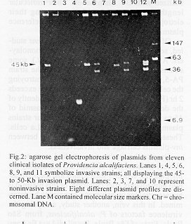
Fig.2: agarose gel electrophoresis of plasmids from eleven clinical isolates of Providencia alcalifaciens. Lanes 1, 4, 5, 6, 8, 9, and 11 symbolize invasive strains; all displaying the 45- to 50-Kb invasion plasmid. Lanes: 2, 3, 7, and 10 represent noninvasive strains. Eight different plasmid profiles are discerned. Lane M contained molecular size markers. Chr = chromosomal DNA. Copyright 1996 Fundacao Oswaldo Cruz
The following images related to this document are available:Photo images[oc96140a.jpg] [oc96140b.jpg] |
| |||||||||
