 |
| About Bioline | All Journals | Testimonials | Membership | News |
|
||||||
|
||||||
Vol. 92(1), Jan./Feb. 1997 Diagnostic Importance of Female External Genital Structure of Phlebotomine Sand Flies (Diptera:Psychodidae) as Observed by Scanning Electron Microscopy J Mukhopadhyay/*, KN Ghosh /*/+
Department of Zoology, University of Calcutta, 35 Ballygunge Circular Road,
Calcutta 700 019, India +Corresponding author. Fax: +1-203-785.4782
Received 27 February 1996.
Code Number:OC97012
Sizes of Files:
    Text: 15.1K
    Graphics: Line drawings (gif) - 375.0K
              Tables (gif) - 18.0K
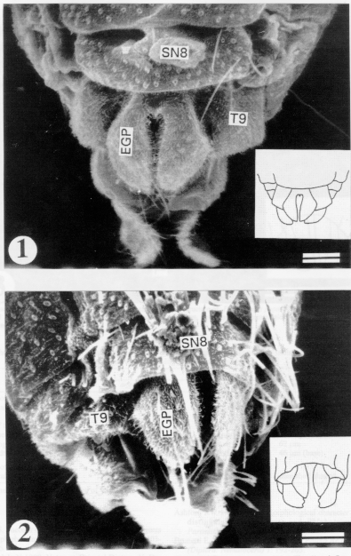
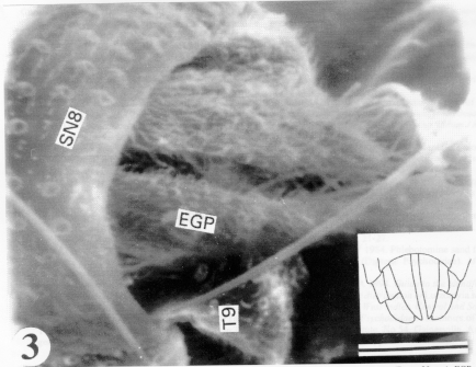
Morphological description of sand flies has remained a neglected area. The different organs used in taxonomy have not yet been described adequately with the scanning electron microscopy (SEM). We have examined the external genital structures of females of three Old World phlebotomine sand flies under SEM and recorded the morphological variations of the organs. We have found the female external genital structures of the three species varied considerably in morphology. The importance of the female external genital structures in sand fly identification is indicated. Key words: Indian sand flies - Phlebotomus - external genital structures - scanning electron microscopy Phlebotomine sand flies are important vectors of many human diseases including leishmaniases (Adler & Theodor 1957). The description of sand flies started in 1786 when an Italian scientist/Naturalist Dr Bonanni described a sand fly. Later, a similar species was described by Scopoli (1786) and it was considered a species of Phlebotomus. However, the description of sand flies in India started in 1908 when Annandale (1908) described P. argentipes. Since then, a series of works on sand flies of different aspects including the morphological descriptions of the Old World sand flies were carried out by the Kala-azar Commission during 1900-1942. Sinton (1923, 1924, 1925, 1927) contributed the majority of the work dealing with morphological description of Indian sand flies. After 1942, there was few reports of the sand fly description and distribution. Mitra and Roy (1953) and Mitra (1959) described sand flies including the morphological variations of P. argentipes, collected from Maharashtra and Kashmir states of India, respectively. Kaul et al. (1973) listed the sand flies in Rajasthan state including a description of a new sand fly, Sergentomyia (Sintonius) sirohi sp. nov. In contrast, most of the work dealing with the morphological descriptions of the New World sand flies were carried out by Young and his coworkers (Young & Perkins 1984, Young & Duncan 1994). The majority of the descriptive studies have dealt with the importance of the male genitalia for identification and, in some cases, a few features of the female mouth parts are considered. Recently, Ashford (1991) indicated the importance of microtrichia as a new taxonomic character to distinguish between Sergentomyia and Phlebotomus. There is no morphological study dealing with the importance of external female genitalia as an identification character, mainly because it is difficult to see their details by light microscopy. However, Sinton (1927) emphasized the importance of the internal female genitalia for the purpose of identification. Recently, Pesson et al. (1994) have shown the importance of female genital armature in separating the females of P. papatasi and P. duboscqi, two very closely related Old World species that are difficult to separate. Here we have used scanning electron microscopy (SEM) to examine the external genital structure of female sand flies of three Old World species and have found that the characters of the external genital plate may have a value for the identification of the species. MATERIALS AND METHODS The females of P. argentipes Annandale and Brunetti and P. papatasi (Scopoli) used in this study were taken from two laboratory colonies (Ghosh et al. 1992). P. major major Annandale was collected from the Dankuni area of West Bengal. The gravid females were allowed to oviposit individually in the laboratory in modified plastic vials to obtain the adults following the method of Ghosh and Bhattacharya (1989). The F1 progeny was identified following the key provided by Lewis (1978) and confirmed to be P. m. major. The adult females of the three species were fixed in 1% glutaraldehyde solution in PBS (pH 7.2) at 4 C. After 2-4 hr, the glutaraldehyde was replaced by 3% glutaraldehyde and kept for 8-12 hr at the same temperature. The specimens were then washed with PBS three successive times to remove the glutaraldehyde from the samples. After that, the samples were passed through ascending grades of ethanol (30%-100%) and were brought to the SEM Unit where ethanol was replaced by isoamyl acetate. The samples were then dried in a critical point drying apparatus (Polaron E500). Afterwards, they were mounted on a metal stub with double sided adhesive tape and were coated with 200-300 A egrees of an Au-Pd alloy in a vacuum evaporator (Edwards S150 sputter coater). Specimens were examined under a Philips SEM (PSEM 500) with tilt angles and photographed. RESULTS
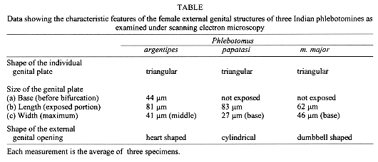
Fig. 3: scanning electron micrograph of the female external genital structure of Phlebotomus papatasi (Bar = 30 mm). EGP: external genital plate; T9: ninth tergite; SN8: eighth sternite. In inset, diagrammatic representation of the external genital structures.
DISCUSSION For most sand flies, the male external genital structures are important characters as they are very unique, having species specific characters. However, in some cases, the males of closely related species have almost identical morphological characters viz., P. papatasi, P. salehi and P. duboscqi. In such situations it is difficult to separate the females which are also morphologically very similar. It has always been a problem to separate the females of these species, particularly P. papatasi and P. duboscqi. So, an easily detectable and reliable female specific diagnostic character is very important for identification and determining the epidemiological role of the insect. Female external genital structures in sand flies are less complicated and are limited to the genital plate. The genital plate is actually a modification of the 9th sternite and its modification probably is a functional need. In the present investigation we have found that the external female genital plate in P. argentipes, P. papatasi and P. m. major are triangular in shape but the triangular shape is quite different having different dimensions at the different sides of the plate (Figs 1-2, Fig. 3). In all of the above three cases as the orientation of the plates are different so is the space in between them, the external genital opening which varies in shape and size. Thus, the external genital structures bear species specific diagnostic characters (Table). Recently, Killick-Kendrick et al. (1994) have shown that the morphology of the genital atria of Kenyan species of Laurroussius bears some important distinguishing characters. It is also established that spermathecae can be utilized for species identification. However, as spermathecae are soft and contractile, there is a high probability that the morphology of the structure may change. Spermathecae of certain species of sand flies may vary considerably owing to contraction and also for the different mounting media (Quate 1962, Lewis & Dyce 1976). On the other hand, the comparatively hard cuticular parts are less vulnerable to changes and so they are more reliable for taxonomic identification. The above result on the morphology of female external genital structure clearly indicate variation among different species as seen here in three Old World sand flies. Similar studies on other Old World sand flies and New World sand flies are necessary for a better understanding of the validity of the female external genital structure as a possible identifying character. The results from the future work with other sand flies is expected to strengthen the foundation that the female external genital structure might be helpful in separating the females of very closely related species. ACKNOWLEDGEMENTS To the Scanning Electron Microscope Unit, Regional Sophisticated Instrumentation Centre, Bose Institute, Calcutta for providing SEM facilities. To Dr M Maroli for his constant help and inspiration during our sand fly work. REFERENCES
Copyright 1996 Fundacao Oswaldo Cruz The following images related to this document are available:Line drawing images[oc97012a.gif] [oc97012b.gif] [oc97012c.gif] |
| |||||||||
