 |
| About Bioline | All Journals | Testimonials | Membership | News |
|
||||||
|
||||||
Vol. 92(1), Jan./Feb. 1997 Specific Identification of Biomphalaria tenagophila and Biomphalaria occidentalis Populations by the Low Stringency Polymerase Chain Reaction Edina Rodrigues Pires, Teofania HDA Vidigal,Horacio MS Teles*, Andrew JG Simpson, Omar S Carvalho/+
Laboratorio de Helmintoses Intestinais, Centro dePesquisas Rene
Rachou-FIOCRUZ, Caixa Postal 1743, 30190-002 Belo Horizonte, MG,
Brasil +Corresponding author. Fax: +55-31-295.3115
Received 6 May 1996
Code Number:OC97021
Sizes of Files:
    Text: 19.5K
    Graphics: Line drawings (gif) - 8.6K
              Photographs (jpg) - 53.5K
              Tables (gif) - 17.8K
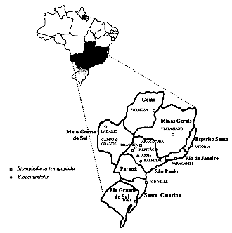
Although Biomphalaria occidentalis and B.tenagophila areindistinguishable on the basis of shell morphology and the majority oftheir genital organs, only the latter is susceptible to infection withSchistosoma mansoni. Thus, the identification of these species is fundamental to epidemiological studies of schistosomiasis. Here we describe a simple and rapid method for differentiating B.tenagophila from B. occidentalis based on low stringency polymerase chain reaction and using a pair of primers specific for the amplification of the 18S rRNA gene. Analysis of the low stringency product profiles of populations of these snails from different geographical regions confirmed this approach as being applicable to the identification of B.tenagophila and B. occidentalis in cases where classical morphology is inconclusive. Key words: polymerase chain reaction - low stringency - Biomphalaria tenagophila - Biomphalaria occidentalis - schistosomiasis Paraense (1981), described Biomphalaria occidentalis, aspecies which could not be differentiated from B. tenagophila by shell characteristics and by the morphology of most genital organs. The same author showed that in the laboratory, the two species are separated by absolute reproductive isolation. The differentiation of the two species can be accomplished only by careful dissection of the male and female genitalia and the demonstration of the presence of a vaginal pouch in B. tenagophila and its absence in B. occidentalis (Paraense 1981). B. occidentalis has never been successfully infected by the trematode Schistosoma mansoni (Paraense & Correa 1982,Coimbra & Engel 1982), and the identification of these species is important for the epidemiology and control of schistosomiasis mansoni. Bailey et al. (1986) used electrophoresis of the hemolymphs of these snails in agarose gel in order to differentiate them. Mascara and Morgante (1995) proposed isoenzyme patterns that might contribute to the identification of these molluscs. An alternative, and indeed very powerful, method to study the genetics of molluscs is the polymerase chain reaction (PCR). A modification of this technique is the random amplification of polymorphic DNA (RAPD), by which complex and informative genomic fingerprints can be readily generated without prior sequence determination (Williams et al. 1990, Welsh & McClelland1990). This technique has already been used in the study of molluscs for the identification of Bulinus species (Langand et al. 1993) and in analysis of genetic variability of B. glabrata populations (Vidigalet al. 1994). A related methodology, described by Dias-Netoet al. (1993),was named low stringency PCR (LS-PCR). This method has been used for sexdetermination in S. mansoni, for the identification of Leptospira serovars (Caballero et al. 1994) and morerecently inidentification of B. glabrata and B. tenagophila(Vidigal etal. 1996). LS-PCR utilizes specific primers under LS conditions, incontrast to AP-PCR (arbitrarily primed PCR) where the choice of primer is arbitrary, although the conditions of the reaction remain the same. The result of complex LS-PCR amplification is a specific fragment defined by the primer used, together with a complex set of other fragments (known as LS products or LSPs) derived from LS interactions of the primers with other sequences in the target genome. Here we have used LS-PCR with specimens of B. tenagophila and B. occidentalis from different geographical regions of Brazil showing that distinct profiles are consistently produced which distinguish these two planorbid species. MATERIALS AND METHODS Snail populations - The studies were undertaken using seven populations of B. tenagophila and six of B. occidentalis from different geographical regions of Brazil (Fig. 1).
All the snails used in the study were reared and maintained at room temperature under identical conditions in aquaria with running water and sterilized earth, sand, and calcium carbonate. The snails were exposed to artificial diurnal lighting for 10 hr and fed with lettuce. In all cases, the snails were identified by means of comparative morphology based on the reproductive organs and shells (Paraense 1975). The snails were examined in order to determine whether they released any type of cercaria and none were found to be infected by S. mansoni or other trematodes. Preparation of DNA - Total DNA was extracted from the foot of the snails basically as described for B. glabrata by Vidigal et al. (1994). Briefly, the foot of each snail was mechanically disrupted in 50mM Tris HCl ph 8.0, 100mM NaCl, 50mM EDTA, 0.5% SDS and incubated with 50 mg/ml proteinase K overnight at 37 C. Following phenol/chloroform extraction and ethanol precipitation, DNA was resuspended in 10 mM Tris-HCl, 1 mM EDTA pH 8.0 and the DNA concentration estimated by comparison with known quantitative standards in 1.5% ethidium bromide stained agarose gels. A one nanogram template DNA was utilized for each reaction of the PCR. DNA amplification by LS-PCR - The protocol used was that previously applied for the study of sex differentiation in S. mansoni (Dias-Neto et al. 1993). DNA samples from each individual were amplified using 1 ng of template DNA. Each reaction was undertaken in a final volume of 10ml containing 0.8 units of Taq DNA polymerase (Cenbiot, RS, Brazil), 200 mM each dNTP, 1.5 mM MgCl2, 50 mM KCl, 10 mM Tris-HCl, pH 8.5, together with 3.2 pmoles of the primers ET1 (5'-GTCCAGACACTACGGGAAT-3') and NS1 (5'-TAGTCATATGCTTGTCTCAG-3'). The primers used are based on the sequence of the 18S rRNA gene from the bivalve mollusc Placopecten available in GenBank (access number X53899). The primers correspond to a conserved region of the gene and show a high degree of homology with human (access number X03205), S. mansoni (access number X53467), fungi (Neurospora crassa, access number X04971) and other 18S rRNA genes. The reaction mix was overlaid with 20 ml of mineral oil and, following an initial denaturation at 95 C for 5 min, was subjected to two cycles through the following temperature profile: 30 C for 2 min for annealing, 72 C for 1 min for extension and 95 C for 30 sec for denaturation followed by 33 cycles where the annealing step was altered to 40 C. In the final cycle, the extension step lasted 5 min. For analysis of the snail amplification products, 3ml of the final reaction mix was applied to the gel. Electrophoresis was undertaken using 4% polyacrylamide gels. Following the separation, the gels were fixed with 10% ethanol/0.5% acetic acid for 5 min and DNA bands revealed by staining with 0.2% silver nitrate for 10 min and reduction with 0.75 M NaOH/0.1 M formaldehyde for 5 min as previously described (Santos et al. 1993, Sanguinetti et al. 1994). RESULTS
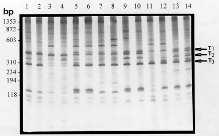
Lanes 1 and 2 are representative of B. tenagophila specimens from Formosa, GO. Lanes 3 and 4: B. tenagophila specimens from RS. Lanes 5 and 6: B. tenagophila specimens from Joinvile, SC. Lanes 7 and 8: B. tenagophila specimens from Aracatuba, SP. Lanes 9 and 10: B. tenagophila specimens from Vitoria, ES. Lanes 11 and 12: B. tenagophila specimens from Paracambi, RJ. Lanes 13 and 14: B. tenagophila specimens from Vespasiano, MG. The LS-PCR amplification products were visualized in a 4% polyacrylamide gel stained with silver.
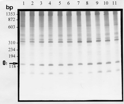
Fig. 3: comparison
of LSP profiles of Biomphalaria occidentalis obtained with the
primer pair NS1-ET1 and 1 ng of DNA template.
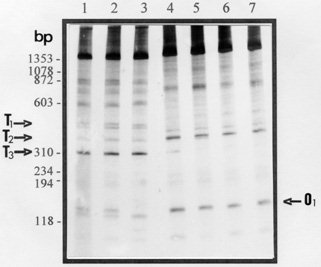
Lane 1 to 3: B. tenagophila specimens from Vespasiano; MG, Vitoria; ES and Joinvile; SC, respectively. Lanes 4 to 7: B. occidentalis specimens from Campo Grande; MS, Ladario; MS; Assis; SP and Dracena; SP, respectively. The LS-PCR amplification products were visualized in a 4% polyacrylamide gel stained with silver.
The identification of B. tenagophila and B. occidentalis is difficult because these species are very similar and cannot be differentiated by shell characteristics or by the morphology of most of the genital organs (Paraense 1981). Thus, alternative approaches are required. Bailey et al. (1986), used hemolymph analyzed by agarose gel electrophoresis in an attempt to separate these species but this approach demonstrated that B. glabrata and B. straminea could not be separated. Mascara and Morgante (1995) demonstrated that isoenzyme analysis may contribute to the identification of these two species of snails. These authors observed that the value of these techniques is questionable for taxonomic studies because quantitative and qualitative variations are associated with the age and size of individual snails. The use of PCR provides a new method for the study of molluscs at the genomic level and has been used with success in the study of the genetic variability of Bulinus and B. glabrata populations (Langand et al. 1993, Vidigal et al. 1994), and in two species of marine gastropods: Littorina saxalitis and L. arcana (Crossland et al. 1993). In the present study, LS-PCR was used as an approach to the identification of B. tenagophila and B. occidentalis. Analysis of the results with specimens obtained from widely separated localities probed with a pair of primers specific for the rRNA gene (NS1-ET1) indeed shows that these two species can be readily distinguished by this technique. It is noteworthy that in both cases, although there are polymorphisms, the LSP profiles are relatively consistent, facilitating identification and suggesting a reduced level of genetic variation in these species as compared with B. glabrata (Vidigal et al. 1994). The profiles of the two species, while allowing identification, are generally quite similar. This is consistent with the close phylogenetic relationship of the two species and, at a practical level, demonstrates that the test should be employed using standards and coelectrophoresis on the same gel to avoid errors. ACKNOWLEDGMENTS To Dr Wladimir Lobato Paraense and Dr Ligia Correa from the Departamento de Malacologia, Instituto Oswaldo Cruz, for providing the snails. To Cristiana Brito, Rosane Sturzeneker, Emmanuel Dias Neto and Patricia Pinto from the Centro de Pesquisas Rene Rachou, by their invaluable advice and discussions, and Dr Alvaro Romanha for the facilities provided in the development of the project. REFERENCES
Work partially supported by Fundacao de Amparo agrave; Pesquisa de Minas Gerais (FAPEMIG) and Coordenacao de Aperfeicoamento de Pessoal de Nivel Superior (CAPES). Copyright 1996 Fundacao Oswaldo Cruz The following images related to this document are available:Photo images[oc97021e.jpg] [oc97021c.jpg] [oc97021d.jpg]Line drawing images[oc97021b.gif] [oc97021a.gif] |
| |||||||||
