 |
| About Bioline | All Journals | Testimonials | Membership | News |
|
||||||
|
||||||
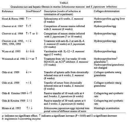
Differential regulation of granuloma size and hepatic fibrosis in schistosome infections Allen W Cheever Biomedical Research Institute, 12111 Parklawn Drive, Rockville, MD 20852
USA and Laboratory of Parasitic Diseases, National Institute of Allergy and
Infectious Diseases, Bethesda, MD 20892-0425 USA
Received 16 April 1997; Accepted 30 June 1997
Code Number:OC97129
Sizes of Files:
    Text: 13.4K
    Graphics: Tables (jpg) - 80.9K
              Line drawing (jpg) - 21.8K
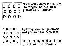
Granuloma size is the variable most frequently used to evaluate the immunopathogenesis of schistosome infections. However, hepatic fibrosis is at the least an equally relevant variable. Hepatic fibrosis and the size of circumoval granulomas are frequently dissociated in experimental murine Schistosoma mansoni and S. japonicum infections. Virtually nothing is known of the immunoregulation of schistosomal hepatic fibrosis. This review notes many of the studies which have found discrepancies in granuloma volume and hepatic fibrosis, attempts to put them in perspective and to evaluate different methods of calculating changes in collagen synthesis or content. Key words: schistosomiasis - granulomatous inflammation - fibrosis In experimental schistosomiasis the degree of hepatic fibrosis often correlates with volume of hepatic granulomas, but there are numerous exceptions to this and the regulation of hepatic fibrosis and granuloma size are clearly at least partly independent. It is clear that when comparing granuloma size and fibrosis that one should consider only those situations in which the duration of infection is the same in the groups being considered and in which only newly formed granulomas are measured, i.e. reactions around eggs containing mature, viable embryos. It is less clear how one should quantify fibrosis. We have generally used the total increase above normal levels of hepatic collagen (determined as the increase in hydroxyproline per liver per egg). This gives a measure of the cumulative increase in total collagen (synthesis minus destruction) at any given time point, but different time points cannot be rigorously compared and rates of synthesis and destruction at specific times after infection are not determined. Collagen synthetic rates should give a better picture at any given moment, but one does not generally know the rate of collagen destruction at that moment. Even if the level of collagenases is known, should one in any case consider constitutive or latent collagenases, besides needing to consider at least type I and type III collagenases? The frequent dissociation of granuloma size and collagen content or synthetic rates is evident in the Table. I find the interpretation difficult when granulomas decrease in size and the collagen or synthetic rate per g granuloma does not change, i.e. the collagen or synthetic enzymes in the granulomas are perhaps decreased but compacted into a smaller volume (Fig.). However, when the granulomas remain the same size and the collagen and synthetic rates per g granuloma decrease (Olds et al. 1989) or when different treatments have different effects (Olds & Kresina 1989), the results seem unequivocal. Similarly the results seem clearly interpretable when total hepatic collagen is considered in relation to egg numbers. Figure:Illustration of an instance in which the
interpretation of dissociation of granuloma size and hepatic fibrosis is
equivocal. The box represents a gram of granulomatous tissue.
There are dissociations between granuloma size and fibrosis in addition to the treatments and comparisons noted in the Table. For example, in Schistosoma mansoni-infected mice granuloma volume does not change with intensity of infection (expressed as worm pairs or total liver eggs) while the fibrosis per egg decreases as infection intensity increases (Cheever 1986). We know nothing of the kinetics of collagen deposition in relation to granuloma size in humans. We do know that in many cases of "active" Symmers' fibrosis that downregulatory anti-idiotypic T cells are absent and proliferation of peripheral blood mononuclear cells is greater than that in asymptomatic hepato-intestinal cases (Montesano et al. 1990). Similar anti-idiotypic T cells downregulate the size of in vitro granulomas formed around schistosome eggs by PMBC of infected patients (Parra et al. 1991). Obviously, it will be important to have surrogate variables of hepatic fibrosis and collagenolysis to follow in infected humans. Past studies suggest that serum procollagen III (Zwingenberger et al. 1988, Mincis et al. 1990, Fayol et al 1991, Shahin et al. 1992) or other serum precursors (Tanabe et al. 1989, Parise & Rosa 1992, Shahin et al. 1995) or in vitro responses of liver from infected patients (Dunn et al. 1979, Monteiro & Borojevic 1995) may be of interest in this regard. Collagen production by in vitro granulomas (Parra et al. 1991) has apparently not yet been examined. The examples cited above and in the Table indicate that granuloma size and hepatic fibrosis are frequently regulated independently. The factors relevant to immune-modulation of granuloma size include CD8^+ suppressor effector cells, CD4^+ suppressor inducer and effector cells, macrophages, cross-regulation of Th1 and Th2 cells, anti-idiotypic antibodies (S. japonicum) and anti-idiotypic t cells (S. mansoni and S. japonicum). Immunoregulation of schistosomal hepatic fibrosis has been less examined and might affect pathways involved in collagen synthesis, cross-linking or collagenase activity. All of these pathways determining granuloma size and fibrosis are presumably affected by the balance of cytokines which may affect reaction size and fibrosis in different ways or to different degrees. For example, cytokines affecting cell recruitment might be expected to affect primarily the size of granulomas. Some treatments noted in the Table, i.e. anti-IL-4 treatment (Cheever et al. 1994), may act directly on enzymes affecting collagen (Postlewaite et al. 1992) as well as through immunoregulatory pathways. REFERENCES
Copyright 1997 Fundacao Oswaldo Cruz The following images related to this document are available:Photo images[oc97129a.jpg] [oc97129b.jpg] |
| |||||||||
