 |
| About Bioline | All Journals | Testimonials | Membership | News |
|
||||||
|
||||||
Leishmania major: Parasite Interactions Suggesting Sexuality Maria Auxiliadora de Sousa/^+, Mirian Claudia de Souza Pereira*, Suzana Corte-Real* Colecao de Tripanosomatideos, Departamento de Protozoologia
*Departamento de Ultraestrutura e Biologia Celular, Instituto Oswaldo Cruz,
Av. Brasil 4365, 21045-900 Rio de Janeiro, RJ, Brasil
Received 30 December 1996; Accepted 10 July 1997
Code Number:OC97142
Sizes of Files:
    Text: 20.1K
    Graphics: Tables (jpg) - 17.1K
              Line drawings and photographs (jpg) - 63.5K
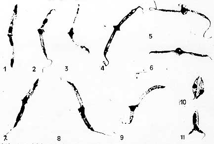
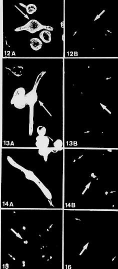
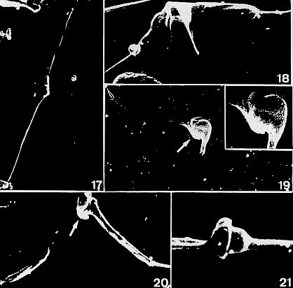
In five experiments, Leishmania (Leishmania) major (MRHO/SU/59/P-strain) grew poorly when seeded in FYTS medium supplemented with 15% fetal calf serum, but presented several peculiar pairs of promastigotes diametrically opposed and attached at their posterior ends (5.8-13.5%). As seen in Giemsa-stained smears, a ring-like line and/or an enlargement, generally occurred at the parasite junction. A close proximity of nuclei, which sometimes were difficult to distinguish from each other, was also observed at this junction. Several of these pairs appeared to be composed of fused cells in which the nuclei could be apparently fused, as shown by fluorescence microscopy to detect beta-tubulin and DNA, and by scanning electron microscopy. Under other culture conditions these pairs were absent or occurred at very low rates (0.2-2.2%). Such pairs differ markedly from longitudinally dividing cells and resemble those described in two other Leishmania species, as well as in Herpetomonas megaseliae and Phytomonas davidi, suggesting steps of a putative sexual process. Key words: Trypanosomatidae - Leishmania major - sexuality - morphology -immunocytochemistry - scanning electron microscopy The occurrence of sexuality in Leishmania has been a subject of interest since the beginning of this century, but several older cytological observations ascribed to sexual events were unconvincing (Rogers 1904, Adie 1921-22, Christophers et al. 1926, Wenyon 1926, Wenrich 1954). This subject was discredited, until Maazoun et al. (1981) reported heterozygous patterns in enzyme electrophoretic variants of three Leishmania species, and their paper was followed by several others describing naturally occurring putative hybrids using biochemical and/or molecular methods (Le Blancq et al. 1983, Le Blancq & Peters 1986, Evans et al. 1987, PagIs et al. 1989, Kelly et al. 1991, Cupolillo et al. 1992, Belli et al. 1994, Piarroux et al. 1994, Dujardin et al. 1995). However, genetic recombination between species or strains of Leishmania has not yet been demonstrated under laboratory conditions (Gradoni et al. 1986, Evans et al. 1989, Panton et al. 1991, Shehata et al. 1991), although Lanotte and Rioux (1990) had recorded by videocinematography fusion of pairs of apposed promastigotes in cultures of two species, and also evidenced the possibility of nuclear fusion by examining these cultures in Giemsa-stained smears. In this paper we describe pairs of apposed promastigotes occurring at relatively high percent in L. (L.) major (P strain) when seeded in a medium described for insect trypanosomatids (Roitman et al. 1972), which we had supplemented with fetal calf serum. Such pairs were studied on Giemsa-stained smears, by fluorescence microscopy to examine the beta-tubulin distribution and nuclear DNA features, as well as by scanning electron microscopy. The frequency of such pairs under other culture conditions was also examined. MATERIALS AND METHODS Promastigotes of a L. (L.) major strain (MRHO/SU/59/P) grown in BHI+LIT medium overlaying blood-agar (Jaffe et al. 1984) were seeded in FYTS medium (Roitman et al. 1972) supplemented with 15% heat-inactivated fetal calf serum (FYTS + 15% FCS), pH 7.0, which was distributed in 4-ml-volumes in 16x150 mm screwcap tubes, and kept at about 27 C. Following the finding in fresh preparations of unusual pairs of apposed cells, Giemsa-stained smears were prepared as previously described (Sousa 1994) for morphological studies. Subsequently, two experiments were performed to compare the frequency of such pairs in FYTS + 15% FCS with those in LIT medium (Chiari & Camargo 1984), LIT/blood-agar and FYTS + 15% FCS/blood-agar. The inoculum was always promastigotes from BHI+LIT/blood-agar cultures, which in the first experiment had no pair of apposed cells, while in the second one presented 0.5%. The cultures were maintained at about 27 C and the percent of pairs of apposed cells was determined daily, from the 2nd to the 7th days, by examining at least 500 cells in fresh preparations. The maximum parasite growth was compared in two culture conditions, FYTS + 15% FCS and LIT/blood-agar, in both cases beginning with about 8.5x10^5 cells/ml. The occurrence of pairs of apposed cells in FYTS + 15% FCS was also verified by using promastigotes from LIT/blood-agar as inoculum (3 assays). Pairs of apposed cells from a 5-day old culture in FYTS + 15% FCS were studied under fluorescence microscopy to examine concomitantly the beta-tubulin distribution and the nuclear DNA. Then, the cells were washed in phosphate buffered saline (PBS), pH 7.2, allowed to adhere to 0.1% poly-L-lysine-coated coverslips, fixed for 5 min at room temperature with 4% paraformaldehyde in PBS and rinsed three times in PBS containing 0.5% Triton X-100. Subsequently, the cells were incubated for 1 hr at 37 C with 1:50 anti-beta tubulin monoclonal antibody (Sigma), rinsed and incubated for 1 hr with 1:25 fluorescein isothiocyanate (FITC)-coupled rabbit anti-mouse IgG; following a 10 min incubation with 2 mg/ml 4,6-diamidino-2-phenylindole (DAPI; Sigma), the coverslips were rinsed in 0.85% NaCl and mounted in 1.4-diazabicyclo-(2.2.2)-octane (DABCO; Sigma). The preparations were observed and photographed with a Zeiss microscope equipped for epifluorescence using selective FITC or DAPI filters. For scanning electron microscopy, cells from a 5-day old culture in FYTS + 15% FCS were adhered to poly-L-lysine coated coverslips as above-described, fixed for 1 hr at 4 C in 2.5% glutaraldehyde in 0.1 M Na-cacodylate buffer, pH 7.2, plus 3.5% sucrose, and subsequently washed in this same buffer. After post-fixation in 1% OsO4 for 30 min, the material was dehydrated in graded series of acetone, dried with CO2 by the critical-point method, mounted on aluminum stubs and coated with a 20 nm-thick gold layer. The samples were examined with a Carl Zeiss DSM 940 scanning electron microscope. RESULTS FYTS + 15% FCS medium was unsuitable for growing L. major promastigotes, whose population increased somewhat more than twice within 48-72 hr, but thereafter gradually declined. However, such cultures presented a relatively high percent (5.8-13.5%) of peculiar pairs of promastigotes, at times very slender, diametrically opposed to each other and attached at their posterior ends, there generally occurring a ring-like line and/or an enlargement (Figs 1-9) sometimes resembling a hat brim (Fig. 5), as seen in Giemsa-stained smears. In such pairs both nuclei were usually in close proximity and in some instances it was difficult to distinguish one from the other, either in Giemsa-stained slides or under DAPI fluorescence (Figs 3-6, 9,) (Figs. 12B-14B, 15, 16). Several pairs seemed composed of fused cells, as suggested either by indirect immunofluorescence of beta-tubulin (Figs 12A, 13A, arrows) or by scanning electron microscopy, which also evidenced the peculiar enlargement and meeting of nuclei at the level of the parasite junction (Figs 17-21). Typically dividing cells could also be seen in FYTS +15 % FCS (Figs 10, 11).
Figures 1-11
Figures 12-16
Figures 17-21
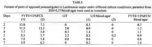
In fresh preparations, the pairs under study were actively moving and presented an uncommon brightness at the level of nucleus. The phenomenon of formation of these pairs in FYTS + 15% FCS medium was reproducible in 5/5 assays, the inoculum being promastigotes either from BHI+LIT/blood-agar or LIT/blood-agar. The percent of these pairs in different culture conditions are given in Table. L. major grew luxuriantly in LIT/blood-agar and to a lesser extent in other media (FYTS + 15% FCS/blood-agar>LIT>FYTS + 15% FCS). Beginning with about 8.5x10^5 promastigotes/ml, the maximum growth in LIT/blood-agar was nearly 6x10^7 cells/ml (5th day), whereas in FYTS + 15% FCS it was around 2x10^6 cells/ml (2nd day). DISCUSSION To our knowledge, in Leishmania species, pairs of promastigotes such as these described herein have not been reported previously. They also differ from those found in another L. major strain during experimental infection in Lutzomyia longipalpis (Walters et al. 1993), which were apposed paramastigotes. These pairs also markedly differ from cells joined laterally during longitudinal binary fission (compare Figs 1-9 with 10 and 11). It is worthy mentioning that although dividing trypanosomatids can occasionally be attached by their posterior ends before complete separation, they neither present any enlargement nor the proximity of nuclei at the level of their junction, as commonly seen here. However, the pairs of promastigotes described by us are somewhat similar to those reported in Herpetomonas megaseliae and Phytomonas davidi (Sousa 1991, 1994), and resemble those formed by fusion previously described in L. infantum and L. tropica (Lanotte & Rioux 1990), since the parasite attachment occurs by the posterior ends, the cells remaining apposed to each other, and on several occasions nuclear fusion appears to take place. Our studies to detect both beta-tubulin and DNA in such pairs, as well as our findings by scanning electron microscopy, strongly suggest cellular fusion in the majority of cases, as well as close contact or even fusion of the nuclei (Figs 12A-21). Taking together our findings and those from the literature, we have considered the possibility that a fusion process to promote nuclear interactions could have generated the pairs here described, this strongly suggesting a sexual process. Our data confirm that in trypanosomatids phenomena can occur in which the cytological bases for genetic recombination are clearly present. The finding of putative hybrids of other L. major isolates (Le Blancq et al. 1983) and of L. major/L. arabica (Evans et al. 1987, Kelly et al. 1991) support the hypothesis of occurrence of genetic exchange in these species, although it has been considered infrequent (Le Blancq et al. 1983, Kelly et al. 1991). In the present study, we verified that the pairs of apposed promastigotes suggesting a sexual process were not common in media other than FYTS + 15% FCS, they usually not being found or occurring in low numbers. It is accepted that genetic recombination mediated by sex is generally a major mechanism promoting diversity within a species, consequently enhancing its chances of survival in a fluctuating environment. As our paper shows close association between two cells and their nuclei, the question arises whether the unsuitable conditions for L. major growth in supplemented FYTS medium could have triggered the formation of such pairs to propitiate some type of genetic exchange. There is much to be done on this subject and studies are planned to explain better the present findings, such as determining the biochemical and molecular structure of this organism, both before and after being cultured in supplemented FYTS. ACKNOWLEDGMENTS To Dr Wladimir Lobato Paraense for reviewing the manuscript, Miss Sheila Medeiros dos Santos for technical assistance and Dr Monika Barth for allowing several photomicrographs to be taken at her laboratory. REFERENCES Copyright 1997 Fundacao Oswaldo Cruz The following images related to this document are available:Photo images[oc97142b.jpg] [oc97142c.jpg] [oc97142d.jpg] [oc97142a.jpg] |
| |||||||||
