 |
| About Bioline | All Journals | Testimonials | Membership | News |
|
||||||
|
||||||
Expression and Function of beta1 Integrins on Human Eosinophils Maria-Cristina Seminario, Bruce S Bochner^+
Department of Medicine, Division of Clinical Immunology, Johns Hopkins
Asthma and Allergy Center, The Johns Hopkins University, Hopkins Bayview
Circle, Baltimore, MD 21224-6801, USA Received 3 September 1997; Accepted 30 September 1997
Code Number:OC97181
Sizes of Files:
    Text: 41.5K
    Graphics: Tables (jpg) -24.3K
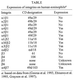
Eosinophils preferentially accumulate at sites of chronic allergic diseases such as bronchial asthma. The mechanisms by which selective eosinophil migration occurs are not fully understood. However, interactions of cell-surface adhesion molecules on the eosinophil with molecular counterligands on endothelial and epithelial cells, and on extracellular matrix proteins, are likely to be critical during the recruitment process. One possible mechanism for selective eosinophil recruitment involves the alpha4beta1 (VLA-4) integrin which is not expressed on neutrophils. Correlations have been found between infiltration of eosinophils and endothelial expression of VCAM-1, the ligand for VLA-4, in the lungs of asthmatic individuals as well as in late phase reactions in the lungs, nose and skin. Epithelial and endothelial cells respond to the Th2-type cytokines IL-4 and IL-13 with selective de novo expression of VCAM-1, consistent with the possible role of VCAM-1/VLA-4 interactions in eosinophil influx during allergic inflammation. Both beta1 and beta2 integrins on eosinophils exist in a state of partial activation. For example, eosinophils can be maximally activated for adhesion to VCAM-1 or fibronectin after exposure to beta1 integrin-activating antibodies or divalent cations, conditions that do not necessarily affect the total cell surface expression of beta1 integrins. In contrast, cytokines like IL-5 prevent beta1 integrin activation while promoting beta2 integrin function. Furthermore, ligation of integrins can regulate the effector functions of the cell. For example, eosinophil adhesion via beta1 and/or beta2 integrins has been shown to alter a variety of functional responses including degranulation and apoptosis. Thus, integrins appear to be important in mediating eosinophil migration and activation in allergic inflammation. Strategies that interfere with these processes may prove to be useful for treatment of allergic diseases. Key words: eosinophil - interleukin - beta1 integrins - allergic diseases Eosinophil accumulation is a distinctive feature of allergic airways inflammation (Bochner et al. 1994). Evidence for a role of eosinophils in the airway inflammation in asthma comes from a variety of studies. The presence of increased numbers of these cells has been demonstrated in bronchial biopsies, bronchoalveolar lavage (BAL) fluid and peripheral blood of patients with asthma. Furthermore, these cells appear to be in an activated state or in the process of degranulation and the levels of their granule proteins have been extensively correlated with clinical symptoms of asthma. Recent studies on the role of eosinophils have focused on the mechanisms by which these cells infiltrate the airways (Resnick & Weller 1993, Bochner & Schleimer 1994). Although the exact mechanisms by which selective eosinophil recruitment occurs remain incompletely defined, leukocyte recruitment is known to result from the interaction of cell-surface adhesion molecules (e.g., selectins, integrins, and immunoglobulin superfamily members) with molecular counterligands on vascular endothelial cells, extracellular matrix (ECM) proteins, epithelial cells and other tissue structures (Carlos & Harlan 1994, Bochner & Schleimer 1997). While other factors determine the phenotype of infiltrating cells, such as cytokines and chemokines (Springer 1995), this chapter will focus on the role of beta1 integrins in eosinophil trafficking and function. Expression of integrins on eosinophils Integrins are plasma membrane receptors composed of alpha and beta heterodimeric transmembrane subunits generated from at least 16 alpha and 8 beta subunits to produce over 20 different receptors (Bochner & Schleimer 1997). Both chains are required for normal receptor expression and for ligand binding. Members of the integrin family mediate cell-to-cell and cell-to-extracellular matrix interactions. Table summarizes the expression of integrins on eosinophils. The predominant integrins on all leukocytes are in the beta2 (CD18) subfamily (Bochner & Schleimer 1997). Granulocytes including eosinophils express the beta2 integrins LFA-1, Mac-1, p150,95 and alpha d beta2 (Grayson et al. 1997). LFA-1 binds specifically to intercellular adhesion molecule-1 (ICAM-1), ICAM-2 and ICAM-3, Mac-1 binds to ICAM-1 and the iC3b product of activated complement, alpha d beta2 recognizes ICAM-3, while cellular ligands for p150,95 are as yet unknown. Because all granulocytes express beta2 integrins, there appears to be no immediate explanation for how they might contribute to selective eosinophil recruitment. However, there are conditions under which eosinophil beta2 integrins, especially Mac-1, may be selectively altered by stimuli such as cytokines [e.g., IL-5 (Walsh et al. 1990)] and chemokines (e.g., eotaxin) (Burke Gaffney & Hellewell 1996). When other integrins are examined, more obvious differences in expression among granulocytes are observed (Georas et al. 1993, Ebisawa et al. 1995). Unlike neutrophils, eosinophils express the alpha4 integrins VLA-4 (alpha4beta1) and alpha4beta7 which mediate binding to VCAM-1, an immunoglobulin superfamily member induced by cytokines on endothelium and epithelial cell lines (Atsuta et al. 1997, Bochner & Schleimer 1997), and to an alternatively spliced domain in fibronectin, CS-1 (Anwar et al. 1994, Matsumoto et al. 1997). The alpha4beta7 integrin binds to the mucosal addressin cell adhesion molecule-1 (MAdCAM-1) that has structural homology to ICAM-1 and VCAM-1 (Walsh et al. 1996, Briskin 1997). Eosinophils also express alpha6beta1 (VLA-6), a ligand for the extracellular matrix protein laminin (Georas et al. 1993, Tourkin et al. 1993). Basophils resemble eosinophils in that they too express alpha4beta1 and alpha4beta7, but instead of alpha6beta1, they express alpha5beta1, another ligand for fibronectin (Saini et al. 1997). Eosinophil-endothelial interactions through beta1 integrins One mechanism of selective eosinophil recruitment involves the beta1 integrin alpha4beta1 (VLA-4), which is expressed on human eosinophils but not on neutrophils. This may be important for allergic inflammatory responses since it is a receptor for VCAM-1, and correlations have been found between infiltration of eosinophils and expression of VCAM-1 in the lungs of patients with asthma as well as in late phase reactions in the lungs, nose or skin (Kyan-Aung et al. 1991, Bentley et al. 1993, Lee et al. 1994, Gosset et al. 1994, Ohkawara et al. 1995, Fukuda et al. 1996). Expression of VCAM-1 also correlated with eosinophil numbers in nasal polyp tissues (Jahnsen et al. 1995, Beck et al. 1996). Resting endothelial cells do not express VCAM-1. However, exposure of endothelial cells to IL-1, TNF, or bacterial endotoxin induces expression of endothelial adhesion molecules, including ICAM-1, E-selectin and VCAM-1. Specific antibodies to ICAM-1 and E-selectin have been shown to inhibit adherence of eosinophils to IL-1 stimulated endothelial monolayers by about 20-30% (Bochner et al. 1991). In contrast, VCAM-1 antibodies are extremely effective at inhibiting eosinophil but not neutrophil adherence (Bochner et al. 1991). Furthermore, anti-VLA-4 antibodies inhibit eosinophil, but not neutrophil, adhesion to IL-1 stimulated endothelium (Dobrina et al. 1991, Walsh et al. 1991). These results indicate that specific induction of VCAM-1 on endothelial cells could selectively promote eosinophil adherence. Eosinophils are predominant at inflammatory sites where Th2-type cytokines, such as IL-4 and IL-13, are prevalent (Hamilos et al. 1996, Rankin et al. 1996). In vitro, both of these cytokines selectively lead to the induction of VCAM-1 expression without any significant effect on the expression of E-selectin or ICAM-1 on endothelial cells (Schleimer et al. 1992, Kaiser et al. 1993, Bochner et al. 1995). Furthermore, incubation of endothelial cells with IL-4 or IL-13 has no effect on neutrophil adhesion but induces eosinophil adhesion in a dose-dependent manner (Schleimer et al. 1992, Bochner et al. 1995). These cytokines can be synergistic and selective in their ability to induce VCAM-1 on endothelial cells. The combination of IL-4 with either IL-1 or TNF results in at least 5-fold higher levels of VCAM-1 surface expression than either cytokine alone, with no induction of ICAM-1 or E-selectin (Iademarco et al. 1995, Ebisawa et al. 1997). Further support for the potential importance of beta1 integrins and their ligands is provided by in vivo studies where the function of these adhesion molecules, or the cytokines that induce their expression, has been blocked. Efforts to antagonize VLA-4, VCAM-1, and IL-4 have all been shown to reduce eosinophil recruitment and allergic airways or cutaneous inflammation in a variety of animal models (Gonzalo et al. 1996, Richards et al. 1996, Lobb 1997, Fryer et al. 1997). Eosinophil-extracellular matrix (ECM) interactions through beta1 integrins After migration through the endothelium, eosinophils come into contact with the proteins of the basement membrane and ECM. The ECM is a complex web of large fibrillar proteins that underlies the endothelium and epithelium and surrounds connective tissue cells. Cellular interactions with ECM proteins can have profound consequences on leukocyte function (Hunt et al. 1997). Eosinophils interact with two ECM proteins, fibronectin and laminin, through two beta1 integrins, namely VLA-4 and VLA-6 (alpha6beta1), respectively. Fibronectin - Fibronectin is encoded by a single gene, but alternative splicing of the primary RNA transcript gives rise to polypeptide diversity that appears to be regulated in a cell type-specific fashion (Walsh & Wardlaw 1997). The IIICS region of fibronectin contains a 25 amino acid site, named CS-1, that contains a sequence (LDV) recognized by VLA-4. Plasma fibronectin lacks the IIICS binding site in at least half of its subunits, whereas tissue fibronectin has it in both subunits. Despite expression of alpha4 integrins on eosinophils, whether they spontaneously attach to fibronectin remains controversial. Some studies have shown that resting eosinophils adhere to fibronectin in a VLA-4-dependent manner and exhibit prolonged survival via autocrine production of cytokines such as GM-CSF (Anwar et al. 1994, Neeley et al. 1994, Walsh et al. 1995). Other studies, however, have found little or no adhesion without prior activation with platelet-activating factor, Mn^++ or a beta1 integrin-activating antibody (Kuijpers et al. 1993, Kita et al. 1996, Matsumoto et al. 1997). A possible explanation for these discrepancies may arise from the fact that eosinophils express the beta2 integrin Mac-1, and engagement through this receptor to a different site on fibronectin, or to the blocking protein (typically albumin), may be occurring. Eosinophils also express alpha4beta7, another ligand for fibronectin (Erle et al. 1994, Walsh et al. 1996). Levels of alpha4beta7 on eosinophils are comparable to those for alpha4beta1. In addition to functioning as a fibronectin ligand, it can also be a ligand for MAdCAM-1 and VCAM-1 (Erle et al. 1994, Walsh et al. 1996). However, alpha4beta7 on eosinophils appears to be relatively inactive, because activation with Mn^++ is required to demonstrate consistent adhesion (unpublished observations). Laminin - Laminin consists of 3 distinct chains coded for by different but related genes. The mechanism by which laminin interacts with cells is complex (Walsh & Wardlaw 1997). It is recognised by different integrin receptors including alpha1beta1, alpha2beta1, alpha3beta1, alpha6beta1 and alpha v beta3, of which only alpha6beta1 appears to be specific for laminin. Eosinophils can adhere to plate-bound laminin; this interaction requires divalent cations and is completely abolished by anti-alpha6 or anti-beta1 antibodies. Indeed, eosinophils were shown by flow cytometry and immunoprecipitation to express alpha6beta1 (Georas et al. 1993). As has been shown for fibronectin, eosinophils cultured on laminin exhibit prolonged survival (Tourkin et al. 1993). Eosinophil-epithelial interactions through beta1 integrins Airway epithelium may also be an active participant in allergic inflammation. Epithelial cells are biologically active, express adhesion receptor proteins, and produce cytokines and chemokines (Polito & Proud 1997). Until recently, only ICAM-1, but not E-selectin or VCAM-1, had been identified in the respiratory epithelium in vitro and in in vivo biopsies from patients with asthma (Bloemen et al. 1993, Fukuda et al. 1996, Stark et al. 1996). However, in the BEAS-2B bronchial epithelial cell line, culture with TNF or IL-1 was found to induce VCAM-1 mRNA and cell surface expression (as well as ICAM-1 expression), while culture with IL-4 induced VCAM-1 but not ICAM-1 expression (Atsuta et al. 1997). Maximal VCAM-1 expression resulted from the combination of TNF and IL-4. Furthermore, TNF treatment increased adhesion of eosinophils to BEAS-2B monolayers and this adhesion was blocked with VCAM-1 antibodies. These findings suggest that cytokine activation can induce expression of VCAM-1 on airway epithelium which can functionally interact with eosinophils through VLA-4. Alterations in function versus expression of beta1 integrins on eosinophils Levels of alpha4beta1 and alpha6beta1 on eosinophils are not altered after migration in vitro or in vivo or after cytokine activation, nor do levels differ among hypodense versus normodense eosinophils or cells from allergic versus nonallergic donors (Georas et al. 1992, 1993, Hansel & Walker 1992, Kroegel et al. 1994). However, in addition to the amount of expression of cell surface adhesion molecules, the functional state of integrins can be regulated, leading to changes in the affinity for counterligand binding without changing the level of cell surface expression (Diamond & Springer 1994). Recent studies have shown that the activation state of integrins can influence cell adhesion and function. The avidity of integrins, not just the total number of molecules expressed, influences cell adhesion and migration (Hunt et al. 1997). While a particular integrin may have more than one ligand, the avidity for each ligand may differ. It has recently been demonstrated that alpha4beta1 integrins on eosinophils exist in a state of partial activation, and can be maximally activated for adhesion to ligands such as fibronectin and VCAM-1 after exposure to manganese or integrin-activating antibodies, conditions that do not affect the total cell surface expression of beta1 integrins (Werfel et al. 1996, Matsumoto et al. 1997). Maintenance of basal levels of beta1 integrin function on eosinophils appears to require tyrosine kinase activity, because reversible downregulation of VCAM-1 adhesion is seen in cells exposed to genistein or tyrphostins (Nagata et al. 1995, Matsumoto et al. 1997). Signaling via beta1 integrins on eosinophils Outside-in signaling - Adhesion molecules are not only involved in adhesive interactions but also in transducing signals from the extracellular to the intracellular compartments and regulating effector functions of the cell (Ginsberg et al. 1992, Clark & Brugge 1995). For eosinophils, ligation of integrins has been shown to alter a variety of functional responses (Dri et al. 1991, Anwar et al. 1993, Tourkin et al. 1993, Neeley et al. 1994, Nagata et al. 1995, Kita et al. 1996). Signaling mechanisms via integrins are still poorly understood. The intracytoplasmic domains of integrins lack kinase or phosphatase activity of their own; they also lack sequence homology with known signaling proteins (Hemler et al. 1994). However, recent reports have shown that integrin engagement, either with ligand or with antibodies, is capable of transducing signals (Miyamoto et al. 1995) and induces the phosphorylation of the tyrosine kinase pp125^FAK (FAK) (Schaller & Parsons 1994). Outside-in signaling is initiated by the b subunit cytoplasmic-domain dependent rearrangement of cytoskeletal components and actin into focal adhesion complexes (FAC), found at areas of cell-ECM interaction (Clark & Brugge 1995). Formation of FAC's in adherent cells is thought to be associated with cell spreading. A predominant FAC's component, FAK, has been shown to physically interact with the cytoplasmic domain of b integrins, which in turn is thought to recruit several signaling molecules to FAC's (Schaller & Parsons 1994). It is not clear whether the cytoskeletal and signaling components found in FAC's associate with integrins in leukocytes. Besides FAK, beta1 integrin interacts directly or indirectly with cytoskeletal proteins (McArthur Lewis & Schwartz 1995, Yamada & Miyamoto 1995, Wahl et al. 1996). In addition to FAK, integrin receptor occupancy leads to the activation of the Src family of tyrosine kinases (Shattil et al. 1994) and the Ras/MAP kinase pathway (Schaller & Parsons 1994). Recently a novel serine/threonine kinase has been reported to associate with the beta1 integrin cytoplasmic domain (Hannigan et al. 1996). The 59 kD protein, known as integrin-linked kinase (ILK), was found to phosphorylate a peptide representing the beta1 integrin cytoplasmic domain and to co-localize with beta1 in focal plaques. Outside-in signaling is also regulated by the a subunit cytoplasmic tails. Those of alpha2 and alpha5 localize predominantly to FAC's and show increased spreading on ECM. In contrast, the expression of the alpha4 cytoplasmic tail correlates with chemo and haptotactic migration, suggesting that alpha4 is responsible for weaker integrin-cytoskeletal interactions (Kassner et al. 1995). This is a potential mechanism by which alpha4beta1, highly expressed in eosinophils, could increase cell motility. Inside-out signaling - The rapidity of inside-out signaling insures that leukocytes can quickly modify their adhesiveness in response to stimuli. This is achieved by changes in integrin functional activity rather than integrin expression on the cell surface. The signaling pathways involved in inside-out signaling are still ill-defined (Hunt et al. 1997). Recent progress in this field has been mainly in T cells. Several activation stimuli have been shown to upregulate integrin function. Treatment of T cells with PMA or the Ca+2 ionophore alpha23187 has been shown to upregulate integrin-mediated T- cell adhesion, indicating that both protein kinase C and Ca+2 are involved in the intracellular signaling events (Shimizu et al. 1990), whereas treatment of Jurkat cells with the serine-threonine phosphatase inhibitor okadaic acid depresses fibronectin adhesion through beta1 integrins (Seminario et al. 1997). Furthermore, cell-surface receptors, called integrin regulators, can induce an inside-out signal. These integrin regulators include the TCR complex, CD2, CD28, CD7 (Chan et al. 1991, Shimizu et al. 1992) and cell surface signaling proteins of the transmembrane-4 superfamily that include CD9, CD53, CD63, CD82 and CD81 (Mannion et al. 1996). Receptors for chemokines have been shown to be a class of integrin regulators for leukocyte interactions with the endothelium (Springer 1990, del Pozo et al. 1995). Chemokine receptors belong to the seven membrane spanning family of G protein coupled receptors (Schall & Bacon 1994, Teran & Davies 1996). Signaling through chemokine receptors is thought to be very rapid inducing integrin activity within minutes. For example, the chemokines RANTES, MCP-1, MIP-1a and MIP-1b have been shown to upregulate beta1 integrin mediated adhesion of T cells (Carr et al. 1996), monocytes (Weber et al. 1996) and eosinophils (Weber et al. 1996b) to counterreceptors and ECM ligands. Conclusions Eosinophils selectively migrate to sites of chronic allergic inflammation where they are thought to be one of the major contributors to the underlying pathology. An initial step in the recruitment of leukocytes is their adhesion to endothelium, followed by transendothelial migration and chemotaxis into tissue parenchyma. Several adhesion molecules have been implicated in eosinophil adherence, however, VLA-4, which is not present in neutrophils, provides a potential mechanism by which specific recruitment occurs. This chapter has reviewed the biology of beta1 integrins on eosinophils and their potential roles in preferential cellular trafficking. Although the exact mechanisms of tissue eosinophilia are yet to be fully defined, it is clear that beta1 integrin-mediated adhesion to tissue structures are important for cellular migration and is capable of altering cellular function. Further elucidation of this process should lead to a better understanding of mechanisms of allergic inflammation, and may ultimately lead to the design of better therapies for allergic diseases. This work was supported by grant HL49545 from the National Institutes of Health and an Underrepresented Minority Investigator in Asthma and Allergy Award to Dr Seminario from the National Institutes of Health and the American Academy of Allergy, Asthma, and Immunology. Dr Bochner was also supported in part by a Developing Investigator Award from the Burroughs Wellcome Fund and a grant from the Office of Naval Research, awarded through the Asthma and Allergy Foundation of America. References
Copyright 1997 Fundacao Oswaldo Cruz - Fiocruz The following images related to this document are available:Photo images[oc97181a.jpg] |
| |||||||||
