 |
| About Bioline | All Journals | Testimonials | Membership | News |
|
||||||
|
||||||
Morphological Features of Trypanosomes from Squirrel Monkeys from the Brazilian Amazon Mariangela Ziccardi^+, Ricardo Lourenco-de-Oliveira
Laboratorio de Transmissores de Hematozoarios, Instituto Oswaldo Cruz, Av.
Brasil 4365, 21045-900 Received 23 September 1996; Accepted 20 August 1997
Code Number:OC98010
Sizes of Files:
    Text: 33.7K
    Graphics: Line drawings and photographs (jpg) - 208.1K
              Tables (jpg) - 258K
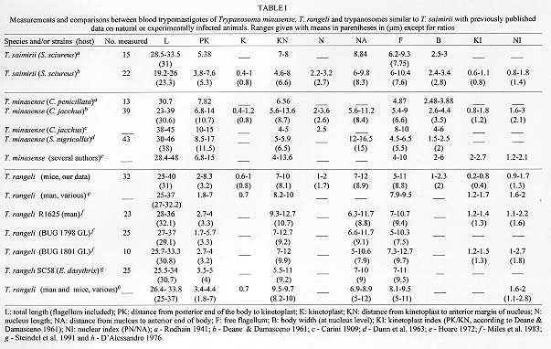
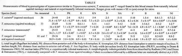
A morphometric analysis of blood trypomastigotes identified as Trypanosoma minasense, T. saimirii, and T. rangeli harbored by squirrel monkeys from the Brazilian Amazon was performed. Additionally, morphological and biological comparative analyses were conducted of T. saimirii-like and T. rangeli development forms from haemoculture and xenodiagnosis. Illustrations are given of blood trypomastigotes as well as of developing flagellates in triatomine and axenic culture. Mean values of blood trypomastigotes of T. saimirii differ statistically from those of T. rangeli in only two out of ten morphological characters measured, and ranges overlapped. The developing forms of T. saimrii-like parasites were essentially identical in both xenodiagnosis and haemoculture to those of T. rangeli. Trypanosomes confirmed as T. rangeli were transmitted to mice by the bites of the great majority of triatomines that fed on T. saimirii-like infected monkeys. We conclude that, based on morphology and on the development in triatomine bugs and haemoculture, T. saimirii should not be considered a distinct species. We therefore propose T. saimirii to be a junior synonym of T. rangeli. Key words: Trypanosoma rangeli - Trypanosoma saimirii - Trypanosoma minasense - trypanosomes - measurements - trypanosomatid flagellates - neotropical primates-culture - xenodiagnosis Nearly all of the surveys on the prevalence of trypanosomes in non-human primates have been based on Giemsa-stained blood films. Although some species of trypanosomes, like Trypanosoma (Schizotrypanum) cruzi Chagas and T. (Megatrypanum) lambrechti Marinkelle, develop blood trypomastigotes that are morphologically very characteristic, identification of other species is often difficult due to the interspecific morphological similarities and intraspecific variability (Dunn et al. 1963, Marinkelle 1966, Dunn 1968). For example, Trypanosoma (Herpetosoma) saimirii Rodhain, because it is poorly characterized, has seldom been identified by most of the authors during surveys of trypanosomes in squirrel monkeys. It is believed that T. saimirii resembles Trypanosoma (Megatrypanum) minasense Chagas in blood trypomastigotes, but the former infects triatomine bugs and develops profusely in Novy, McNeal and Nicolle (NNN) medium, while T. minasense does not (T. minasense may be cultured under special condictions, but not only in NNN, see Ziccardi et al. 1996). When Rodhain (1941) described T. saimirii he seemed to be much more influenced by biological features than morphological ones in the distinction between this parasite and T. minasense. To learn more about the morphological characteristics of blood trypomastigotes as well as the multiplying forms of these parasites we performed a morphometric analysis. In addition, we discuss the taxonomic status of T. saimirii based on both morphological and biological features. MATERIALS AND METHODS The parasites analyzed were found in isolates as well in blood smears from 165 squirrel monkeys during the trypanosome survey conducted by Ziccardi and Lourenco-de-Oliveira (1997). Details about the collection sites as well as procedures for haemoculture and xenodiagnosis are available in Ziccardi and Lourenco-de-Oliveira (1997). A light microscope was used to observe trypomastigotes in Giemsa-stained thin blood smears from two squirrel monkey species, Saimiri sciureus and Saimiri ustus, from the Amazon (Ziccardi & Lourenco-de-Oliveira 1997), and from marmosets, Callithrix penicillata (Geoffroy), from FelixlAndia, State of Minas Gerais. T. (Tejeraia) rangeli Tejera trypomastigotes were analyzed morphometrically in thin blood smears of naturally infected squirrel monkeys as well as from a marmoset and from mice experimentally infected with isolates of this parasite from squirrel monkeys from Balbina and Samuel. A standard strain of T. rangeli (stock R1625-CDC) was used for comparisons (Miles et al. 1983). A sample of these blood trypomastigotes, as well as multiplying forms from haemoculture and xenodiagnosis (triatomine bugs), was sketched using a Leitz Dialux 20 EB microscope with a camera-lucida. Classification of T. rangeli in the subgenus Tejeraia follows Anez (1982). Morphometric analysis was performed according to Hoare (1972). T. cruzi was not included in the analysis. Statistical analysis was done using a t-Student test. In groups where values showed a great variability the non-parametric test of Mann-Whitney was used. The differences were considered to be significant when p £ 0.01. In Table III, because we had a non-normal distribution, data were transformed by logarithm. RESULTS AND DISCUSSION One of the most widely distributed species of trypanosome in Neotropical monkeys is T. minasense. This trypanosome was originally described by Chagas (1908) from the blood of a marmoset, C. penicillata, from Lassance, State of Minas Gerais, Brazil. Carini (1909) found trypomastigotes similar to those of T. minasense in the blood of a Callithrix jacchus (Linnaeus) purchased in Rio de Janeiro. He redescribed the species in much greater detail and included its physical dimensions (Table Ic). Rodhain (1937a) found trypanosomes similar to T. minasense in the blood of a squirrel monkey, S. sciureus, from Brazil. Although he noted that these flagellates were smaller than those reported by Carini (1909), he conditionally identified them as T. minasense. In his first trials with the trypanosome from S. sciureus, he obtained a transitory infection in a splenectomized rat inoculated with the infected blood, but he did not succeed in infecting five other rats, three mice, a hamster, a Rhesus monkey or a marmoset (C. penicillata). In spite of the scanty parasitemia in the blood of squirrel monkeys, he obtained haemocultures in NNN medium. Later, Rodhain (1937b, 1941), suspicious of the size differences in comparison with T. minasense, decided to feed fleas, ticks and hematophagous Hemiptera on the infected squirrel monkey. Unlike T. minasense, the trypanosome from S. sciureus successfully infected Pans-trongylus megistus (Burmeister) and Cimex lectularius Linnaeus. Since the squirrel monkey trypanosome multiplied readily in NNN medium and in triatomine bugs, while that of the marmoset, T. minasense, besides being larger, appeared to be incapable of infecting such insects or of being cultured, Rodhain (1941) decided to describe the squirrel monkey parasite as a new species, which he called T. saimirii. Studying and measuring 15 blood try-pomastigotes (Table Ia) from two squirrel monkeys, Rodhain (1941) concluded that T. saimirii had a thinner posterior end than that of the T. minasense, which is also gradually tapered, and that its kinetoplast was frequently elongated, in the form of a short rod. Otherwise, it was morphologically similar to the marmoset trypanosome. According to Rodhain (1941) the nucleus is oval and situated at the junction between the middle and anterior thirds and there are frequently vacuoles near the nucleus. The undulating membrane is well developed, with several folds. The end of the flagellum sometimes presents a punctiform thickening, which is also displayed by T. minasense (Carini 1909, Rodhain 1937a, Deane & Damasceno 1961). Deane and Damasceno (1961), studying trypanosomes found in the blood of four S. sciureus and comparing them to those found in a C. jacchus, corroborated the observations by Rodhain (1941). They observed that T. saimirii had smaller dimensions than T. minasense ( Table Ib). In the present paper, T. minasense is distinguished from the other trypanosome species from blood smears by taking into account several morphological features which included mean measures used by previous workers (Table I). However, among the several analyzed parameters, the body length and width, the distance from posterior end of the body to kinetoplast and the shape of the posterior end of the body were given more weight for species identification. In contrast to both T. rangeli and T. saimirii (Table I) the posterior end of the body (PK) in T. minasense is usually large but not gradually tapered to a point and the cytoplasm is stained deep blue. But, even in the blood of its original host (marmosets) T. minasense sometimes displays a posterior end as short as 5 um (Fig. 2, Table I, Table II). Notwithstanding, these trypomastigotes with a short PK displayed all other features of typical T. minasense. Our morphometric measurements of T. minasense from marmosets (Table II) agree with those observed by Carini (1909), Rodhain (1941), Deane and Damasceno (1961) and Dunn et al. (1963) (Table I).
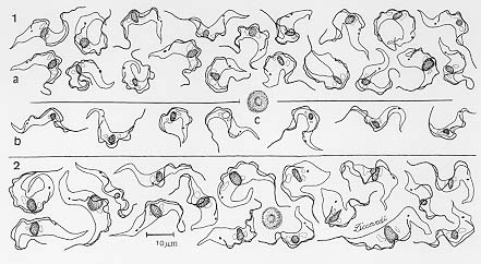
Fig. 1: trypomastigotes found in thin blood smears from naturally infected squirrel monkeys from Balbina and Samuel: a: Trypanosoma minasense; b: T. saimirii-like; and c: a red blood cell from Saimiri sciureus for comparison. Fig. 2: T. minasense found in thin blood smears from naturally infected marmosets, Callithrix penicillata, from FelixlAndia, Minas Gerais. A marmoset red blood cell is show for comparison.
Figure 3: Trypanosoma rangeli. Parasites found in thin blood smears from a: naturally infected squirrel monkey, Saimiri ustus; b: a marmoset, Callithrix jacchus, experimentally infected with haemoculture from S. sciureus; c: mice, experimentally infected with isolates from squirrel monkeys from Balbina and Samuel (Ziccardi & Lourenco-de-Oliveira 1997).
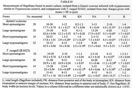
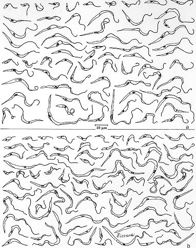
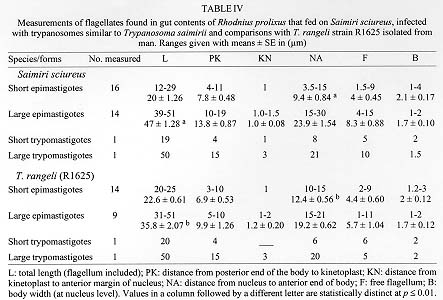
Both T. minasense and T. saimirii display a degree of morphological variation (Table I, II) that makes their identification difficult, particularly in thick blood smears. For example, there are trypomastigotes in the blood of marmosets and squirrel monkeys, respectively, that are smaller than those described for T. minasense and larger than those considered to be T. saimirii (Deane & Damasceno 1961, Dunn et al. 1963 and see Figs 1, 2). Although the dimensions are larger in the marmoset trypanosome than in the trypomastigotes found in squirrel monkeys (identifiable as T. saimirii), the minimum and maximum measurements for the former generally overlap those of the latter, a fact that was also noted by Dunn et al. (1963). The trypomastigotes we identified as T. rangeli (Table II and Fig. 3a) were typical forms, similar in dimensions and feature to those described by other authors in human and non-human primates, rodents, and marsupials, both naturally and artificially infected (Table I and Fig. 3b,c; Groot et al. 1951, Herbig-Sandreuter 1957, Deane 1958, D'Alessandro 1976, Miles et al. 1983, Steindel et al. 1991, Urdaneta-Morales & Tejero 1992). The T. rangeli trypomastigotes we detected in the blood of squirrel monekys and marmosets had a mean length of 30.9 um, were narrower (width of 1.9 um) and, had a cytoplasm paler than those belonging to both T. minasense and T. saimirii-like. The kinetoplast was generally closer to the posterior end (around 3.8 um from the posterior end) and 7.8 um from the nucleus (Table II and Fig. 3a). Blood trypomastigotes of T. saimirii and T. rangeli were statistically different only in nucleus length and in body width. These results show that indeed there is a higher morphological similarity between T. saimirii and T. rangeli than between T. saimirii and T. minasense (Table II). We were faced with a range of forms, some typical of T. minasense and T. rangeli, others similar to T. saimirii, and still others intermediate, often in the same animal (Table II). Squirrel monkeys harbored trypanosomes that lacked diagnostic parameters to be distinguished. This wide range of forms was also reported by Dunn et al. (1963). Sometimes, the parameters to distinguish T. saimirii from the above mentioned species can not be established except arbitrarily. The morphometry and the general features did not assure the identification of this parasite. The pleomorphism previously described in T. rangeli (Herbig-Sandreuter 1957, Hoare 1972, D'Alessandro 1976, Miles et al. 1983, Steindel et al. 1991, Urdaneta-Morales & Tejero 1992) also includes blood trypomastigotes that are wider (0.9-5.1 um), longer (9.3-39 um), and sometimes with a greater distance between the posterior end and the kinetoplast (0.5-8 um), with a well-developed undulating membrane and a vacuoled, densely stained cytoplasm. Such robust or "mature" forms of T. rangeli, with a large PK, have dimensions and feature that are similar to those of trypanosomes identified as T. saimirii in the blood of squirrel monkeys (Tables I, II). It is likely that the trypomastigotes from the blood of squirrel monkeys that have been identified by others as T. saimirii correspond to either (1) T. minasense, considering its pleomorphism (resulting from the length of the infection or interaction with a different host (Ziccardi 1995) or, (2) the "mature" forms of T. rangeli. Indeed, the morphometry of T. saimirii-like blood trypomastigotes differs statistically from those of T. rangeli in only two out of ten characters analyzed. This conclusion strengthens the hypothesis made by Deane and Damasceno (1961). They got, as did Rodhain (1941), positive haemocultures (in NNN) as well as xenodiagnosis (triatomine bugs) from squirrel monkeys infected with what they called T. saimirii (trypomastigotes resembling T. minasense, but with smaller dimensions). They did not find metacyclical forms of the parasite in these invertebrates nor did they determine the form of transmission (i.e., inoculative or contaminative). However, Deane and Damasceno (1961) did not rule out the possibility that the forms observed in culture and in the triatomine might be of another trypanosome, not detected by direct examination (Giemsa-stained blood smears), but nevertheless circulating in the blood of the squirrel monkeys they examined. But what are the multiplying forms supposely belonging to T. saimirii? The developing forms found in haemoculture of squirrel monkeys in whose blood we had found only parasites identifiable as T. saimirii were essentially identical to those of T. rangeli (strain R1625), both in the general features and morphometry ( Fig. 4, Table III). The haemoculture of primates supposedly infected with T. saimirii were always positive and profuse, with the predominance of long and slender epimastigotes. The statistical analysis of measures of cultured forms isolated from S. sciureus supposedly infected with T. saimirii, and of those of T. rangeli (strain R1625), showed that among short and large epimastigotes the only significant difference is the length of free flagellum. In short trypomastigotes the differences are PK, KN and in the length of the free flagellum, and ranges overlapped. This shows a morphometric similarity between these parasites (Table III).
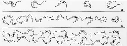
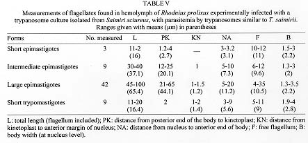
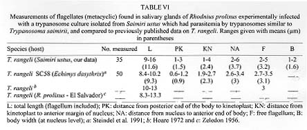
Figure 4 : forms found in haemoculture. a: Trypanosoma rangeli R1625; b: parasites isolated from a naturally infected squirrel monkey, Saimiri sciureus from Balbina, with parasitemia by trypanosomes similar to T. saimiriiThe flagellates found in the gut of triatomine bugs fed on squirrel monkeys, in whose blood only T. saimirii-like parasites had been detected, were also essentially identical to those of T. rangeli ( Table IV, Fig. 5a). When haemocultures of T. saimirii-like infected primates were injected in the hemocoel of R. prolixus, both the hemocoel and the salivary glands became infected (Ziccardi & Lourenco-de-Oliveira 1997) with flagellates also indistinguishable from those belonging to T. rangeli (Table V, Table VI and Fig. 5b,c; Zeledon 1956, Hoare 1972, Cuba-Cuba 1973, Steindel et al. 1991). Trypanosomes confirmed as T. rangeli were transmitted to mice by the bites of the great majority of triatomines that we fed on a T. saimirii-like infected monkey (Ziccardi & Lourenco-de-Oliveira 1997). Indeed, the multiplying forms of T. saimirii in both the triatomine bugs and axenic culture illustrated and/or described by Rodhain (1937a), Deane and Damasceno (1961) and Lourenco-de-Oliveira (1988) resemble to those of T. rangeli (see Herbig-Sandreuter 1957, Zeledon 1966 apud Hoare 1972, Cuba-Cuba 1973).
Figure 5 : forms found in triatomine bugs (Rhodnius prolixus) that fed on squirrel monkeys Saimiri sciureus, and in adult of R. prolixus injected in the hemocoel with samples of haemoculture from S. sciureus and S. ustus. The squirrel monkeys are naturally infected, with parasitemia by trypanosomes similar to T. saimirii: a: gut contents; b: hemolymph and c: salivary glands.Even though T. saimirii has developing forms essentially identical to those of T. rangeli in triatomine bugs, it is believed that the infection by T. saimirii is restricted to the gut while in T. rangeli the infection reaches the hemocoel and the flagellate invades the salivary glands. However, several strains of T. rangeli, mainly those from long-term culture in axenic media, may fail to invade the hemocoel of triatomine bugs and subsequently are not transmitted by the bite to susceptible hosts (Coutinho & Nussenzweig 1952, Tobie 1961, Zeledon 1965, Hoare 1972, Cuba-Cuba 1973, D' Alessandro 1976). If the invasion and infection of the hemocoel by T. rangeli is eventual, depending on several circunstances, such as the parasite strain and the triatomine bug species, this biological event does not assure the distinction between T. rangeli and T. saimirii. P. megistus was the only triatomine species used by Rodhain (1937b) in his experimental infections with T. saimirii. However, it is belived that in P. megistus, as well as in some Triatoma species, that T. rangeli infects only the gut, with invasion of the hemocoel rare, and the parasite has never been found in their salivary glands (Coutinho & Nussenzweig 1952, Zeledon & Blanco 1965, Hoare 1972, Cuba-Cuba 1973). The invasion of the triatomine hemocoel by T. rangeli may not occur until 30 days after an infective blood meal (Hoare 1972, Cuba-Cuba 1973). This period may not have been taken into account by the authors (Rodhain 1941, Deane & Damasceno 1961) in their examination of xendodiagnosis of squirrel monkeys infected with T. saimirii-like parasites. Both T. saimirii and T. rangeli experimentally infect the bed bug C. lectularius, as well as may display non-metacyclic trypomastigotes in the gut of triatomine (Rodhain 1937b, 1941, Herbig-Sandreuter 1957, Deane 1958, Zeledon & Blanco 1965, Hoare 1972, Cuba-Cuba 1973). In conclusion, there are neither reliable morphological nor biological differences between T. rangeli and T. saimirii in the developmental cycle in the invertebrate hosts nor in axenic culture. The flagellates found in haemocultures and in xenodiagnosis of squirrel monkeys displaying parasitemia by T. saimirii are actually developing forms of T. rangeli. Besides, most of xenodiagnosis and haemo-culture of squirrel monkeys infected with T. minasense was positive, and the respective developing forms were actually found to belong to T. rangeli (Ziccardi & Lourenco-de-Oliveira 1997). However, T. minasense does not develop in triatomine bugs and its developing forms in culture media are rather distinct from those of T. rangeli (Dias & Campos-Seabra 1943, Deane & Damasceno 1961, Ziccardi et al. 1996). Those primates were therefore considered to have mixed infections of T. minasense and T. rangeli. Wild animals may be simultaneously infected with more than one trypanosome species, although only one may be detected in blood smears. Therefore, the parasite developing in xenodiagnosis or/and haemoculture may not belong to the same species found in blood smears. This possibility was not often taken into account, and mixed infections have already led some authors to describe new species, e.g., T. sanmartini Garnham and Gonzales-Mugaburu (Deane 1969, Hoare 1972, Marinkelle 1976). This was probably the case in T. saimirii. That is, in view of the morphological and biological features of T. saimirii in both vertebrate and invertebrate hosts discussed above and its resemblance to T. rangeli (although also to T. minasense in some blood trypomastigotes), we conclude that in his description of T. saimirii, Rodhain (1941) actually worked with squirrel monkeys infected with T. rangeli or with both T. rangeli and T. minasense. Indeed, T. rangeli has been the most frequent trypanosome detected in squirrel monkeys from Brazil and other countries in the Americas and mixed infections of T. minasense and T. rangeli have often been reported in these primates (Dunn el al. 1963, Ayala 1964, Marinkelle 1966, Baker 1972, Deane et al. 1972, Hoare 1972, D' Alessandro et al. 1986, Sullivan et al. 1993, Ziccardi & Lourenco-de-Oliveira 1997). Results of on-going biochemical analysis using SDS-PAGE show a great similarity with the parasite growing in the haemoculture of T. saimirii-like infected squirrel monkeys to T. rangeli and that T. minasense has a particular peptideme quite distinct from other assayed trypanosome species (unpublished data). We concluded that based on morphology and on the development in the triatomine bugs and haemoculture, T. saimirii cannot be considered a distinct species. We therefore propose T. saimirii as junior synonym of T. rangeli. ACKNOWLEGMENTS To Dr Pedro Cabello for the guidance on statistical procedures, Dr R Wilkerson and Dr LP Lounibos for critical review of the manuscript and Teresa F Silva for the aid with the illustrations. REFERENCES
Copyright 1998 Fundacao Oswaldo Cruz - Fiocruz The following images related to this document are available:Photo images[oc98010g.jpg] [oc98010j.jpg] [oc98010f.jpg] [oc98010i.jpg] [oc98010b.jpg] [oc98010d.jpg] [oc98010h.jpg] [oc98010e.jpg] [oc98010c.jpg] [oc98010a.jpg] |
| |||||||||
