 |
| About Bioline | All Journals | Testimonials | Membership | News |
|
||||||
|
||||||
Eimeria peltocephali n. sp., (Apicomplexa:Eimeriidae) from the Freshwater Turtle Peltocephalus dumerilianus (Chelonia:Pelomusidae) and Eimeria molossi n. sp., from the Bat, Molossus ater (Mammalia:Chiroptera) R Lainson/^+, RD Naiff*
Departamento de Parasitologia, Instituto Evandro Chagas, Caixa Postal 691,
66017-970 Belem, PA, Brasil Received 26 July 1997; Accepted 5 August 1997
Code Number:OC98015
Sizes of Files:
    Text: 25.4K
    Graphics: Line drawings and photographs (jpg) - 192.9K
              Tables (jpg) - 163.6K
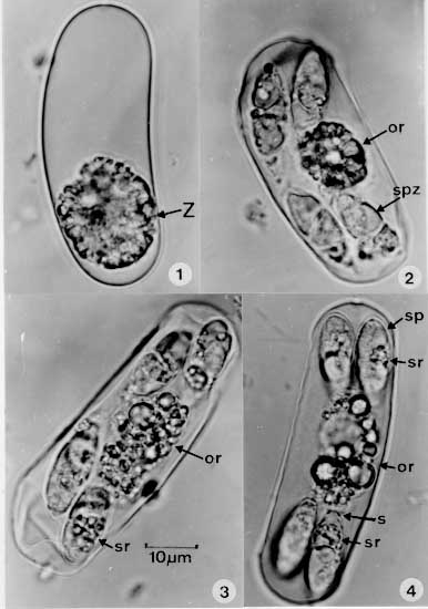
The oocyst is described of Eimeria peltocephali n.sp. from faeces of the freshwater turtle Peltocephalus dumerilianus from Barcelos, State of Amazonas, Brazil. Sporulation is exogenous and fully developed oocysts are elongate, ellipsoidal or cylindrical, frequently curved to a banana-shape, 54.4 x 19.1 (37.5-68.7 x 18.7-20.0 um), shape-index 2.8 (1.8 -3.9). The oocyst wall is a single thin, colourless layer about 1 um thick, with no micropyle. There is a bulky oocyst residuum, at first spherical to ellipsoidal, 19 x 16 (16.2-26.2 x 16-21.5um) , but becoming dispersed on maturation. There are no polar bodies. The sporocysts, 19.1 x 6.8 (17.5 -21.2 x 6.2-7.5 um), shape- index 2.8 (2.3-3.2), are usually disposed in pairs at each end of the oocyst, and bear an inconspicuous Stieda body in the form of a flat cap. The sporozoites are elongate and slightly curved around the residuum. No refractile bodies were seen. Eimeria molossi n.sp., is described from the molossid bat Molossus ater. Sporulation is exogenous and the mature oocysts are predominantly broadly ellipsoidal, 23.4 x 17.5 (18-30 x 15-22.5 um), shape-index 1.3 (1-1.6). The oocyst wall is about 2 um thick, and of three layers: an inner thin, colourless one and two outer layers which are thicker, yellowish-brown, prominently striated and in close apposition. There is no micropyle or oocyst residuum, but one and occasionally two polar bodies are usually present. Sporocysts are ellipsoidal, 10.2 x 7.5 (10-12.5 x 7.5 um), shape-index 1.4 (1.3-1.7) with an inconspicuous Stieda body. Endogenous stages are described in the epithelial cells of the small intestine. Key words: Eimeria peltocephali n. sp. - Eimeria molossi n. sp. - turtles - Peltocephalus dumerilianus - bat - Molossus ater During a scientific expedition to Barcelos, on the river Rio Negro, State of Amazonas, north Brazil (0.58' S: 62.57' W) in January l996, one of us (RDN) had the opportunity to collect material from a number of freshwater turtles when these were being killed and sold in the local market A hitherto unrecorded species of Eimeria was encountered in the faeces of 9 out of 18 adult specimens of the "cabecudo", Peltocephalus dumerilianus (Schweigger, 1812), and faecal material from a number of "irapucas", Podocnemis erythrocephala (Spix, 1824) was found to contain scanty coccidian oocysts which failed, however, to sporulate. The parasite from P. dumerilianus is described below. Coccidial oocysts found in faecal samples from 17 of 38 adult specimens of the bat Molossus ater Geoffroy 1805, captured in the suburbs of Manaus, State of Amazonas, Brazil, are considered to be those a new species of Eimeria. Descriptions are given of the immature and mature oocysts, and of endogenous stages of the parasite seen in sections of the small intestine. MATERIALS AND METHODS Faecal material removed from the rectum of each animal was gently triturated in 2% (w/v) aqueous potassium dichromate ( K2Cr2O7) and maintained at room temperature (23 -24 C). Fifty oocysts and 30 sporocysts of the Eimeria sp. from Peltocephalus, and 100 oocysts and 50 sporocysts of the parasite from M. ater were measured by normal light microscopy with a x100 neofluar objective, x 8 eyepieces and an ocular micrometer. Photomicrographs were prepared using a Zeiss Photomicroscope III and Kodak TMX 100 film. All measurements are in um and are given as means, with the range in parentheses, followed by the shape-index (ratio of length/width). The small intestine of each bat was fixed in buffered 10% formaldehyde for the subsequent preparation of histological sections, cut at 4 um and stained with haematoxylin and eosin. Unfortunately, work conditions and the large size of the turtles did not permit a similar treatment for the intestines of these animals. RESULTS Eimeria peltocephali n. sp. (Figs 1-4 and Fig. 24)
Description: with the characters of the genus Eimeria Schneider, 1875. Oocysts elongate, frequently in the form of a gently curved cylinder with rounded ends, 54.4 x 19.1 (37.5-68.7 x 18.7-20), shape-index 2.8 (1.8-3.9). Oocyst wall a single, colourless layer approximately 1 thick and with no micropyle. Oocyst residuum at first a semi-spherical mass of globules which measures a mean of 19 x 16 (16.2-26.2 x 16-21.5) in the unsporulated oocyst, and frequently becomes dispersed when this is mature: no polar bodies are produced. Sporocysts 19.1 x 6.8 (17.5-21.2 x 6.2-7.5), shape index 2.8 (2.3-3.2), very frequently located in pairs towards the ends of the oocyst. Sporocyst residuum bulky and composed of a mixture of fine and slightly larger granules. Sporocyst wall very delicate and bearing an inconspicuous cap-like Stieda body, apparently without a sub-Stieda body. The sporozoites occupy almost the entire length of the sporocyst and are slightly recurved around the residuum. No refractile bodies were visible under ordinary light-microscopy. Type host: the freshwater turtle, Peltocephalus dumerilianus (Schweigger, 1812) (Reptilia: Chelonia: Pelomusidae). Local name "cabecudo". Location in host: uncertain, but with the failure to detect parasites in the gall-bladder of infected animals, it is most probably in the intestine. The oocysts were described from the faeces. Sporulation: exogenous. Sporulation time not recorded. Type locality: Barcelos, State of Amazonas, north Brazil (0.58' S: 62.57' W). Prevalence: of 18 turtles examined, 9 were infected. Pathogeniciy: unknown. Etymology: the specific name is derived from the generic name of the host, Peltocephalus. Eimeria molossi n. sp. (Figs 5-13, 14-23 and 25)
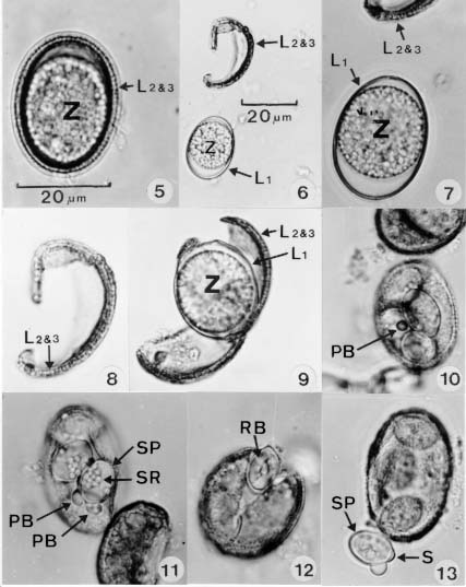
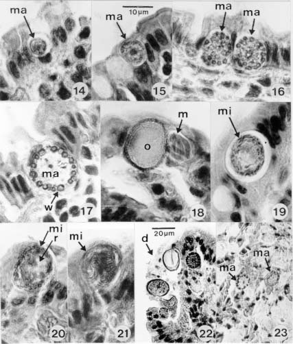
Description: with the characters of the genus. Oocyst predominantly broadly ellipsoidal, sometimes subspherical, 23.4 x 17.5 (18-30 x 15-22.5), shape index 1.3 (1-1.6). Intact oocyst wall about 2, and with three layers. An inner one which is thin, colourless and unstriated, and two outer layers which are thicker, yellowish-brown, prominently striated and closely contiguous. The two outer layers may be lost, so that the oocyst then appears smooth, colourless and thin-walled. There is no micropyle. Formation of the four sporocysts leaves no oocyst residuum, but most oocysts have a conspicuous spherical to ellipsoidal polar body of about 1.9; on rare occasions two polar bodies may be present. Sporocysts broadly ellipsoidal, 10.25 x 7.5 (10-12.5 x 7.5), shape-index 1.4 (1.3-1.7), with the very fine wall bearing a small, nipple-like Stieda body. No sub-Stieda body could be detected. Sporocystic residuum composed of from 4 to 12 relatively large spherules lying between the two sporozoites, which lay in "head-to-tail" fashion, occupy the entire sporocyst, and are usually recurved on themselves. At least one refractile body is present (seen with difficulty). Host: the bat Molossus ater Geoffroy 1805 (Chiroptera:Molossidae). Location in host: epithelium of the ileum, with all stages positioned between the brush-border and the host cell nucleus, which is usually grossly displaced or destroyed by the larger parasites. Endogenous stages: in histological sections, the six mature meronts seen had a mean measurement of 12.3 x 9.3 (11-14 x 8-10) and produced an estimated 8-12 merozoites measuring 6 x 1 (Fig.18). Young macrogametocytes are at first spherical (Figs 14-16), becoming ellipsoidal with growth and finally reaching about 18 x 14, when the wall-forming glycoprotein granules become very conspicuous and may measure up to 2 in diameter (Fig. 17). The oocyst wall is fully developed before the oocysts are shed into the gut lumen (Fig.18).
Mature microgametocyes seen in sections (Figs 19-21) averaged 15.8 x 11.8 (15.5-17 x 11-12), and shed > 50 microgametes measuring about 3 x 0.5. There is a bulky residual body of about 10 x 8. Sporulation: exogenous. Sporulation time was not determined, but it was noted that many oocysts (sometimes as many as 70% of a given faecal specimen) failed to sporulate. Locality: Manaus, State of Amazonas Prevalence: 17 of the 38 bats examined (44.7%) were infected. Pathogenicity: there were no outward signs of disease in the infected animals. Histological sections of the ileum of heavily infected bats, however, showed epithelial damage presumed to be caused by the parasite (Fig. 22), and endogenous stages were commonly seen together with sloughed epithelial cell debris in the gut lumen (Fig. 23). Etymology: the specific name is derived from the generic name of the host, Molossus.
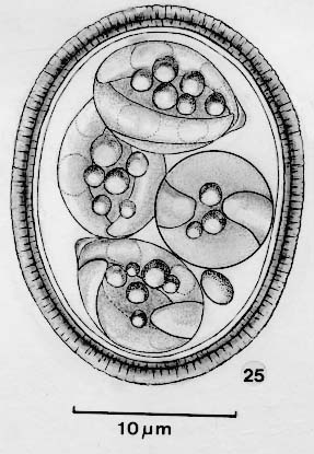
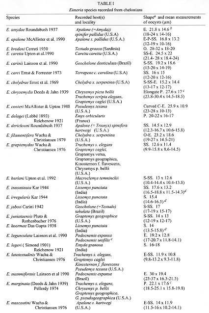
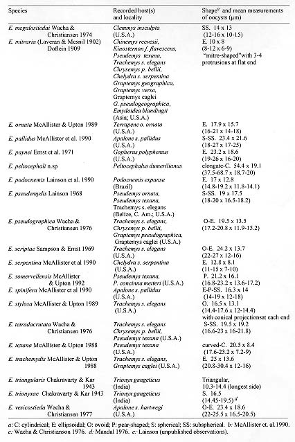
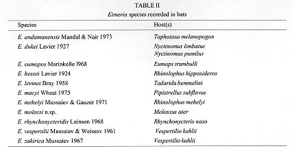
Figure 25: Line-drawing of a mature oocyst of Eimeria molossi n.sp., from the bat Molossus ater. DISCUSSION Of the 44 previously named species of Eimeria in chelonids ( Table I (A) and Table I B), the oocysts of E. peltocephali n.sp. most resemble those of E. texana and E. cooteri (McAllister & Upton, 1989), which are also elongate-cylindrical. They are, however, very much larger (mean 54.4 x 19.1 versus 20.5 x 8.4 for E. texana and 25.9 x 10.9 for E. cooteri). The sporocysts of E. peltocephali are elongate (19.1 x 6.8), while those E. texana are ovoid (8.1 x 4.7). Although elongate, the sporocysts of E. cooteri (14.9 x 5.3) differ in the possession of a strangely elongated Stieda body capped by tiny, knob-like thickenings. From their own and other authors' studies, McAllister and Upton (1989) concluded that "....most, but not all, of the turtle Coccidia from aqueous environments in North America are not particularly species specific", and that "....most species of coccidia in the Testudines are specific at the family level" (McAllister et al. 1990). A glance at Table I certainly supports this view, with some Eimeria species recorded in three (E. marginata and E. tetradacrutata), four (E. graptemydos and E. lutotestudinis) or even an astonishing eight different genera of chelonians (E. mitraria). For records of such multiplicity of hosts, reference may be made to McAllister and Upton (1988, 1989b, 1992), McAllister et al. (1990a, 1991) and Wacha and Christianson (1976, 1979). Much less is known about the host range and prevalence of the coccidia of chelonians in the neotropics and the Old World. It would be strange, however, if a similar situation does not exist in these regions. As far as we are aware, 13 different specific names have been allocated to the genus Eimeria found in bats. Of these, however, E. viridis (Labbe 1893) Reichenow 1921 was clearly considered as a nomen nudum by Pellerdy (1974) due to what appears to be a confused description of more than one parasite, while E. myotis and E. plecoti Gottschalk 1969 must also be regarded as nomina nuda, because their description was restricted to unsporulated oocysts, the true nature of which is clearly questionable. Of the remaining ten species (Table II), Eimeria molossi n.sp., is readily differentiated from E. andamanensis, E. hessei, E. levinei, E. mehelyi, E. rhynchonycteridis, E. vespertilii and E. zakirica, which all have a smooth, unstriated oocyst wall, and from E. dukei which has a large oocystic residuum. Morphology of the oocyst of E. molossi n.sp., most closely approaches that of E. eumopos and E. macyi, both of which have a roughish, striated oocyst wall. The oocysts of E. eumopos, however, are substantially larger (35 x 28, range 34-36 x 27-28 versus 23 x 17, range 18-30 x 15-22), and the oocyst wall has only two layers. Mature meronts of E. molossi n.sp. are small and produce only from 8-12 merozoites, whereas Marinkelle described those of E. eumopos as measuring up to 98 x 62 (globidial schizogony?), with a thick wall and containing up to 350 merozoites. Finally, the oocysts of E. macyi are smaller than those of E. molossi n. sp., (19 x 17.6 versus 23 x 17 ), more inclined to a spherical shape (shape-index 1 versus 1.3), and have a wall of only one layer. Furthermore, its sporocysts have a much more prominent Stieda body and possesses a very conspicuous sub-Stieda body, not seen in the sporocysts of E. molossi. ACKNOWLEDGEMENTS To Constancia Maia Franco and Walter M Campos, Instituto Evandro Chagas, Belem, for technical assistance; to Dr Suely Marques, Museu Paraense Emilio Goeldi, Belem, for the identification of bats and advice regarding chiropteran taxonomy; and to Dr WE Magnusson, Instituto Nacional de Pesquisas da Amazonia, Manaus, for identification of the turtles. REFERENCES
Copyright 1998 Fundacao Oswaldo Cruz - Fiocruz The following images related to this document are available:Photo images[oc98015d.jpg] [oc98015a.jpg] [oc98015h.jpg] [oc98015g.jpg] [oc98015c.jpg] [oc98015b.jpg] [oc98015e.jpg] [oc98015f.jpg] |
| |||||||||
