 |
| About Bioline | All Journals | Testimonials | Membership | News |
|
||||||
|
||||||
Morphometric Analysis of McCoy Cells Inoculated with Cerebrospinal Fluid from Patients with Rabies Yeda L Nogueira
Servico de Virologia, Instituto Adolfo Lutz, Av. Doutor Arnaldo 355,
01246-902 Sao Paulo, SP, Brasil Received 18 December 1997; Accepted 22 May 1998
Code Number:OC98099 To demonstrate the potential of McCoy cells for the isolation of rabies virus from the cerebrospinal (CSF) fluid of a patient with a diagnosis of rabies, McCoy cells were inoculated with CSF from a patient with a clinical diagnosis of rabies and investigated in terms of morphometric aspect using the JAVA analysis system for the quantification of the increased size of infected cells compared to noninfected cells. The cells were also examined in terms of specific staining for the diagnosis of rabies by the method of Sellers for the observation of intracytoplasmic inclusions and by specific immunofluorescence staining for rabies virus. Infected cells showed changes in cell permeability and morphologic modifications which differed significantly compared to normal cells (P<0.001) when analyzed by the Mann-Whitney and Kruskal-Wallis tests. Intense activity of the endoplasmic reticulum was also observed, as indicated by the presence of intracytoplasmic inclusions visualized by specific staining. The present study demonstrated the isolation of rabies virus from the CSF of a patient with rabies, showing that McCoy cells can be used for the laboratory diagnosis of patients suspected to have rabies. Key words: image analysis - McCoy cell line - rabies virus - cerebrospinal fluid - morphometry - patient with rabies diagnosis
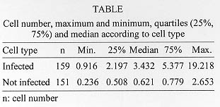
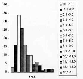
Rabies is a disease that attacks the central nervous system (CNS) with 100% of lethality and is transmitted to humans by contagion with domestic animals such as dogs and cats, and also with wild animals such as foxes, wild dogs, coatis, opossum and bats within other mammalians. Bats represent the major source of infection in humans and are currently considered to be the most important source of rabies transmission in the United States (CDC 1995a, b) and it is the second most important in Brazil (Uieda 1993). In Brazil, the clinical and laboratory diagnosis of rabies in humans may be undernotified, however, according to the National Health Foundation (Ministry of Health 1996), 47 cases of human rabies were notified. Due to the neurologic signs and symptoms presented by patients with rabies, the disease is not always identified immediately since many tropical diseases of unknown origin exist in our country and the signs of encephalitis may be confused with those of other encephalitis of diverse viral origin which present disorders of neurologic metabolism. The diagnosis of human rabies is almost always clinical and confirmed post mortem due to the need to use a fragment of brain tissue to perform laboratory tests, impossible when the patient is alive. The use of cerebrospinal fluid (CSF) for the isolation of rabies virus is not common and is not always easy since the concentration of virus is higher in the cerebellum, hippocampus, thalamus, and hypothalamus and lower in the cerebral cortex (Fedaku et al. 1982). However, a high sensitivity of McCoy cells to rabies virus has been observed in several studies (Nogueira 1987, 1992a, b). The investigator also observed the process of viral replication in these cells, which increased in size until they practically burst and released the virus into the extracellular medium. Furthermore, in each serial passage of the virus through McCoy cells the viral titer increased geometrically. In view of the reports cited before, the present investigation was undertaken to study the morphometric alterations of infected and noninfected cells by image analysis using the JAVA system in order to identify the relationship between increased cell size and reproductive capacity of viral particles for human diagnostic purposes. MATERIALS AND METHODS Cell culture - The McCoy cells used were obtained from the Cell Culture Section of the Adolfo Lutz Institute. The cell is catalogued in the American Type Culture Collection CRL 1696 (ATCC 1995). Isolation of rabies virus - CSF from a patient admitted to the Emilio Ribas Hospital in 1994 whose causa mortis was identified as rabies was used. The virus was isolated in McCoy cells according to described methods (Nogueira 1992a,b). CSF (0.5 ml) was inoculated on a plate containing 2 to 3 ml of a confluent monolayer of McCoy cells. The morphologic changes of the cells were observed until the occurrence of the cytopathic effect. Identification of the isolated virus - The virus was identified after the second serial passage through McCoy cells by specific direct immunofluorescence using an anti-nucleocapsid antibody manufactured by the Pasteur-Merieux Institute and anti-rabies virus total protein antibodies (anti-challenge virus standard - CVS), both labeled with fluorescein isothiocyanate. The results were observed under an inverted IM-35 Zeiss microscope with epifluorescent illumination. Observation of morphologic changes - Morphologic changes were observed by staining according to the method of Sellers, which permitted the visualization of intracytoplasmic inclusions similar to Negri corpuscles (Negri -Luzzani 1905). The images of these cells were analyzed with a videocamera coupled to the inverted IM-35 microscope and the results were stored in the data bank of the JAVA System video analysis software version 2.1 (Jandel Scientific, CA, USA). The software permitted the measurement and calculation of cell area (um^2). Statistical analysis - The areas of infected and noninfected cells were analyzed with the JAVA image analysis system and the resulting data bank was downloaded into the EXCEL 8.0 and EPI INFO 6.02 programs. The Kolmogorov-Smirnov test (Siegel 1956) was applied to determine whether the values for the areas of infected and noninfected cells followed a normal distribution pattern, with the level of significance set at p<0.05. Since the distributions obtained were not normal, the use of parametric tests for comparison of infected and noninfected types was excluded. Thus, the Mann-Whitney and Kruskal-Wallis tests (Siegel 1956) were used for statistical analysis of the distribution profiles, with the level of significance set at p<0.001. It should be pointed out that the two tests are evaluated in a satisfactory manner by the specialized literature, with no loss of quality in the differential analysis of the profiles delineated. RESULTS Table summarizes the variation in cell size according to the percentiles obtained for each set of cells (infected and noninfected). The Kolmogorov-Smirnov test showed that the distribution of the two sets did not fit a normal curve at the p <0.05 level. The difference between the two profiles was then determined by the Mann-Whitney and Kruskal-Wallis tests and showed a significantly more elevated pattern (p<0.001) for the measurements of the area of infected cells compared to noninfected cells. Table also shows the maximum and minimum cell size values for the two populations. It can be seen that the maximum values of infected cells were about 20 times the minimum value, in contrast to noninfected cells, whose maximum values were 11 times the minimum value. Even so, the maximum value of noninfected cells was in the interval between the first quartile (25%) and the median compared to infected cells, i.e., below the median. Fig. 1 illustrates the distribution of the values for the areas separated into ranges, with most infected cells being concentrated in the 2.1-3.0 um^2 range, while the modal value of the noninfected cells was in the 0.01-1.0 um^2 range (figure not shown).
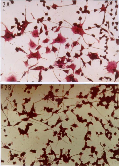
Fig. 2 shows the morphometric changes and the differences in size between infected and noninfected cells, which are clearly visible. Fig. 2A also shows the changes in cell permeability and the intracytoplasmic inclusions similar to Negri corpuscles, a typical formation in cerebral tissue infected with rabies virus, denoting intense activity of the endoplasmic reticulum.
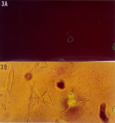
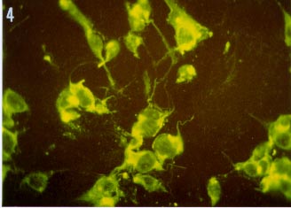
Fig. 3A and B shows the immunofluorescence reaction specific for rabies virus using an anti-nucleocapsid antibody conjugated with fluorescein isothiocyanate. Fig. 4 also shows a rabies virus-specific immunofluorescence reaction using an anti-CVS conjugate. The reaction shows some infected cells soon after the rupture of the cell membrane (cell lysis) when viral particles are released. Cells with fluorescent viral particles can also be observed adhering to the cell membrane before the latter breaks, demonstrating the replication of rabies virus in McCoy cells.
Fig. 4: immunofluorescence reaction with anti-CVS (Challenge Virus Standard) conjugate showing the cellular rupture (cell lyses) of McCoy cell (inoculated with cerebrospinal fluid from the patient) and the virus-release to extracellular compartment (epifluorescent microscopy, original magnification 200X), IM-35 Zeiss microscope.
The replication of animal viruses in mammalian cells provokes profound changes in cell morphology which will result in cell lysis (Carrasco 1987, Paez & Esteban 1987, Ugli et al. 1987, Carrasco et al. 1989). The mechanisms involved in this process of cell lysis are still unknown. We know that the change in cell membrane permeability occurring during viral replication (poliomyelitis, Semliki Forest and influenza viruses) is related to these mechanisms, which are attributed to different factors such as the action of viral proteins that bind to the membrane distorting the lipoprotein layer, the increase in phospholipase activity or the increase in Ca^2+ ion entry into the cells (Iruzun et al. 1993, 1995). According to these investigators, the free Ca^2+ concentration in the cell may participate in the induction of the cytopathic effect and in the morphogenesis of the viral particle in cytomegalovirus infection. The observation of the cytopathic effect in rabies virus infection in mammalian cell culture is not common. Rabies virus is considered to cause persistent or chronic infection without causing cell lysis (Wiktor & Clark 1972, Villareal & Holland 1976, Montagnon et al. 1985). However, previous reports (Nogueira 1992a, b) have demonstrated the presence of a constant cytopathic effect when rabies virus is inoculated into McCoy cells. The present results show statistically significant differences in the morphometry of infected and noninfected cells (p<0.001 by the Mann-Whitney and Kruskal-Wallis test). Another difference observed was the activity of the endoplasmic reticulum, the structure in which the polypeptides that will form the viral proteins like glicoproteins and that will be transported to the cell membrane are synthesized (Carrasco 1987, Carrasco et al. 1989). However, we do not know the exact mechanism involved in the increase in cell permeability with the formation of giant cells and its relation to the intense synthesis of viral proteins. This increase in the synthesis of rabies virus proteins with a geometric increase in virus titers during replication in McCoy cells permits an easy isolation of rabies virus from biological materials and fluids, including those containing a low viral load such as the CSF. On the basis of the present study, it is possible to perform laboratory diagnosis of rabies by inoculating the CSF of patients suspected to have the disease into McCoy cells, thus permitting a diagnosis in living patients. Acknowledgements To Dr A Flammand (Centre National de la Recherche Scientifique, France) for giving me the conjugates. To Dr SLD Gotlieb and Dr JLF Antunes for the helpful comments and help with the statistical methods and Mr A Longatto Filho for giving access to the image analysis workstation. REFERENCES
Human Rabies - California 1995b. MMWR Morb Mortal Wkly Rep 45: 353-356.
Copyright 1998 Fundacao Oswaldo Cruz - Fiocruz The following images related to this document are available:Photo images[oc98099b.jpg] [oc98099e.jpg] [oc98099a.jpg] [oc98099d.jpg] [oc98099c.jpg] |
| |||||||||
