 |
| About Bioline | All Journals | Testimonials | Membership | News |
|
||||||
|
||||||
Vol. 93(6): 741-744 Eimeria minasensis n. sp. (Apicomplexa: Eimeriidae) in the Domestic Goat Capra hircus, from Brazil Andréa C Silva/+, José D Lima* Departamento de Parasitologia, IPTSP, Universidade Federal de Goiás,
74001-970 Goiânia, GO, Brasil Received 28 January 1998; Accepted 13 July 1998
Code Number:OC98204
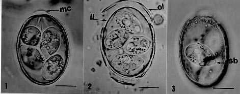
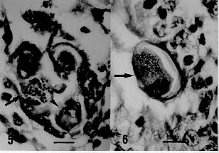
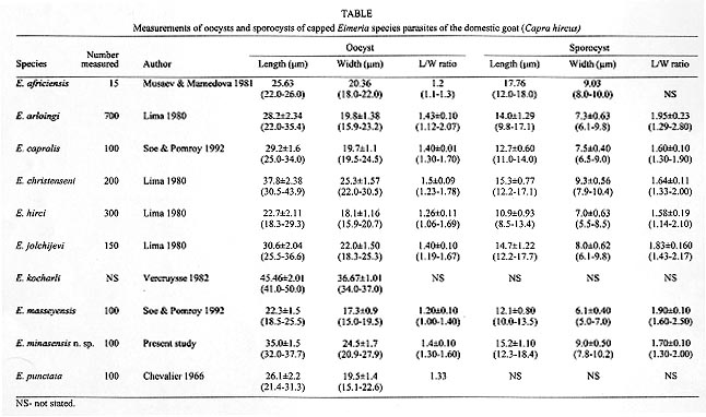
Eimeria minasensis n. sp. is described in the domestic goat Capra hircus from Brazil. Oocysts ellipsoidal are 35 x 24.5 (32-37.7 x 20.9-27.9) µm. Sporocysts elongate-ellipsoid are 15.2 x 9 (12.3-18.4 x 7.8-10.2) µm, with a Stieda body at the narrow end. Oocyst wall smooth and bilayered; outer layer about 1.2 (0.8-1.6) µm and colorless; inner layer about 0.5 (0.4-0.8) µm and dark-brown. Micropyle, a mound-shaped micropylar cap 1,6 x 8,9 (0,8-2 x7-10,2) easily dislodged; one or more oocyst polar granules present. Oocyst residuum absent. Sporocyst residuum present, composed of many scattered granules. Sporozoites elongate, lying lengthwise, "head to tail" in the sporocysts; one or two refractile globules are usually visible. Sporulation time was 120 hr at 27oC, prepatent period, 19 to 20 days and patent period 15 to 25 days. Gamonts, gametes and oocysts present in cecum and colon. Prevalence was 12.8% (6/47) in goats from Minas Gerais, Brazil. Key words: Eimeria minasensis n. sp. - goat - coccidia - Brazil The number of Eimeria species considered to be parasites of the domestic goat (Capra hircus) is variable and controversial, and depends upon the acceptance of the validity of some species (Levine & Ivens 1970, Musaev 1970, Pellérdy 1974, Lima 1979, Musaev & Mamedova 1981, Norton 1986). Several species considered as parasites of both goat and sheep were not able to infect one or other of those hosts in cross transmission studies (Levine & Ivens 1970, Lima 1979a). Levine (1988) listed 13 species as true parasites of goats. Later, Soe and Pomroy (1992) described three new species of Eimeria as parasites of goats in New Zealand. A new species of Eimeria from the domestic goat found in the State of Minas Gerais, Brazil, is described in this paper. MATERIALS AND METHODS Fecal samples of adult goats from the municipalities of Esmeraldas (15 samples) and Sete Lagoas (32 samples), State of Minas Gerais, Brazil, were examined for coccidia. Positive samples were mixed with 2.5% (w/v) potassium dichromate (K2Cr2O7), filtered to remove coarse debris, spread in a thin layer in covered Petri dishes and allowed to sporulate at room temperature (about 25oC), for a week. To determine some biological parameters, five goats were experimentally infected, at different times, with oocysts of the new species as described below. The goats used for these infections were separated from does immediately after birth, and raised under coccidia-free conditions, in individual cages kept in closed rooms with restricted access. Fifty sporulated oocysts of the new species were collected with a micropipette, using a dissecting microscope, and their identity confirmed by light microscopy; and given per os to a seven-mo-old goat, in order to build up an inoculum for further experiments. A three-mo-old kid was inoculated with 105 sporulated oocysts obtained from the previously inoculated goat. Feces were collected daily and samples containing oocysts were allowed to sporulate as described above. After sporulation, pure cultures of oocysts of the new species were stored at 4oC for futher inoculations. This kid was killed 23 days after inoculation, tissues samples were fixed in 10% buffered formalin solution, embedded in paraffin, sectioned at 3-5 µm, stained with haematoxylin-eosin and examined using light microscopy to determine the site of infection. Three, one-to-three-mo-old kids were inoculated with 105 sporulated oocysts. To determine sporulation time, prepatent and patent periods, fecal samples were collected daily, mixed with 2.5% K2Cr2O7 solution, and incubated at 27oC. Stages of sporulation were checked at 24 hr intervals. Sporulation was considered to be completed when no additional increase in the percentage of sporulated oocysts was observed. Sporulated oocysts were examined after flotation with Sheather's sugar solution. One hundred oocysts and 100 sporocysts were measured with an ocular micrometer. All measurements are presented as mean ± SD followed by the range in parentheses, and the shape-index (ratio of length/width). RESULTS Eimeria minasensis n. sp. (Figs 1-3, 4) Figs 1-3: photomicrographs of sporulated oocysts of Eimeria minasensis n. sp., recovered from the feces of goats in Brazil. Bar = 10 µm; mc: micropylar cap, ol: outer layer; il: inner layer; sb: Stieda body. Fig. 4: Eimeria minasensis n. sp. Line drawing of a sporulated oocyst. Oocysts ellipsoidal, 35±1.5 (32-37.7) x 24.5±1.7 (20.9-27.9) µm, shape-index 1.4±0.1 (1.3-1.6). Oocyst wall smooth and bilayered; outer layer about 1.2±0.2 (0.8-1.6) µm and colorless; inner layer about 0.5±0.1 (0.4-0.8) µm and dark-brown. Micropyle present. An easily dislodged mound-shaped micropylar cap present, colorless, 1.6±0.2 (0.8-2) µm high and 8.9±0.7 (7-10.2) µm wide. One or more oocyst polar granules present, oocyst residuum absent. Sporocysts elongate-ellipsoid 15.2±1.1 (12.3-18.4) x 9±0.5 (7.8-10.2) µm, with a Stieda body at the narrow end; shape-index 1.7±0.1 (1.3-2). Sporocyst residuum present, composed of many scattered granules. Sporozoites elongate, lying lengthwise, "head to tail" in sporocysts; they usually contain one or two refractile bodies. Type host: Capra hircus (Linnaeus, 1758) (domestic goat). Type locality: municipalities of Esmeraldas and Sete Lagoas counties, Minas Gerais, Brazil. Site of infection: gametogony in cecum and colon (Figs 5, 6). Figures 5-6: Sexual stages of Eimeria minasensis n. sp. in the cecum of the domestic goat. Fig. 5: macrogamete (arrow) and microgamont (head). Fig. 6: oocyst (arrow). Bar = 10 µm. Sporulation time: 120 hr at 27oC. Prepatent period: 19 to 20 days. Patent period: 15 to 25 days. Prevalence: oocysts found in 20% (3/15) and in 9.3% (3/32) of goat feces examined in the municipalities of Esmeraldas and Sete Lagoas, respectively. Type-material: phototypes of oocysts deposited in the United States National Parasite Collection no. 87296. Etymology: the name is derived from the first name of the State (Minas Gerais) where the species was found. DISCUSSION Among the 16 accepted species of Eimeria from goats (Levine 1988, Soe & Pomroy 1992), E. minasensis n. sp. differs from E. alijevi, E. apsheronica, E. caprina, E. caprovina, E. charlestoni, and E. ninakohlyakimovae by having a micropylar cap. Of the capped species (Table), E. africiensis, E. arloingi, E. capralis, E. hirci, E. masseyensis, and E. punctata are considerably smaller than E. minasensis. E. jolchijevi is smaller than E. minasensis and has a typically urn shaped oocyst. E. christenseni and E. kocharli are larger than E. minasensis. In addition, E. arloingi and E. christenseni, which somewhat resemble E. minasensis, have known life cycles with sexual stages restricted to the small intestine of the host (Sayin et al. 1980, Lima 1981), whereas gamonts, gametes and oocysts of E. minasensis were found in cecum and colon of goat. ACKNOWLEDGMENT To Mr Humberto Borém for the line drawing. This work was sponsored by CNPq and Fapemig. REFERENCES
Copyright 1998 Fundacao Oswaldo Cruz - Fiocruz The following images related to this document are available:Photo images[oc98204c.jpg] [oc98204b.jpg] [oc98204a.jpg] [oc98204d.jpg] |
| |||||||||
