 |
| About Bioline | All Journals | Testimonials | Membership | News |
|
||||||
|
||||||
Mem Inst Oswaldo Cruz, Rio de Janeiro, Vol. 94(4),Jul./Aug. 1999: pp 505-508 Sensitivity of a Vacuum Aspiratory Culture Technique for Diagnosis of Localized Cutaneous Leishmaniasis in an Endemic Area of Leishmania (Viannia) braziliensis Transmission Gustavo Adolfo Sierra Romero+, Raimunda Nonata Ribeiro Sampaio, Vanize de Oliveira Macêdo, Philip Davis MarsdenU Núcleo de Medicina Tropical, Universidade de
Brasília, Campus Universitário, Asa Norte, 70919-970,
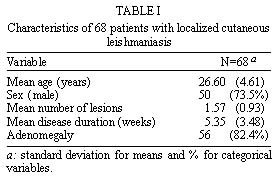
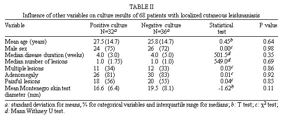
Brasília, DF, Brasil Received 23 October 1998 Code Number:OC99093 Sixty eight patients with localized cutaneous leishmaniasis from an area with Leishmania (Viannia) braziliensis transmission had cultures performed with a modified Marzochi´s vacuum aspiratory puncture technique to establish sensitivity and contamination rate with this new method. Overall sensitivity of three aspirates was 47.1%; (CI95% 39.4; 59.4) significantly greater than the sensitivity of a single one aspirate. Fungal contamination was observed in 6/204 (2.9%) inoculated culture tubes. We recommend that this useful technique should be adopted as routine for primary isolation of L. (V.) braziliensis from localized cutaneous ulcers. Key words: Leishmania (Viannia) braziliensis - culture sensitivity - fungal contamination - vacuum aspiratory technique Culture sensitivity for diagnosis of cutaneous leishmaniasis caused by Leishmania (Viannia) braziliensis (L.V.b.) has shown variable results depending on quality of culture media (Shaw & Lainson 1981, Cuba et al. 1986), collection technique (Evans 1989) and other characteristics such as disease duration and previous use of anti-leishmanial drugs (Cuba et al. 1984). Marzochi et al. (1993) described a new collection technique using direct inoculation of culture tubes containing NNN media with a liquid phase through a skin puncture system with five to ten mililiters vacuum. We modified the culture medium using a smaller amount of an unenriched liquid phase composed of isotonic saline solution with gentamicin, resuming 15 ml vacuum in each tube. We used the same aspiratory puncture system with commercial needles but the aspiration was modified by allowing air to come into the tubes when pulling the needle out to obtain a larger amount of inoculum. We studied the sensitivity of this method for diagnosis of cutaneous leishmaniasis in a group of patients with suspected leishmanial ulcers and a positive Montenegro´s skin test in a rural area of the State of Bahia, Brazil where L.V.b. transmission is endemic (Llanos-Cuentas et al. 1984, Rosa et al. 1988). MATERIALS AND METHODS Culture medium was prepared using blood agar base no. 2 (DIFCO cod. 0696-17) with 15% defibrinated rabbit blood which was added after fusion of agar at 50oC. Gentamicin (100 m g/ml) and 5-fluorocytosine (100 m g/ml) were added and the mixture was distributed in 10 ml glass tubes (VacutainerR). Tubes were covered with rubber caps and vacuum restored aspirating 15 ml with a 20 ml syringe using a 22 gauge needle. A liquid phase composed of 0.3 ml isotonic saline with gentamicin (100 m g/ml) was added immediately before the aspiratory puncture by injection through the rubber cap with a 1 ml syringe and a 22 gauge needle. All injection procedures were performed through the rubber cap after desinfecting it with 0.2% iodinated alcohol. The aspiration puncture was made with the commercial VacutainerR needle holder dispositive and 21 gauge needles for vacuum blood extraction. The puncture site was cleaned with 0.2% iodinated alcohol and local anesthesia was induced with 0.3 ml lidocaine (2%) injected with a 1 ml syringe with a 13 gauge needle. The procedure was performed through intact skin close to the ulcer border at a 20o angle with a rotatory movement and finally was pulled out allowing air come into the needle with the aspirated material. The material which adhered to the glass wall of the tube was washed toward the solid phase using the liquid phase. The procedure was repeated at the same place three times in three separate tubes which were labeled as first, second and third aspirates. When patient had more than one ulcer only one was aspirated, usually the one with the most recent onset. Culture tubes were kept at 22-28oC and were observed every day using an inverted microscope for 28 days. When positive, new tubes were inoculated to obtain parasites for cryopreservation. Culture aspiration and observation of leishmanial growth were performed by one researcher, thus avoiding inter-observer variations. All isolates were characterized with monoclonal antibodies at Instituto Evandro Chagas, Belém, State of Pará, Brazil. Eligible patients were those with less than six cutaneous ulcers with two to 20 weeks of evolution who had a positive Montenegro´s skin test. Pregnant women, children under eight, patients with more than five lesions, history of previous cutaneous lesions with typical scars suggestive of old leishmaniasis, mucosal involvement or history of anti-leishmanial treatment were excluded. Lesion number for inclusion was chosen to be less than six to differentiate clearly the localized clinical syndrome from disseminated cutaneous leishmaniasis (Carvalho et al. 1994). Montenegro´s skin test was performed with 0.1 ml L. (Leishmania) amazonensis (MHOM/BR/86/BA 125) antigen (250 m g/ml). Reaction of 5 mm diameter or greater at 48-72 hr after intradermic injection were recorded as positive. All patients were treated in a basic health unit at Corte de Pedra district, Presidente Tancredo Neves municipality, State of Bahia, Brazil with conventional drugs recommended by the Ministry of Health. The study was part of a research project aproved by de Ethics Comitee of the University of Brasília. Group comparisons were processed using Epi Info 6.04 software. RESULTS Sixty eight patients were eligible among 131 attended at the Corte de Pedra´s unit from August to November 1996. All were agricultural workers living in the cocoa production area in the southeast region of Bahia. Table I shows the characteristics of the sample. Sixty six percent had one lesion, 16.2% two lesions, 13.2% three lesions and 4.4% more than three. Cultures were positive in 32 patients (47.1% CI95% 39.4; 59.4). The mean time to obtain a positive result (at least one culture tube) was 9.8 days (2-26 days; CI95% 8.8; 10.8). Eighty one percent of positive culture tubes were positive in the first two weeks of observation. Nineteen patients out of 32 (59.3%) had a positive result in only one culture tube (four in the first aspirate, six in the second and nine in the third one). Eleven patients (34.3%) had a positive culture in two tubes (five in the first and second aspirates and six in the second and third aspirates) and two patients had three positive aspirates (6.2%). Results among different aspirates of the same patient had a poor concordance. The Figure shows the dynamics of parasite growth during the 28 days observation time. The sensitivity was 14.7%, 27.9% and 26.5% for the first, second and third aspirates respectively. Fungal contamination was observed in cultures from five patients. There was contamination of one tube in four patients and two tubes in one. Contamination rate was 2.9% (6/204 inoculated tubes). The mean time to develop contamination was 16 days (10-22 days; SD=5.2) Figure: Cummulative percentage of positive cultures for the 1st, 2nd, and 3rd aspirates during the 28 days of observation. * All aspirates curve corresponds to cummulative percentage of patients with at least one positive result among the three aspirates. There was no influence of age, sex, number of lesions, multiple lesions, presence of adenomegaly, painful lesions or the Montenegro´s skin test diameter over the chance of a positive culture ( Table II ). Disease duration was slightly longer in patients with negative culture but the difference did not achieve statistical significance. All isolates were characterized as L.V.b. (data not shown). All patients had a confirmed parasitological diagnosis by at least one of the following methods: histopathological sections, hamster inoculation and imprint of skin biopsy material or lymph node aspiration (data not shown). DISCUSSION Reports of prospective studies to assure sensitivity of culture for the diagnosis of cutaneous leishmaniasis frequently involve patients with infections due to diverse species, disease acquired at different geographic regions and a wide variety of clinical forms (Weigle et al. 1987, Barral et al. 1987). Problems arise when these results are used to guide diagnostic interventions in areas with transmission of other species and different clinical presentation. This is the first study to define sensitivity of a modified original culture technique under field conditions in a significant sample of a prospectively evaluated population, describing the most important clinical characteristics of the cutaneous leishmanial disease. We established that the vacuum aspiratory technique is reasonably sensitive. Culture of three aspirates significantly improved sensitivity. The contamination rate in our study was very low (2.9%) compared to 28.6% in the original proposal (Marzochi et al. 1993). The greater sensiivity could be explained by modifications of culture medium specially those of low-volume liquid phase which was not enriched as previously recommended by Shaw and Lainson (1981). Cuba et al. (1984) showed that contamination with fungi was an important technical problem which affected 27.5% of cultures using the same culture medium. This observation indicates that the vacuum aspiration procedure could be crucial to diminish contamination rate. We suggest that this modified vacuum aspiratory puncture technique could be adopted as routine for L.V.b. primary isolation from localized cutaneous ulcers. ACKNOWLEDGEMENTS To Dr Edna Ishikawa for identification of our stocks
with monoclonal antibodies, Dr César Cuba Cuba for comments
about culture modifications, Dr Roque Almeida for the Montenegro
antigen preparation and Mr Tércio Pereira Rodrigues for
technical assistance during the cryopreservation procedure. REFERENCES
Copyright 1999 Fundacao Oswaldo Cruz - Fiocruz The following images related to this document are available:Photo images[oc99093a.jpg] [oc99093b.jpg] [oc99093c.jpg] |
| |||||||||
