 |
| About Bioline | All Journals | Testimonials | Membership | News |
|
||||||
|
||||||
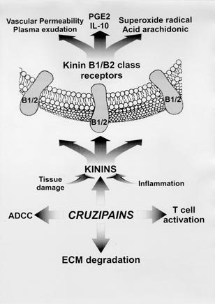
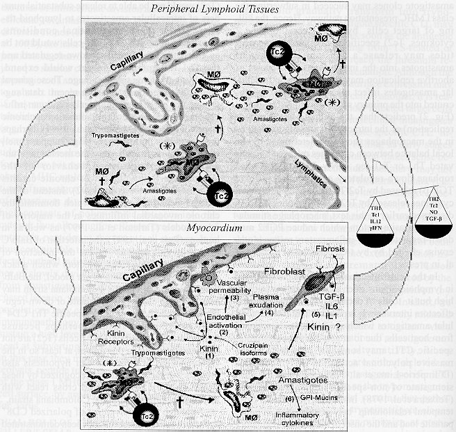
Mem Inst Oswaldo Cruz, Rio de Janeiro, Vol. 94, Suppl. I: pp. 51-63, 1999 A Role for Extracellular Amastigotes in the Immunopathology of Chagas Disease Julio Scharfstein+, Alexandre MorrotLaboratório de Imunologia Molecular, Instituto de Biofisica Carlos Chagas Filho, Universidade Federal do Rio de Janeiro, 21944-970 Rio de Janeiro, RJ, Brasil Corresponding author. Received 9 June 1999 Code Number:OC99136 In spite of the growing knowledge obtained about immune control of Trypanosoma cruzi infection, the mechanisms responsible for the variable clinico-pathological expression of Chagas disease remain unknown. In a twist from previous concepts, recent studies indicated that tissue parasitism is a pre-requisite for the development of chronic myocarditis. This fundamental concept, together with the realization that T. cruzi organisms consist of genetically heterogeneous clones, offers a new framework for studies of molecular pathogenesis. In the present article, we will discuss in general terms the possible implications of genetic variability of T. cruzi antigens and proteases to immunopathology. Peptide epitopes from a highly polymorphic subfamily of trans-sialidase (TS) antigens were recently identified as targets of killer T cell (CTL) responses, both in mice and humans. While some class I MHC restricted CTL recognize epitopes derived from amastigote-specific TS-related antigens (TSRA), others are targeted to peptide epitopes originating from trypomastigote-specific TSRA. A mechanistic hypothesis is proposed to explain how the functional activity and specificity of class I MHC restricted killer T cells may control the extent to which tissue are exposed to prematurely released amastigotes. Chronic immunopathology may be exacerbated due the progressive accumulation of amastigote-derived antigens and pro-inflammatory molecules (eg. GPI-mucins and kinin-releasing proteases) in dead macrophage bodies. Key words: cysteine proteinases - kinins - T cells - immunopathology - Trypanosoma cruzi - Chagas disease DYNAMICS OF IMMUNE RESPONSE EARLY IN T. CRUZI INFECTION Host resistance to microbial infection integrates two major and overlapping defense systems, innate and adaptive immunity. Intracellular pathogens can quickly relay activation signals that stimulate non-specific humoral and cellular effector responses in the infected host. Assisted by these innate defense responses, the rate of microbial growth is delayed for several days, while the adaptive branch of immunity gets prepared to confront the pathogen on the long term. The infection can only persist when the organisms succeed to counteract the selective pressure imparted by immune effector cells and/or antibodies. In the case of chronic viral infections, immune subversion is often targeted against intracellular pathways involved in antigen processing and/or presentation by class I MHC molecules. In the case of T. cruzi, the mechanisms which enable their persistent growth in mammalian tissues were not characterized. As discussed later in this text, there are reasons to think that the molecular diversity of T. cruzi organisms may affect the dynamics of tissue and organ involvement. This is supported by recent evidences showing that acute infection with parasite stocks pertaining to different genotypic groups induce distinct histopathology patterns in acutely infected mice (de Diego et al. 1998) Recently confirmed by research in genetic epidemiology (Souto et al. 1996, Brisse et al. 1998), the concept that T. cruzi has a multi-clonal descent was earlier proposed on the basis of isoenzyme (Miles et al. 1978, Romanha et al. 1979, Ready & Miles 1980, Tibayrenc et al. 1986) and fingerprint analysis of k-DNA (Morel et al. 1980). While not excluding the importance of host genetics as a determinant of host susceptibility in vivo (Trischman et al. 1978), studies performed with laboratory strains of T. cruzi (Brener 1965, Andrade & Andrade 1966, Mello & Brener 1978) and also with parasite clones (Postan et al. 1986, de Diego et al. 1998, Macedo & Pena, 1998) suggested that the variable expression of Chagas disease may caused, at least to some extent, by the genetic and biological diversity of the T. cruzi clones which circulate in sylvatic and domestic reservoirs. In spite of the poor knowledge about the innate responses which metacyclic trypomastigotes stimulate in wound tissues, this system is fully operative by the time the first cycles of intracellular infection are completed. Once released from disrupted cells, the trypomastigotes spread the infection via the bloodstream and/or lymphatics. At this early stage of infection, a wide range of non-phagocytic host cells can be invaded by the trypomastigotes, but host cell target preference can differ markedly from one parasite clone to another due to the variable composition and expression levels of their cell surface adhesion molecules, some of which are highly polymorphic (Affranchino et al. 1989, Colli 1993, Tackle & Cross 1991, Schenckman et al. 1994, Giordanno et al. 1994, Pereira et al. 1996, Salazar et al. 1996) or due to differential signalling ability of the invading parasite clones (Ming et al 1995, Burleigh and Andrews 1998). The survival strategies of the T. cruzi clones which preferentially invade macrophages are not well characterized. In hosts that are innately resistant, these parasite subpopulations must inhibit macrophage activation or somehow defend themselves from their microbicidal machinary. As true for other intracellular pathogens, innate immunity against T. cruzi depends on the release of g -IFN by NK cells (Aliberti et al. 1996, Cardillo et al. 1996). In genetically resistant strains, the onset of this T cell independent pathway depends on IL-12 production by activated macrophages and appears to be stimulated by tGPI-mucins, a potent class of pro-inflammatory molecules expressed by trypomastigotes and by amastigotes (Camargo et al. 1997). Synergized by TNF-a (Munoz-Fernandez et al. 1992), g -IFN induces a heightened state of microbicidal activation of macrophages in genetically resistant animals, at least so during the first days of infection. The mechanisms by which activated macrophages ultimately exert their anti-parasite activity is somewhat controversial, but there are indications the production of nitric oxide (NO) metabolites is critically involved (Gazzinelli et al. 1992). As for the innately susceptible inbred mice strains, their response to infection is dominated by the macrophage down-regulatory cytokines IL-10 or TGF-b (Silva et al. 1992, Gazzinelli et al. 1992). Interestingly, recent evidences suggest that factors leading to the accumulation of cyclic AMP by macrophages may down-regulate the pro-inflammatory response which tGPI-mucins otherwise stimulate in such cells (Procopio et al. 1999); under these conditions, tGPI-mucins can upregulate IL-10 expression by macrophages, thereby inhibiting the synthesis of both IL-12(p40) and TNF-a . It is thus conceivable that T. cruzi clones that have a relatively stringent preference for mononuclear-phagocytic cells (macrophagic) may actively convert macrophages from innately resistant animals into a susceptible target. Interestingly, macrophage activation in genetically susceptible mice infected by myotropic strains is higher than in resistant strains (Russo et al. 1989). In contrast to parasite clones that preferentially invade macrophages, those that invade non-phagocytic cells in the first few days of infection inevitably kill the target cells within 5-6 days. The necrosis caused by some myotropic strains can be extensive, sometimes involving multiple tissues and organs (Lenzi et al. 1996, Cotta de Almeida et al. 1977). Inflamed tissues are exposed to high amounts of parasite antigens, either released by extracellular parasites or leaked from killed organisms. Once captured by immature dendritic cells, these antigens are transported to the proximal draining lymph nodes. After upregulating their MHC molecules, the matured dendritic cells present the MHC-bound peptide antigens to naive CD4+ and CD8+ T cells. Depending on the genetic background of the individual, antigen load and cytokine balance, the functional characteristics of CD4+ T cells primed by dendritic cells can be rapidly polarized under the influence of type 1 or type 2 stimulating cytokines (IL-12 and IL-4, respectively). Driven by inflammatory chemokines, these circulating CD4+ Th1 and CD8+ T (Tc1) memory cells attach to the vascular adhesins expressed by activated endothelial cells (Kumar & Tarleton 1998) and are recruited into the inflamed tissues. Upon antigen-stimulation, they secrete g -IFN and TNF-a (Russo et al. 1988). and/or directly kill the infected targets by apoptosis parasite tissue clearance is gradually accompanied by a down-regulatory T cell response mediated by lymphocytes from the Th2 subset. Although the involvement of polarized CD8+ cells from the Tc2 subset has not been shown in T. cruzi infection, in other settings they were shown to produce IL-4, IL-5 and IL-10; while sustaining the capacity to act as cytotoxic T cells upon re-stimulation (Cerwenska et al. 1998). As discussed later on, the Tc2 effectors, if indeed present (Bahia-Oliveira et al. 1998), may play an important role in the regulation of the anti-parasite immune response in Chagas' disease. RELATIVE ROLES OF T CELLS AND ANTIBODIES IN ADAPTIVE IMMUNITY After two decades of intense investigation, the relative contribution of B or T cells in acquired resistance was clarified (Tarleton et al. 1992, Kumar & Tarleton 1998). Mice with B cell deficiency showed increased mortality rates at late stages of infection, indicating that antibody production, although secondary in importance to cellular immunity, is nonetheless important to acquired resistance as suggested by early studies performed by Krettli and Brener (1982). Interestingly the B cell deficient mice showed a delayed rise in the acute parasitaemia, suggesting that antibodies may actually enhance parasite virulence in early stages of infection. Depending on the isotype and antibody specificities stimulated early in the antibody response, opsonization may occur, thereby enhacing the uptake of the parasite by macrophages (Lages-Silva et al. 1987). At times when macrophage activation is transiently blocked by Th2-type of cytokines (refer to the abortive cycles of amastigote replication, later in this text), opsonization may increase rather than diminish the parasite load. In contrast to B cell knockouts, mice lacking class I or class II MHC-restricted T cells died during the acute phase of the infection with the Brazil strain. Not surprisingly, the infection was even worsened in animals that were deficient in both class I and class II MHC expression. Interestingly, the tissue inflammatory responses were absent in mice with the MHC-II class deficiency and the animals succumbed to infection due to high parasite load. Taken together, these studies have demonstrated that effector CD4+ and CD8+ T cells are critically involved in the acute control of T. cruzi infection. Similar processes may occur in humans, given the evidence that HLA-A2+ chagasic patients (indeterminate form) often display antigen-specific CD8+ CTL in their peripheral blood. The molecular mechanisms of target cell killing was recently investigated in infected mice (Kumar & Tarleton 1998). These authors showed, somewhat unexpectedly, that mice with targeted deletion of genes encoding for perfurin or granzyme B could control T. cruzi (Brazil strain) infection as efficiently as wild type animals. Their data suggest that class I MHC restricted CD8+ T cells may kill T. cruzi infected targets using the Fas/Fas L interaction system, or alternatively engage TNF-a and IFN-g cytokines in this process. T. cruzi PROTEASES AS FACTORS OF VIRULENCE AND PATHOGENICITY There correlation between focal immunopathology and the presence of parasite remnants in tissues has been extensively docuneted over the years (Andrade ZA, this volume). Ribeiro dos Santos and Hudson (1980) were the first to propose that the passive adsorption of T. cruzi antigens onto certain types of non-infected host cells may renderi them susceptible to by-stander antibody-dependent cellular cytotoxicity (ADCC). In addition to being possibly involved in the peripheral neuropathy which occurs in the acute phase (Koberle et al. 1968), ADCC may be possibly involved in the microangiopathy observed in acutely infected dogs (Andrade et al. 1994). Although the molecular basis of microvascular pathology is still unknown, recent studies on the cysteine-proteinases of the cruzipain family (Cazzulo et al. 1989, Murta et al. 1990, Eakin et al. 1992, Meirelles et al. 1992, McKerrow et al. 1995) may shed light on such processes. Originally identified because of their antigenic properties in chagasic patients (Scharfstein et al. 1983, 1986, Murta et al. 1990, Gazzinelli et al. 1990, Arnholdt et al. 1993, Morrot et al. 1997), the members of the cruzipain family consist of a heterogeneous group of closely related isoforms (Lima et al. 1994) encoded by about 130 genes (Campetella et al. 1992). The expression of these proteinases is upregulated when the invading trypomastigotes transform into amastigotes (Tomas & Kelly 1996), this process being accompanied by increased cell surface expression of the enzymes. It is thus conceivable that these amastigotes, once released into interstitial spaces, may degrade the adjacent extracellular matrix (ECM) in adjacent tissues (unpublished observations). By doing so, the parasites may prevent or disrupt ECM-interactions with TNF-a , chemokines and other inflammatory mediators involved in the recruitment and/or activation of immune cells T cells (Gillat et al. 1996). Cruzipain (and/or other minor isoforms) are worth studying in this context, because they were recently identified as antigenic deposits in sites of myocardial inflammation in autopsies from patients with severe chronic cardiomyopathy (Morrot et al. 1997). Given that cruzipain (and/or other isoforms) are stable and enzymatically active in neutral-acid pH, we reasoned that their half-life in extracellular tissues would likely depend on the levels of host proteinase inhibitors which permeate inflammatory sites. The immunological implications of cruzipain interactions with a 2-Macroglobulin, a non-specific plasma proteinase inhibitor were previously studied in our laboratory (Morrot et al. 1997). In view of previous evidences indicating that CD4+ T cells from chagasic patients promptly respond to cruzipain (Arnholdt et al. 1993), we investigated if functional inactivation by a2M could influence the efficiency of antigen presentation and processing by human monocytes. Our studies revealed that monocytes engage a highly endocytic scavenger receptor expressed by monocytes (CD91 or LRP/a 2MR) to rapidly internalize a 2M-cruzipain complexes. The enhanced endocytic uptake of cruzipain-complexes ultimately favor increased intracellular processing and presentation of cruzipain peptides to CD4+ T cells (Morrot et al. 1997). Because a 2M binds to cell surface proteinases from amastigotes (Coutinho et al. 1997), it is conceivable that the multifunctional LRP/a2MR scavenger receptor may promote the uptake of amastigotes coated with a 2M ligated to cruzipain, in ways that are reminiscent of mechanisms recently described for the macrophages' mannose receptor (Khan et al.1995). In both cases, the efficiency of antigen-presentation of amastigotes epitopes to T cells may be potentiated. In short, our studies illustrate how interactions between parasite proteinases and host inhibitors may integrate elements of innate defense systems with those in charge of acquired immunity. As a follow-up from the studies involving a 2M, we turned our attention to kininogen, a member of the cystatin superfamily of cysteine proteinase inhibitors (Barret et al. 1986). In addition to the cystatin-like domains, these multifunctional glycoproteins can participate in the activation of the intrinsic pathway of coagulation (high molecular weight kininogen) and modulate platelet activation by thrombin. Important to our discussion, kininogens (high or low molecular weight) is the parent substrate molecule from which bradykinin or lysil-bradykinin is released, upon proteolytic cleavage either by plasma or tissue kallikrein (Bhoola et al. 1992). Kinins are short-lived peptides engage distinct subtypes of G-coupled kinin receptors in a wide spectrum of biological processes, such as modulation of neuronal activity (Higashida et al. 1990), cell proliferation and vascular permeability (Bhoola et al. 1992), smooth muscle contraction or relaxation (Monbouli & Vanhoutte 1995). Depending on the host cell, kinin signalling is transduced by receptors that are either expressed constitutively (B2) or are induced (B1) during anoxia or noxious stimuli (Burch & Kyle 1992, Regoli 1980). The initiative to investigate the relationship of T. cruzi with the kinin system was initially motivated by enzymatic specificity studies carried out with cruzipain 2 (Lima et al. 1994), a minor isoform whose recombinant form was only recently expressed in S. cerevisae. After noting that its substrate specificity somewhat resembled that of tissue kallikrein, we were able to demonstrate that these isoforms could act as kinin-releasing enzyme (Del Nery et al. 1997), although with different efficiency. On a first look, cruzipain's ability to process kininogen seemed paradoxical because this protease is highly sensitive to inhibition by the tight-binding cystatin domains displayed in both high or low molecular weight forms (Stoka et al. 1995, Scharfstein et al. 1995). Clues to understand the mechanism of kinin-release emerged when the Ki's obtained for kininogen were determined for different isoforms of cruzipain: interestingly, we observed that r-cruzipain 2 was markedly less sensitive to inhibition by kininogen as compared to cruzain (Lima et al. in preparation) the archetype from the cruzipain family (McKerrow et al. 1995). Our data suggest that the kininogenase activity of T. cruzi cysteine-proteinases might have evolved due to structural diversification of the catalytic site of some isoforms, such as cruzipain 2. Notably, in the same study (Del Nery et al. 1997) we also showed that cruzipain could generate bradykinin indirectly, that is, by converting plama prekallikrein in active kallikrein. In a parallel study, we used intravital microscopy to verify if the topical application of purified cruzipain on the hamster cheek pouch could stimulate increases in vascular permeability (Svensjo et al. 1997). Potent responses were indeed observed, suggesting that the hemoflagellate may be able to use cruzipain (and/or isoforms) to generate kinins upon contact with endothelial cells. Ongoing studies in mice with targeted deletion of kinin-receptors should reveal if these kinin-releasing proteinases may facilitate T. cruzi migration across non-fenestrated capillaries. The realization that T. cruzi may use cruzipain isoforms to generate pro-inflammatory kinins upon contact with kinin-receptor bearing cells (Fig. 1) raised questions regarding their role in pathogenesis. More recently, its was shown that bradykinin stimulation of the B2 subtype of kinin-receptor from guinea pig macrophages leads to stimulation of superoxide radical, arachidonic acid and PGE2 (Bockmann & Paegelow 1998). It will be interesting to know if kinins released by T. cruzi may trigger PGE2 production by macrophages. As discussed earlier in this article, factors leading to cyclic AMP accumulation, eg. PGE2, may stimulate IL-10 synthesis in macrophages that had been exposed to tGPI-mucins (Procopio et al. 1999). The role of T. cruzi-induced kinin release in the pathogenesis of chronic heart disease is also worth exploring, the main assumption being that microvascular lesions caused by infection-associated events (Andrade et al. 1994) may slowly accumulate over the years, thereby creating conditions for the exacerbation of chronic inflammation and fibrosis (Morris et al. 1990, Rossi 1990). In a recent study, Higuchi et al. (1999) have reaffirmed the potential importance of microvascular pathology: in a detailed analysis of post-necropsy heart specimens from chronic chagasics by three-dimensional confocal microscopy, these authors observed abnormalities of the cardiac microcirculation as well as in the interstitial matrix patterns of myocardial tissues. Peculiar lesions, often manifested as arteriolar dilatation and capillary vessel tortuosity, were described, being thus far vaguely attributed to fibrosis and/or to as yet uncharac-terized lesions induced by the parasites. Apart from initial studies looking at effects of desialilation by TS enzymes (Libby et al. 1986), little is known about the biochemical basis of T. cruzi interaction with endothelial cells (Morris et al. 1990). As discussed earlier in this section, the realization that cruzipain-isoforms are capable of releasing kinins offers a new experimental framework to investigate the pathophysiological consequences of long-term stimulation of myocardial capillaries by vasoactive kinins (Fig. 1). Our studies in the cheek pouch suggest that cruzipain molecules released/leaked from merely a few parasites may suffice to generate vasoactive kinins, thereby provoking plasma exudation across the nearest capillaries (Fig. 2, right panel). Similar processes, perhaps involving NO released by activated endothelial cells, may underlie the lesions observed earlier in infection, when the parasites are released next to kinin-receptor bearing cells, situated in the proximity of autonomous nervous system ganglia. THE CLASS I MHC PATHWAY OF ANTIGEN PRESENTATION IN T. cruzi INFECTED CELLS During microbial infection, host class I or class II molecules encoded by polymorphic MHC genes bind to antigenic peptides (T epitopes) produced by the invasor. Once associated with the appropriate MHC allele product, the peptide: MHC complex is targeted to the cell surface of the infected cell, where it is "presented" to naive or effector T cells. The intracellular pathways that generate P antigenic peptides by proteolytic processing and then enable MHC loading are collectively referred as antigen processing and presentation system. In the past years, the multiple checkpoints. That are targeted by pathogen-derived factors, viruses in particular, were characterized (Ploegh H 1999). In general terms, the viral products may target elements controlling (i) antigen-proteolysis by proteosomes, (ii) peptide transport to the ER (iii) MHC assembly and/or sorting and (iv) surface expression of class I MHC:peptide complexes. The recent characterization of the specificity of killer T cells directed against T. cruzi should pave the way for elucidation of subversion tactics used by this pathogen. As shown in mice, the analysis of HLA-A2-restricted CTL specificities in the peripheral blood of chagasic patients has identified different members of a subfamily of TS_related antigens as targets: (Wizel et al. 1997, Low et al. 1997): two from amastigote-derived antigens, ASP-1 and ASP-2, and one trypomastigote-specific antigen, TSA-1. Teleologically, the usage of a vast array of polymorhic T. cruzi proteins as parent substrates for the proteosomal proteases makes sense because it should increase opportunities for epitope loading of the highly diverse MHC-class I products that exist in outbred populations. In other words, had the antigen structural variability involved exclusively monomorphic single genes, the parasite clones which display a variant sequence would tend to swarm most individuals from a genetically outbred population. Thus, structural variability imparted by multi-copy polymorphic genes may be advantageous to host-parasite equilibrium because it reduces the excessive risk associated with the growing diversification of parasite sub-populations. It is conceivable that some opportunistic T. cruzi clones may be able to subvert class I and/or class II presentation pathways. In a provocative study, Kahn and Wlekinski (1997) presented data suggesting that antigen variation of T. cruzi cell surface proteins can diminish the efficiency of anti-parasite T cell responses. It was suggested that the simultaneous expression of individual antigens from polymorphic TS subfamily may limit the availability of processed epitopes from each antigen below the threshold level required to stimulate a protective IFN-g response against the parasite. It will be interesting to know if similar mechanisms may allow for parasite escape from detection by class I-MHC restricted killer T cells. Admittedly, however, antigen-variation alone may not allow the parasites to escape from the antigen-presentation system of host cells from genetically outbred individuals (e.g. dogs and humans), because they count with a more diversified array of MHC allelic products. The parasite may need to use multiple mechanisms, possibly acting synergistically with antigen variation, to escape from immune detection during infection in humans. T. cruzi CLONAL DIVERSITY AND AMASTIGOTE EVASION MECHANISMS The selective pressure which the killer T cells exert on the intracellular amastigotes must be very strong in genetically outbred individuals, because class I MHC-restricted CTLs would be more effective by attacking the target before the intracellular amastigotes are transformed into the more aggressive trypomastigotes. Conversely, one might expect that amastigotes must possess special resources to counteract the highly protective amastigote-specific CTL responses. Future attempts to elucidate the mechanims of evasion of T. cruzi must take into account the finding that rates of intracellular division and transforming efficiency of amastigotes vary significantly among different clones (Dvorak & Hyde 1973). Indeed, the measurements of some of these in vitro parameters in a wide collection of T. cruzi stocks permitted their classification into the three main genetic groups, thus showing a strong correlation with genetic distances evaluated by multilocus enzyme electrophoresis (MLEE) or random amplification by polymorphic DNA (RAPD) methods (Revollo et al. 1998). Here we advance the proposition that the phenotypic changes associated with these major genetypes will differentially affect the function of the class I antigen-processing machinary of infected target cells. For example, it is possible that the levels of processed epitopes generated from parasite antigens (from a given parasite clone) might be controlled by parasite-factors that are likewise encoded by other polymorphic genes. The developmentally regulated cysteine-proteinases (cruzipain-issoforms), discussed later in this text, merits investigation in this context, because some of these proteases are targeted to the cell surface of amastigotes. Given the indications that they are stable and active at neutral pH (Murta et al. 1990) and that their substrate specificity may differ (Lima et al. 1994), it will be worth examining if their differential expression by different parasite clones may control the quality and/or quantity of processed epitopes derived from amastigote surface antigens (e.g. ASP-1/2). In principle, these parasite proteinases may act by cleaving the dominant parasite antigens prior to their unfolding, thus either accelerating or blocking the subsequent processing of a given antigen by the proteosomes. Although entirely speculative, this mechanism illustrates how the interplay of products encoded by two different polymorphic gene system (the structurally and functionally diverse cruzipain isoforms and the TS-related antigens) may improve the parasite's ability to resist immune attack. Of course, the amastigote may also engage non-specific mechanisms of evasion (by acting on constitutive or inducible proteosomal sub-units, degradation of TAP, down-regulation of adhesion molecules etc.) to down-regulate class I MHC cell surface molecules. Ultimately, some of these hypothetical pathways may converge, thus further elevating the threshold required for amastigote-epitope presentation by class I MHC molecules. Perhaps subversion by antigen variation (Kahn & Wlekinski 1997) may be further optimized if the amastigotes succed reducing their metabolism (for example, by slowering rates of intracellular division, as would be the case of latent forms), because these maneuvers may also decrease the availability of substrate-antigen in the host cytosol. Alternatively, higher rates of amastigote division and of metabolic activity may be accompanied by upregulation of cruzipain expression (or other proteases), hence allowing for enhanced proteolysis of TS-antigens and/or inactivation of host cell factors implicated in essential APC functions. DUAL ROLE OF TRYPOMASTIGOTE-SPECIFIC KILLER T CELLS Since the amastigote division proceeds asynchronously (Dvorak & Hyde 1973), the amastigotes which succeed at subverting the class I antigen presentation pathway should gradually transform into trypomastigotes. The trypomastigote specific CTL may then attack the target cells as soon as the epitopes (derived from TS-related or other dominant antigens) generated in the cytossol reach the threshold required for class I MHC presentation. Although the rates of MHC-loading and surface expression may vary from one cell to another, it is reasonable to predict that the target cells still harbors some intracellular amastigotes at the time of attack by the trypomastigote-specific killer T cells. This time-course make sense because the antigen presentation machinary should not be anymore functional at the very late stage of pseudocyst formation. We may therefore deduce that host cell killing at a relatively early stages of trypomastigote production is a suitable compromise with the host, because in this case, the release of just a few trypomastigotes should suffice to ensure long term survival of both host and parasites. As a corollary, we may predict that the extent to which tissues are exposed to prematurely released amastigotes is variable, and dependent on the interplay between interactions involving class I MHC-allelles (host genetic) and the highly diverse surface antigens from T. cruzi. (parasite genetic make up). This fundamental tenet, leads to the following testable assertions: (1) killer T cells which recognize amastigote epitopes are the most effective effectors of class I MHC-protective immune responses (this assertion has obvious implications for vaccine design); (2) amastigote clones can suceed at subverting the class I pathway of antigen presentation exist, but the phenotype is linked to host MHC-haplotype; (3) expression of the pathogenic phenotype deoends on the activity of CD8+ killers which recognize trypomastigote specific epitopes (TS-related antigens?) at a later stage of intracellular development (implications for vaccine design). We postulate that the deposition of high numbers of nonmotile amastigotes in tissues may be crucial for the onset of apoptosis of effector cells from the Th1 subset (and Tc1). In addition, the amastigotes may also induce non-specific supression by stimulating cells (Th2 or Tc2) to produce TGF-b or IL10 in the inflamed tissues. In summary, we propose that trypomastigote-specific killer cells may play a multiple role in Chagas disease: they are critically important for protection, they may indirectly contribute to Th2-down regulation (to discussed below), and may be promote immunopathology by inducing the premature release of pro-inflammatory amastigotes in adjacent tissues. ABORTIVE-CYCLES OF AMASTIGOTE REPLICATION: ROLE OF class I MHC KILLER T CELLLS On a first impression, the ability of CTL to kill non-phagocytic targets which harbor intracellular trypomastigotes would seem to be a redundant process, because most of the infective parasites should be anyway released into the extracellular fluids upon host cell burst. However, target cell death by apoptosis should be advantageous to the host (Andrade et al. 1999), inasmuch as this mechanism would spare host tissues from the detrimental effects of necrosis that would otherwise occur if the target cells collapse due to excessive numbers of parasites. Since mutual destruction is not a fruitful evolutionary strategy, it is unlikely that host cell apoptosis also destroys all the intracellular parasites. Once released to extracellular spaces, the trypomastigotes tend to move away from the primary foci of infection, while the non-motile extracellular amastigotes [which resist C' mediated lysis (Iida et al. 1989) tend to accumulate in the proximity of the primary site of infection (Fig. 2) . Of necessity, there is only one obvious pathway that may permit the survival of these extracellular amastigotes: the maintenance of a supressive environment, determined by TGF-b or IL10, or alternative mediators. If spared from death, the amastigote may leave the parasitophorous vacuole and lodge in the cytosol, where they actively proliferate (Ley et al. 1988). As previously discussed, we postulate that some amastigote clones may succeed in subverting the class I MHC presentation pathway. Hence, the killing of target cells by CTL (TC2) specific for trypomastigote peptides may release a substantial number of amastigotes into the interstitium. New cycles of abortive replication may ensue, once other macrophages are recruited and likewise infected. In short, our model (Fig. 2) predicts that the ultimate fate (death or replication) of the intracellular amastigotes lodged in the macrophages should be determined by the local balance between cytokines released by activated Th1 (eg. IFN-g , TNF-a ) or Th2 (TGF-b or IL10) or by equivalent cytokines released by Tc1/Tc2 CD8+ lymphocytes. As discussed earlier in this text, macrophage stimulation by parasite-factors that induce PGE2 may down-regulate the pro-inflammatory response induced by t-GPI-mucins (Procopio et al. 1999). In the acute infection, the tissue parasite load in non-phagocytic cells from lymphoid organs, such as the spleen, is initially high but decreases when cellular immune effectors intervene. It is possible that the extracellular amastigotes released from host cells may somehow contribute to the massive polyclonal activation of lymphocytes (D'Imperio-Lima et al. 1985) and/or for the transient state of non-specific immunosuppression (Teixeira et al. 1978) observed. As already mentioned, there is a close temporal relationship between the drop in tissue parasite load and the onset of these immunoregulatory changes in peripheral lymphoid tissues (Dos Reis 1997). Given the evidences that the activity of CD8+ CTL effectors is critically involved in the parasite clearance from non-phagocytic cells in various tissues, perhaps the killer T cells may indirectly play a role in the aforementioned regulatory abnormalities. As already discussed (Fig. 2, left panel), target cell death by trypomastigote-specific CTL may lead to the premature release of some intracellular amastigotes and/or trypomastigotes in the interstitium. Consistent with predictions from the our model, Caulada-Benedetti et al. (1998) have recently reported that macrophages exposed to live amastigotes activate both lymph node and spleen CD4+ and CD8+ T cells. By extension, we suggest that the pool of spleen CD4+ T cells which undergo activation-induced cell death (AICD) upon in vitro stimulation with anti-CD3 (Dos Reis et al. 1995) may be enriched with amastigote-specific CD4+ T cells. As argued by these authors, the elimination of activated Th1-type cells by PCD may favor the polarization of Th2-type responses at late stages of the acute infection (Dos Reis 1997). Perhaps amastigote-derived antigens can exponentially build up in lymphoid tissues due to successive rounds of abortive infection induced by trypomastigote-specific killer T cells (Tc2). If true, CD8 cells from this subset should indirectly control the induction of anergy, clonal deletion and/or Th2-dependent down-regulation of amastigote-specific CD4+ Th1 lymphocytes. As a corollary, we may predict that individual animals or patients which exhibit deficits in trypomastigote-specific CTL (Tc2) should not be able to release substantial numbers of intracellular amastigotes to lymphoid tissues. As a result of such deficits, amastigote-specific CD4+ Th1 cells would not be as efficiently eliminated or down-regulated, perhaps allowing for the expansion of the Th1 inflammatory subset, thereby aggravating acute and chronic immunopathology. These general predictions are consistent with data suggesting that early immunological events can influence the development of inflammation at chronic stages of infection (Mariano et al. 1999). Perhaps the mechanistic hypothesis outlined here (Fig. 2, lef panel) may offers a framework to study the mechanisms underlying the contrasting behavior of the Colom-biana-Balb/C model of chronic carditis (Ribeiro dos Santos et al. 1992). Instead of the CD8+ T cells (anti-parasite) which dominate the chronic myocardial infiltrates in the majority of mice models (Tarleton et al. 1997) and in human as well (Reis et al. 1997) the infection of Balb/C mice with Colombiana favors the induction of autoreactive CD4 T cell effectors which attack cardiac tissues. According to the tenets of our model, the Balb/C animals infected by the Colombiana strain may have failed to efficiently eliminate or down-regulate cross-reactive amastigote_specific Th1 CD4 cells in lymphoid tissues, perhaps due to a primary deficit in the function of trypomastigote-specific CD8 T cells (Tc2). It will be interesting to know if the putative self-epitopes recognized by these CD4 T cell clones are structurally homologous to amastigote antigens from the Colombiana strain. Of course, other regulatory pathways may be of course involved in the Th1/Th2 polarization observed in chagasic infection. For example, it has been recently proposed that the conversion of Tc1 into Tc2 suppressors depends on the NO releasing activity of macrophages, which is in turn stimulated by Th1 lymphocytes. It is believed that the cytokine profile of Tc2 may ensure that target cell apoptosis proceeds without the characteristic necrosis and tissue injury associated with the activity of CTL from the Tc1 subset (Kolb & Kolb-Bachofen 1998). In a recent study of PBMC responses, Bahia-Oliveira et al. (1998) observed that type 2-cytokines predominate in the indeterminate stage of the human disease, while type 1 cytokines are more frequently observed in symptomatic individuals. It will be important to know if Tc2 and Tc1 contribute to these fluctuations, and assess their relationship to the distribution of cellular infiltrates in the myocardial tissues (Higuschi et al. 1993). ROLE OF CD8+ KILLERS IN THE EXACERBATION OF CHRONIC IMMUNOPATHOLOGY Andrade et al. (1997) have recently demonstrated that the canine model accurately reproduces the main features of the indeterminate form of human Chagas disease. They suggested that the heart lesions in the indeterminate stage of the disease are caused by self-limited cycles of focal inflammatory changes, probably determined by subtle shifts in the immunoregulatory activity of effector T lymphocytes. The canine model does not seem to support an obvious role for microvascular lesions in this stage of the disease. In contrast to the progressive nature of he chronic carditis observed in mice, the transition from indeterminate to chronic heart disease in dogs is not gradual. In humans, the distribution of cytokine-producing cells revealed that Th2 type cells are usually associated with higher deposition of parasite antigens (Higuchi et al. 1993, Reis et al. 1997). Our model predicts a role for pathogenic clones in the exacerbation of chronic immunopathology. The pre-requisites for the onset of these pathological processes are: (i) infected cells in some tissues reservoirs sustain the growth of amastigote clones which can occasionally subvert the class I antigen presentation pathway (in other words, the amastigote-speciic killer T cells cannot effectively recognize and attack the infected target cells while the intracellular amastigotes multiply); (ii) these intracellular amastigotes transform into trypo-mastigotes; (iii) early after transformation, the trypomastigotes can successfully load class I MHC molecules with their T cell epitopes; (iv) the infected target cells are then killed by trypomastigote-specific CTL; (v) extracellular amastigotes (which are ultimately responsible for the exacerbation of immunopathology) are released in the interstitium; (vi) the extracellular amastigotes can invade and proliferate in suppressed macrophages; (vii) trypomastigote-specific killer cells from the Tc2 subset induce successive rounds of abortive infection in suppressed macrophages, causing the progressive deposition of amastigote antigens and pro-inflammatory factors inside macrophage apoptotic bodies (Morrot et al. 1997). Here the concept of a "pathogenic" clone is defined in the operational sense: it refers to an opportunistic population of intracellular amastigotes that somehow exploits antigen variation to subvert the class I presentation of target cells which express a given MHC-haplotype. In other words, a pathogenic clone isolated from one individual (outbred species) would not necessarily be more harmful to individuals that express a different MHC haplotype. As argued earlier in this article, subversion of class I pathway of antigen presentation in inbred mice may be a relatively easier process because these animals count with a limited number of alleles to compensate for the extreme antigen variability of the intracellular parasites. We may thus predict that, in immunocompetent chagasic patients (also in the canine model), the "pathogenic" parasite subpopulations were either eliminated due to the powerful selective pressure exerted by amastigote-specific killer T cells, or are present at very low levels somewhere in the body. The pathogenic clones should preferentially thrive in suppressed type of macrophages because these targets are sensitive to the modulatory influence of type-2 cytokines originating from polarized CD4 or CD8 effector T cells, which predominate in the indeterminate stage of the disease (Bahia-Oliveira et al. 1998). Our model predicts that myocardial inflammation in genetically oubred individuals may be exacerbated when these tissues become colonized by pathogenic clones. From where would they come from? As already mentioned, extracellular amastigotes may infect tissue macrophages, using to this end the alternative pathway of invasion (Ley et al. 1988). Organs that usually maintain macrophages in a relatively supressed state (eg. adrenal glands, Teixeira et al. 1977) may allow for the outgrowth of these potentially pathogenic amastigote clones. In a period of a relatively profound immunosuppression, perhaps the threshold required for amastigote-epitope presentation by suppressed macrophages is no longer reached. This would allow for their escape from recognition by amastigote-specific killer T cells. Alternatively, the amastigotes would escape if the susceptible host exhibits a transient deficit of amastigote-specific killer T cells or, of trypomastigote-specific CD8+ function (Tc1). Once released into the bloodstream, the "pathogenic" clone may reach the heart tissues by traversing the blood capillaries which already express adhesion molecules (Zhang & Tarleton 1996). After lodging inside a cardiac fiber, or in a tissue macrophage, they again subvert the class I MHC presentation, avoiding recognition by amastigote-specific killer T cells. At this stage, the cardiac tissues may be already dominated by effector trypomastigote-specific Tc2 killer T cells and/or by effectors from the Th2 subset. Successive rounds of invasion and abortive infection of supressed macrophages would cause the progressive accumulation of amastigote-antigens and/or their pro-inflammatory products, such as GPI-mucins, inside dead macrophages (opoptotic bodies). In organs that are highly vascularized, such as the heart, cruzipain-isoforms secreted or leaked by T. cruzi amastigotes can process kininogen, thus releasing pro-inflammatory kinins. By acting on adjacent endothelial cells, these vasoactive peptides may induce plasma exudation, thereby expanding the inflammatory response, and contributing to interstitial fibrosis. The chemokines released by tissues exposed to tGPI-mucins and other pro-inflammatory mediators might also recruit autoreactive CD8+ or CD4+ lymphocytes (Cunha-Neto et al. 1996) into the myocardium, thus initiating the expanding wave of chronic immunopathology. ACKNOWLEDGMENTS To Dr George dos Reis for helpful comments made while reading this manuscript. Supported by FUJB, Faperj and Pronex II. REFERENCES Affranchino JL, Ibanez CF, Luquetti AO, Rassi A, Reyses MB, Macina RA, Aslund L, Pettersson U, Frash ACC 1989. Identification of a Trypanosoma cruzi antigen that is shed during the acute phase of Chagas' disease. Mol Biochem Parasitol 34: 221-228. Aliberti JC, Cardoso MA, Martins GA, Gazzinelli RT, Vieira LQ, Silva JS 1996. Interleukin-12 mediates resistance to Trypanosoma cruzi in mice and is produced by murine macrophages in response to live trypomastigotes. Infect Immun 64: 1961-1967. Andrade SG, Andrade Z 1966. Estudo histopatologico comparativo das lesões produzidas por 2 cepas do Trypanosoma cruzi. O Hospital 70: 1268-1278. Andrade ZA, Andrade SG, Correa R, Sadigursky M, Ferrans VJ 1994. Myocardial changes in acute Trypanosoma cruzi infection: ultrastructural evidence of immune damage and the role of microangiopathy. Am J Pathol 144: 1403-1411. Andrade ZA, Andrade SG, Sadigursky M, Wenthold RJ Jr, Hilbert SL, Ferrans VJ 1997. The indeterminate phase of Chagas' disease: ultrastructural characterization of cardiac changes in the canine model. Am J Trop Med Hyg 57: 328-336. Andrade ZA, Zhang YuZX, Andrade SG, Takeda K, Sadigursky M, Ferrans VJ 1999. Apoptosis in a canine model of acute Chagasic myocarditis. J Mol Cell Cardiol 581-596. Andrade LO, Machado CMS, Chiari E, Pena SDJ, Macedo AM 1999. Role of parasite and host genetic backgrounds in the differential tissue tropism of Trypanosoma cruzi strains (this volume). Arnholdt AC, Piuvezam MR, Russo DM, Lima AP, Pedrosa RC, Reed SG, Scharfstein J 1993. Analysis and partial epitope mapping of human T cell responses to Trypanosoma cruzi cysteinyl proteinase. J Immunol 15: 3171-3179. Bahia-Oliveira LM, Gomes JA, Rocha MO, Moreira MC, Lemos EM, Luz ZM, Pereira ME, Coffman RL, Dias JC, Concado JR, Gazzinelli G, Correa-Oliveira R 1998. IFN-gamma in human Chagas' disease: protection or pathology? Braz J Med Res 31: 127-131. Barret AJ, Rhawlings ND, Davies ME, Machleidt W, Salvesen G, Turk V 1986. In AJ Barret & G Salvesen (eds), Proteinase inhibitors: cysteine-proteinase inhibitors of the cystatin superfamily, p. 515-569. Elsevier, Amsterdam. Bhoola KD, Figueroa CD, Worthy K 1992. Bioregulation of kinins: kallikreins, kininogens, and kininases. Pharmacol Rev 44: 1-80. Bockmann S, Paegelow I 1995. Bradykinin receptors and signal transduction pathways in peritoneal guinea pig macrophages. Eur J Pharmacol 291: 159-165. Bockmann S, Mohrdieck K, Schmidt H, Zundorf G, Paegelow I 1998. Differential sensitivity of macrophages to bradykinin. Naunyn-Schmiedeberg's Arch Phamarcol 357: 151-158. Brener 1965. Comparative studies of different strains of Trypanosoma cruzi. Ann Trop Med Parasitol 59: 19-26. Burch RM, Kyle D 1992. Recent developments in the understanding of bradykinin receptors. Life Sci 50: 829-838. Burleigh B, Andrews NW 1998. Signaling and host cell invasion by Trypanosoma cruzi. Curr Opin Microbiol 1: 461-465. Camargo MM, Almeida IC, Pereira ME, Ferguson MA, Travassos LR, Gazinelli RT 1997. Glycosylphos-phatidylinsitol-anchored micin-like glycoproteins isolated from Trypanosoma cruzi trypomastigote initiate the synthesis of proinflammatory cttokines by macrophages. J Immunol 158: 5890-5901. Cazzulo J, Cousi R, Raimondi A, Wernstedt C, Hellman U 1989. Further characterization and partial amino acid sequence of a cysteine protease (cruzipain) from Trypanosoma cruzi. Mol Biochem Parasitol 33: 33-42. Campetella O, Henrickson J, Aslund L, Frasch ACC, Petterson U, Cazzulo JJ 1992. The major cysteine protease (cruzipain) is encoded by multiple polymorphic tandemly repeated organuzed genes located in different chromossomes. Mol Biochem Parasitol 50: 225-234. Cardillo F, Voltarelli, JC, Reed SG, Silva JS 1996. Regulation of Trypanosoma cruzi infection in mice by gamma interferon and interleukin 10: role of NK cells. Infect Immun 64: 128-134. Cerwenka A, Carter LL, Reome, JB, Swain, SL, Dutton RW. 1998. In vivo persistence of CD8 polarized T cell subsets producing type 1 or type 2 cytokines. J Immunol 161: 97-105. Colli W 1993. Trans-sialidase: a unique enzyme activity discovered in the protozoan Trypanosoma cruzi. FASEB J 7: 1257-1264. Cotta de Almeida V, Bertho AL, Villa verde DM, Savino W 1997. Phenotypic and functional alteration of thymic nurse cells following acute T. cruzi infection. Clin Immunopath 82: 125-132. Coutinho CMLM, Cavalcanti, GH, van Leuven, Araujo-Jprge, TC. 1997. Alpha-2 Macroglobulin binds to the surface of Trypanosoma cruzi. Parasitol Res 83:144-150. Cunha-Neto E, Coelho V, Guilherme A, Fiorelli N, Stolf N, Kalil J 1996. Autoimmunity in Chagas' disease-identification of cardiac myosin-B13 Trypanosoma cruzi protein cross-reactive T cell clones in heart lesions of a chronic Chagas' cardiomyopathy patient. J Clin Inv 98: 1709-1712. de Diego JA, Palau MT, Gamallo C, Penin P 1998. Relationships between histopathological findings and phylogenetic divergence in Trypanosoma cruzi. Trop Med Int Health 3: 222-233. Del Nery E, Juliano MA, Lima APCA, Scharfstein J, Juliano L 1997. Kininogenase activity by major cysteinyl proteinase (Cruzipain) from Trypanosoma cruzi. J Biol Chem 272: 25713-25718. D' Imperio Lima MR, Joskowicz M, Coutinho A, Kipnis T, Eisen H 1985. Very large and isotypically atypical polyclonal plaque-forming cell responses en mice infected with Trypanosoma cruzi. Eur J Immunol 15: 201. Dos Reis GA 1997. Cell-mediated immunity in experimental Trypanosoma cruzi infection. Parasitol Today 13: 335-341. Dvorak JA, Hyde TP 1973. Trypanosoma cruzi: interaction with vertebrate cells in vitro. Individual interactions at the cellular and subcellular levels. Experim Parasitol 34: 268-283. Eakin AE, Mills AA, Harth G, McKerrow JH, Craik CS 1992. The sequence, organization and expresion of the major cysteine protease (cruzain) from Trypanosoma cruzi. J Biol Chem 267: 7411-7420. Gazzinelli RT, Leme VCM, Cançado JR, Gazzinelli G, Scharfstein J 1990. Identification and partial characterization of T. cruzi antigens recognized by T cells and immune sera from chagasic patients. Infec Immuni 58: 1437-1444. Gazzinelli RT, Oswald IP, James SL, Sher A 1992. IL-10 inhibits parasite killing and nitrogen oxide production by IFN-gamma-activated macrophages. J Immunol 148: 1792-1796. Gillat D, Cahalon L, Hershkoviz R, Lider O 1996. Interplay of T cells and cytokines in the context of enymatically modified extracellular matrix. Immunol Today 17: 16-20. Giordano R, Chammas R, Veiga SS, Colli W, Alves MJ 1994. An acidic component of the heterogeneous Tc-85 protein family from the surface of Trypanosoma cruzi is a laminin binding glycoprotein. Mol Biochem Parasitol 65: 85-94. Higuchi ML, De Brito T, Reis MM, Barbosa A, Bellotti G, Pereira Barreto AC, Pileggi F 1993. Correlation between Trypanosoma cruzi parasitism amd myocardial inflammatory infiltrate in human chronic chagasic myocarditis: light microscopy and immunohistochemical findings. Cardiv Pathol 2: 101-106. Higuschi ML, Fukasawa S, De Brito T, Parzianello LC, Bellotti G, Ramires JAF 1999. Different microcirculatory and intestinal matrix patterns in idiopathic dilated cardiomyopathy and Chagas' disease: a three dimensional confocal microscopy study. Heart 81: 1-6. Iida K, Whitlow NM, Nussenzweig V 1989. Amastigotes of Trypanosoma cruzi escape destruction by terminal complement components. J Exp Med 169: 881-891. Kahn SJ, Wleklinski M 1997. The surface glycoproteins of Trypanosoma cruzi encode a superfamily of variant T cell epitopes. J Immunol 159: 4444-4451. Kahn SJ, Wleklinski M,Aruffo A, Farr A, Coder D, Kahn M 1995. Trypanosoma cruzi adhesion to macrophages is facilitated by the mannose receptor. J Exp Med 182:1243-1258. Koberle F 1968. Chagas' disease and Chagas' syndromes: the pathology of American trypanosomiasis. Adv Parasitol 6: 63-116. Kolb H, Kolb-Bachofen V 1998. Nitric oxide in autoimmune disease: cytotoxic or regulatory mediator? Immunol Today 19: 556-561. Krettli AU, Brener Z 1982. Resistance against Trypanosoma cruzi is associated to anti-living trypomas-tigote antibodies. J Immunol 128: 2009-2012 Kumar S, Tarleton RL 1998. The relative contribution of antibody production and CD8+ T cell function to immune control of Trypanosoma cruzi. Parasite Immunol 20: 207-216. Lages-Silva E, Ramirez LE, Krettli AU, Brener Z 1987. Effect of protective and non-protective antibodies in the phagocytosis rate of Trypanosoma cruzi blood forms by mouse peritoneal macrophages. Parasite Immunol 9: 21-30. Lenzi HL, Oliveira DN, Lima MT, Gattass CR 1996. Trypanosoma cruzi: paninfectivity of CL strain during murine acute infection. Exper Parasitol 84: 16-27. Ley V, Andrews NW, Robbins ES, Nussenzweig V 1988. Amastigote of Trypanosoma cruzi sustain na infective cycle in mammalian cells. J Exp Med 168: 649-659. Libby P, Alroy J, Pereira MEA 1986. A neuraminidase from Trypanosoma cruzi removes sialic acid from the surface of mammalian myocardial and endothelial cells. J Clin Invest 77: 127-135. Lima APCA, Tessier DC, Thomas DY, Scharfstein J, Storer AC, Vernet T 1994. Identification of new cysteine protease gene isoforms in Trypanosoma cruzi. Mol Biochem Parasitol 67: 333. Low HP, Santos MAM, Wizel B, Tarleton RL 1997. Amastigote surface proteins of Trypanosoma cruzi are targets for CD8+ cytotoxic T lymphocytes. J Immunol (in press). Macedo AM, Pena S 1998. Genetic variability of Trypanosoma cruzi: implications for the pathogenesis of Chagas' disease. Parasitol Today 14: 119-124. Mariano CR, Dímperio Lima MR, Grisotto MG, Alvarez JM 1999. Influence of acute-phase parasite load pathology, parasitism, and activation of the immune system at the late chronic phase of Chagas' disease. Infect Immun 67: 308-18. McKerrow JH, McGrath ME, Engel JC 1995. The cysteine protease of Trypanosoma cruzi as a model for antiparasite drug design. Parasitol Today 11: 279-282. Meirelles MN, Juliano L, Carmona E, Silva SG, Costa EM, Murta AC, Scharfstein J 1992. Inhibitors of the major cysteinyl proteinase (GP57/51) impair host cell invasion and arrest the intracellular development of Trypanosoma cruzi in vitro. Mol Biochem Parasitol 52: 175-184. Melo RC, Brener Z 1978. Tissue tropism of different Trypanosoma cruzi strains. J Parasitol 64: 475-482. Miles MA, Souza A, Povoa M, Shaw JJ, Lainson R, Toye PJ 1978. Isozymic heterogenity of Trypanosoma cruzi in the first autochtonous patients with Chagas' diosease in Amazonian Brazil. Nature 272: 819-821. Ming M, Ewen ME, Pereira, MEA. 1995. Trypanosoma invasion of mammalian cells requires activation of the TGFb signaling pathway. Cell 82: 287-296. Mombouli JV, Vanhoutte PM 1995. Kinins and endothelial control of vascular smooth muscle. Annu Rev Pharmacol Toxicol 35: 679-705. Morel C, Chiari E, Camargo EP, Mattei DM, Romanha AJ, Simpson L 1980. Strains and clones of Trypanosoma cruzi can be characterized by restriction endonucleases fingerprint of kinetoplast DNA molecules. Proc Natl Acad Sci USA 77: 6810-6814. Morrot A, Dudley KS, Higuchi ML, Reis M, Pedrosa R, Scharfstein J 1997. Human T cell response against the major cysteine proteinase (cruzipain) of Trypanosoma cruzi: role of the multifunctional alpha2-macroglobulin receptor in antigen presentation by monocytes. Inter Immunol 9: 825-834. Morris SA, Tanowitz H, Wittner M, Bilezikjan JP 1990. Pathophysiological insights the cardiomyopathy of Chagas' disease. Circulation 82: 1900. Murta AC, Persechini PM, Padron T de S, de Souza W, Guimaraes JA, Scharfstein J 1990. Structural and functional identification of GP57/51 antigen of Trypanosoma cruzi as a cysteine proteinase. Mol Biochem Parasitol 43: 27-38. Munoz-Fernandez M, Fernandez MA, Fresno M 1992. Synergism between tumor necrosis factor-alpha and interferon-gama on macrophage activation for the killing of intra cellular Trypanosoma cruzi throught a nitric oxide-dependent machanism. Eur J Immunol 22: 301-307. Pereira ME, Zhang K, Gong Y, Herrera EM, Ming M 1996. Invasive phenotype of Trypanosoma cruzi restricted to a population expressing trans-sialidase. Infect Immun 64: 3884-3892. Postan M, Cheever AW, Dvorak JA, McDaniel JP 1996. A histopathological analysis of the course of myocarditis in C3H/He mice infected with Trypanosoma cruzi clone Sylvio-X10/4. Trans R Soc Trop Med Hyg 80: 50-55. Procopio DO, Teixeira MM, Camargo M, Travassos LR, Ferguson MA, Almeida IC, Gazzinelli RT 1999. Differential inihibitory mechanism of cyclic AMP on TNF a and IL12 synthesis by macrophages exposed to microbial stimuli. British J Pharmacol (in press). Ready PD, Miles MA 1980. Delimitation of Trypanosoma cruzi zymodemes by numerical taxonomy. Trans R Soc Trop Med Hyg 74: 238-242. Regoli D 1980. Kinins. Brit Med Bull 43: 270-280. Reis MM, Higuchi ML, Benvenuti LA, Aiello VD, Gutierrez PS, Bellotti G, Pileggi Fulvio 1997. An in situ quantitative immunohistochemical study of cytokines and IL-2R+ in chronic human chagasic myocarditis: correlation with the presence of myocardial Trypanosome cruzi antigens. Clin Immunol Immunopathol 83: 165-172. Revolo S, Oury B, Laurent J, Barnabe C, Quesney V, Carriere V, Noel S, Tibayrenc M 1998. Trypanosoma cruzi: impact of clonal evolution of the parasite on its biological and medical properties. Exp Parasitol 89: 30-39. Ribeiro dos Santos R, Hudson L 1980. Trypanosoma cruzi: binding of parasite antigens to mammalian cell membranes. Clin Exp Immunol 40: 36-41. Ribeiro dos Santos R, Rossi MA, Sauss JL, Silva JS, Savino W, Mengel J 1992. Anti-CD4 abrogates rejection aand reestablishes long-term tolerance to syngeneic newborn hearts grafted in mice chronically infected with Trypanosoma cruzi. J Exp Med 175: 28-39. Romanha AJ, Da Silva Pereira AA, Chiari E, Kilgour V 1979. Isoenzyme pattern of cultured Trypanosoma cruzi: changes after prolonged subculture. Comp Biochem Parasitol 62B: 139-142. Rossi MA 1990. Microvascular changes as a cause of chronic cardiomyopathy in Chagas' disease. Am Heart J 120: 233-236. Russo M, Starobinas N, Minoprio P, Coutinho A, Hontebeyrie-Joskowicz M 1988. Parasitic load increases and myocardial inflammation decreases in T. cruzi-infected mice after inactivation of helper T cells. Ann Inst Pasteur/Immunol 139: 225-236. Russo M, Starobinas N, Ribeiro dos Santos R, Minoprio P, Hontebeyrie-Joskowicz M 1989. Susceptible mice present higher macrophage activation than resistant mice during infections with myotropic strains of Trypanosoma cruzi. Parasite Immunol 11: 385. Salazar NA, Mondragon A, Kelly JM 1996. Mucin-like glycoprotein genes are closely linked to members of the trans-sialidase super-family at multiple sites in the Trypanosoma cruzi genome. Mol Bioch Parasitol 78: 127-136. Scharfstein J, Abrahamson M, Souza-Palatnik CB, Barral e Silva A, Silva IV 1995. Antigenicity of cystatin-binding proteins from parasitic protozoan: detection by a proteinase-inhibitor based capture immunoassay (PINC-ELISA). J Immunol Methods 182: 63-72. Scharfstein J, Rodrigues MM, Alves CA, de Souza W, Previato JO, MendonçaPreviato L 1983. Trypanosoma cruzi: description of a highly purified surface antigen defined by human antibodies. J Immunol 131: 972976. Scharfstein J, Schechter M, Senna M, Peralta JM, MendonçaPreviato L, Miles MA 1986. Trypanosoma cruzi: characterization and isolation of a 57/51000 molecular weight surface glycoprotein (GP57/51) expressed by epimatigotes and bloodstream trypomatigotes. J Immunol 137: 1336-1341. Schenckman S, Eichinger D, Pereira MEA, Nussenzweig V 1994. Structural and functional properties of Trypanosoma cruzi trans-sialidase. Annu Rev Microbil 48: 499-523. Silva JS, Morrissey PJ, Grabstein KH, Mohler KM, Anderson D, Reed SG 1992. Interleukin 10 and IFN-gama regulation of experimental Trypanosoma cruzi infection. J Exp Med 175: 169-174. Stoka V, Nycander M, Lenarcic B, Labriola C, Cazzulo JJ, Bjork I, Turk V 1995. Inhibition of cruzipain, the major cysteine proteinase of the protozoan parasite, Trypanosoma cruzi, by proteinase inhibitors of the cystatin superfamily. FEBS Letters 370: 101-104. Svensjo E, Cyrino FZ, Juliano L, Scharfstein J 1997. Plasma leakage induced in postcapillary venules by the major cysteine-proteinase from Trypanosoma cruzi and its modulation by H1-blocker mepyramine. Microvasc Res 54: 93-97. Takle GB, Cross GAM 1991. An 85-kilodalton surface antigen gene family of Trypanosoma cruzi encodes polypeptides homologous to bacterial neuraminidases. Mol Bioch Parasitol 48: 185-198. Tarleton RL, Koller BH, Latour A, Postan M 1992. Susceptibility of beta 2-microglobulin-deficient mice to Trypanosoma cruzi infection. Nature 26, 356(6367): 338-340. Teixeira ARL, Teixeira G, Macedo V, Prata A 1978. Acquired cell-mediated rimmunodepression in acute Chagas' disease. J Clin Inv 62: 1132-1141. Teixeira VDPA, Hial V, Gomes RADS, Castro ECC, Reis MG, Rodrigues MLP, Guimarães JV, Rei MA 1997. Correlation between adrenal central vein parasitism and heart fibrosis in chronic chagasic myocarditis. Amer J Trop Med Hyg 56: 177-180. Tibayrenc M, Ward P, Moya A, Ayala FJ 1986. Natural population of Trypanosoma cruzi, the agent of Chagas' disease, have a complex multiclonal structure. Proc Natl Acad Sci USA 83: 115-119. Tomas AM, Kelly JM 1996. Stage_regulated expression of cruzipain, the major cysteine-proteinase of Trypanosoma cruzi is independent of the level of RNA. Mol Biochem Parasitol 76: 91-103. Trischman T, Tanowitz H, Wittner M, Bloom B 1978. Trypanosoma cruzi: role of the immune response in the natural resistance of inbred strains of mice. Exp Parasitol 45: 160-168. Wizel B, Nunes M, Tarleton RL 1997. Identification of a Trypanosoma cruzi trans-sialidase family member as a target of protective CD8+ Tc1 responses. J Immunol 159: 6120-6130. Wizel B, Palmieri M, Mendoza C, Arana B, Sideney J, Sette A, Tarleton R 1998. Human infection with Trypanosoma cruzi induces parasite antigen-specific cytotoxic T lymphocyte responses. J Clin Invest 102: 1062-1071. Zhang L, Tarleton RL 1996. Persistent production of inflammatory and anti- inflammatory cytokines and associated MHC and adhesion molecule expression at the site of infection and disease in experimental Trypanosoma cruzi infections. Exper Parasitol 84: 203-213. Copyright 1999 Fundacao Oswaldo Cruz - Fiocruz The following images related to this document are available:Photo images[oc99136b1.jpg] [oc99136a.jpg] [oc99136b.jpg] |
| |||||||||
