 |
| About Bioline | All Journals | Testimonials | Membership | News |
|
||||||
|
||||||
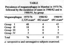
Mem Inst Oswaldo Cruz, Rio de Janeiro, Vol. 94,Suppl. I: pp. 329-330, 1999 Longitudinal Radiological Study of the Esophagus in Chagas Disease Cleudson CastroNúcleo de Medicina Tropical e Nutrição, Universidade de Brasília, Caixa Postal 4517, 70919-970 Brasília, DF, Brasil Fax : +55-61-273.2811. E-mail: tropical@unb.brReceived 9 June 1999 Code Number:OC99194 Key words: esophagus - Chagas disease - radiological study - X-ray Studies on the esophagus of patients with Chagas disease have traditionally been conducted using X-rays. The introduction of 70 mm radiography (Prata et al. 1967), for the study of the esophagus in endemic diseases opened new horizons for the use of X-rays in research on megaesophagus in large numbers of chagasic patients in endemic regions. Our group has been using this method since 1967 on populations in São Felipe and Caatinga do Moura (State of Bahia), Mambaí (State of Goiás), and Água Comprida (State of Minas Gerais). Blood tests for Chagas disease were conducted on 2,820 patients from Mambaí in 1975/76 by means of three different methods, by two reference laboratories, yielding positive results in 34.4% of cases. These subjects also underwent X-rays of the esophagus using 70 mm film: single radiographs of the esophagus were obtained immediately after ingestion of 75 ml of barium sulphate solution, and a second X-rays taken 1 min later, both with the patient in a right anterior oblique position. Interpretation of the X-rays was performed according to the criteria of Rezende et al. (1960), in different periods and under blind-test conditions. The first radiologic study in Mambaí was conducted in 1975/76, and included 2,329 patients aged 4 to 87, of which 1,145 were male and 1,184 female, being 1,006 seropositive (Castro et al. 1987). Seventy-six (3.2%) subjects presented with megaesophagus, of which 71 (7%) were seropositive and 5 (0.37%) seronegative. Of the 76 with megaesophagus, 47 (61.8%) were male and 29 (38.1%) female. According to the classification of Rezende et al. (1960), 48 (63.1%) were group I; 18 (23.7%) group II; 5 (6.6%) group III; and 5 (6.6%) group IV. Among those with chagasic infection, the prevalence of megaesophagus increased with the age of the patients, especially past the age of 30, and attained 21.5% in subjects above 59. From 1980 to 1982, 558 patients were reexamined in Mambaí by means of esophagus x-rays (Castro et al. 1992). Including only patients tested in both periods (1975/76 and 1980/82; average six years of study), and discarding illegible films, 494 patients aged 4 to 79 (average 24.7%) entered the study. Two hundred and twelve (43%) were seropositive. Of these, 201 presented with a normal esophagogram in 1975/76, of which four (2%) subsequently developed megaesophagus (all four were classified as group I). Among the eleven who had already presented megaesophagus in 1975/76, one group I patient and one group II patient progressed to group II and IV, respectively, which represents a 2.8% (6/212) rate of megaesophagus progression among the seropositive population. This six-year progression study revealed an incidence of megaesophagus of 0.33% among the seropositive subset. Four patients with megaesophagus classified as group I in 1975/76 presented normal X-rays in 1980/82, suggesting an apparent remission of this esophageal condition. The X-rays of sixteen subjects _ of which 75% were seropositive _ bordered on normal. In 1988/91, esophagus X-rays were performed on 1,098 subjects (Castro et al. 1994). Cataloging the subjects who did previous X-rays and discarding illegible films, remained 731 cases, among which 382 (52.3%) were seropositive. Of these, 223 had been tested in 1980/82. The average time of study was 13.2 years (1975/76 to 1988/91). Male subjects accounted for 350 (47.9%) and female subjects, 381 (52.1%). The incidence of megaesophagus in this population was 7.9% (55/691); 14.2% (49/345) among seropositive and 1.8% (6/337) among seronegative. The progression of megaesophagus among seropositive patients was 21.7% (35/161) in male and 16.6% (34/204) in female subjects. This study shows a 1.1% yearly incidence of megaesophagus among seropositive subjects. These data suggest that the rate of megaesophagus development increases with time of infection. In this study, approximately 46% of the megaesophagus cases analyzed remained stable, making it impossible to predict which patients will suffer disease progression. Three subjects who had received specific treatment developed megaesophagus. Despite the fact that the municipality of Mambaí was sprayed in 1980 and has been under surveillance ever since, those patients who were already infected at that time continue to develop megaesophagus. Table shows the prevalence of megaesophagus in Mambaí in 1975/76, followed by the incidence of cases in the 1980/82 and 1988/91 periods. REFERENCES Castro C, Macêdo V, Rezende JM, Prata A 1992. Estudo radiológico longitudinal do esôfago, em área endêmica de doença de Chagas, em um período de seis anos. Rev Soc Bras Med Trop 25: 225-230. Castro C, Macêdo V, Rezende JM, Prata A 1994. Estudo radiológico longitudinal do esôfago, em área endêmica de doença de Chagas, em um período de 13 anos. Rev Soc Bras Med Trop 27: 227-233. Castro C, Rezende JM, Camargo M, Prata A, Macêdo V 1987. Prevalência da esofagopatia chagásica no município de Mambaí, Goiás - Brasil. Rev Soc Bras Med Trop 20: 13-17. Prata A, Almeida F, Macêdo V 1967. Estudo comparativo sobre o esvaziamento do esôfago pela abreugrafia entre uma área endêmica de doença de Chagas e outra de esquistossomose, p. 39. III Congresso da Sociedade Brasileira de Medicina Tropical, Salvador, BA. Rezende JM, Lauar KM, Oliveira AR 1960. Aspectos clínicos e radiológicos da aperistálse do esôfago. Rev Bras Gastroenterol 12: 247-262. Copyright 1999 Fundacao Oswaldo Cruz - Fiocruz The following images related to this document are available:Photo images[oc99194a.jpg] |
| |||||||||
