 |
| About Bioline | All Journals | Testimonials | Membership | News |
|
||||||
|
||||||
Indian Journal of Occupational and Environmental Medicine, Vol. 9, No. 3, September-December, 2005, pp. 118-123 Original Article Toxic effect of lead on human spermatozoa: A study among pigment factory workers Naha N, Chowdhury AR1 Department of
Physiology, College of
Medical Sciences,
Chitwan, Nepal, India;
1Industrial Toxicology
Division, Regional
Occupational Health
Center (E), (ICMR),
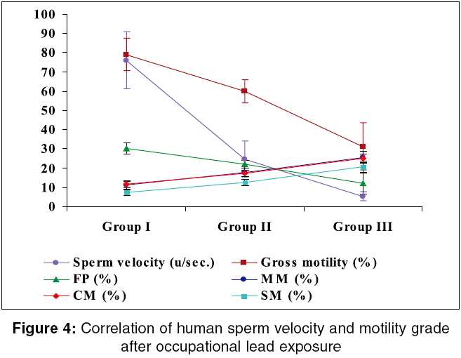
Kolkata, India Code Number: oe05027 Abstract Occupational lead exposure caused male reproductive impairment, but information on spermatozoa activity, motility, and maturation is limited. In the above perspective, spermatozoa morphology, motility, activity, and nutritional status in lead exposed workers (7-15 years exposure) were assessed. Low sperm velocity, gross, and forward progressive motility with high stationary motile spermatozoa revealed lowering of sperm cell activity after exposure (P < 0.001), which was supported by higher seminal fructose and reduced sperm ATPase activity. Lowering of seminal plasma total protein with concomitant increase in free amino acid was prevalent as exposure increased (P < 0.001), suggesting disturbance in cellular nutritional status. Prolonged liquefaction time, reduced semen volume, viscosity, seminal plasma protein, fructose, and cholesterol level among workers indicated accessory sex gland dysfunction after occupational lead exposure (P < 0.001). Deterioration of sperm density and morphology was associated with high blood and semen lead of workers (P < 0.001) leading to infertility without altering FSH, LH, and testosterone level.Keywords: Blood lead, Lead exposed workers, Semen biochemistry, Semen lead, Sperm morphology Introduction The diminution of semen quality due to occupational exposure of heavy metals is a major health concern in the globe.[1],[2],[3]Lead exposure and moderate lead absorption produces alteration in fertility with decreased production in spermatozoa in the battery factory workers probably due to the direct toxic effect of lead on germinal epithelium of testis during spermatogenesis. [4],[5],[6]Sperm velocity and morphology were necessary for fertility potential of the persons [7] and significant reduction in total motile sperm proportion, forward progression, and sperm kinetics as well as abnormality of sperm structures was predominant after exposure.[8],[9] Blood lead level was also inversely correlated with sperm count and viability.[9] Reduction in sperm motility, count, density, and low antioxidant profile along with increase incidence of sperm abnormality and sperm membrane lipid per oxidation was prevalent after occupational lead exposure.[10],[11] The positive correlation between heavy metal lead and cadmium in the seminal plasma of oligo, astheno, teratospermia group was found by Kasperczyk et al.[12] The gradual decline of semen cholesterol with decreasing sperm count was also evidenced from the earlier study.[13] Lowering of seminal plasma fructose was observed in normal subjects when compared with azoospermic patients as reported by Srivastava et al.[14], but Videla et al. reported contradictory result in case of normal, azoo, and oligo spermic subjects, suggesting the failure of germinal line of testis were possible only when the infection or androgen deficiency was prevalent.[15] Interference of inorganic lead to the hypothalamic-pituitary-gonadal axis, semen characteristics, and accessory gland function was prevailed among the working population[16],[17],[18], while some other reports revealed significant decrease in human semen quality after lead exposure without altering the reproductive hormone level. [19],[20],[21] However, very little information is available on sperm motility, activity, and maturation in case of lead exposed factory workers. Therefore, the present study was carried out to compare the morphology, nutritional status, motility, and activity of spermatozoa between lead exposed pigment factory workers and nonoccupationally exposed control subjects. Materials and methods The study was conducted in lead based pigments factories in Kolkata. Prior to study, the clearance was obtained from the ethical committee (ICMR, Government of India). In this cross-sectional study 50 nonoccupationally exposed control subjects (group I) and 95 exposed workers of active reproductive age, 55±5 kg weight and 160±5 cm height were randomly selected. They were divided into two groups depending on duration of exposure: (1) low exposed group with 7-10 years exposure for 8 h/day (Group II: n = 30) and (2) high exposed group for 8 h/day exposure over a period of more than 10-15 years (Group III: n = 50). Using interview technique as a tool for data collection, detail information of the subjects were recorded on the predesigned proforma that includes age, educational level, socio-economic status, working schedule, duration of exposure, use of protective devices, smoking, and other addiction history, marital status, and number of children, use of contraceptive devices, history of disease of the individual subjects, and his family. Semen samples were collected from the subjects of the three groups in a clean, dry, sterilized, wide mouth, well stopper glass vial by masturbation after 2-5 days of abstinence.[22] One part of semen was stored at -20°C in lead free storage vial for lead content analysis. About 2 ml of morning, fasting, venous blood was also stored at -20°C in lead free EDTA vial for metal analysis. Physical characteristics of semen was analyzed after liquefaction of the sample.[23] Sperm density, motility with four different grades, sperm head morphology, and corresponding morphometry was measured at 400 ´ magnification (Model: CH20i, Olympus, India).[22] Sperm velocity track was determined following the standard laboratory technique[24] using nomogram.[25] After liquefaction, whole semen was centrifuged at 800 g for 10 min and seminal plasma was separated. Seminal plasma total protein[26], fructose[27], free amino acid[28], and cholesterol[29] were measured spectrophotometrically (model: DU 64, Beckman spectrophotometer, USA). Serum level of FSH[30], LH[31], and testosterone[32] were assayed following the standard protocol as supplied through the respective ELISA kits. Lead was estimated in whole blood and semen of the subjects of the three groups at 283.3 nm using atomic absorption spectrophotometer (model: GBC AVANTA AAS, software Version 1.33, Australia) attached with GF 3000 graphite furnace.[33] Hormone assay and metal analysis are essential for exploring the in-depth mechanism of action of lead on spermatozoa of the exposed workers. The data obtained from control and exposed groups were compared and one-way anova and Scheffe′s F -test were carried out for level of significance following the computer based statistical software SPSS, Version 10.0 for Windows (SPSS Inc. USA).Results [Table - 1] showed the distribution of subjects according to the questionnaire. Majority of the subjects of groups I (18.75%), II (19.35%), and III (16.12%) were in the age of 35 years. The mean age for group I was found to be 36.13±1.06 years, while that for group II was 36.61±0.95 years, and 37.16±1.05 years for group III. All the subjects belonged to lower strata (100%) according to modified Kuppuswamy′s socio-economic classification.[34] There was no reproductive disease history among the working and control population, but infertility was significantly high among the working population (40%, n = 38). Smoking, alcohol consumption, and use of gutkha/panparag as well as minor electric shock during working were predominant among the battery factory workers of groups II and III in comparison with the matched control subjects (group I). [Table - 2] described the semen profile of the subjects according to extent of occupational lead exposure after adjusting for the confounding factors namely age, socio-economic status, duration of exposure, smoking habits, use of alcohol, and gutkha/panparag as well as absteinence period. Seminal viscosity was significantly decreased in-group III compared with group I ( P < 0.001), but not in-group II. Prolonged liquefaction, reduced semen volume, and sperm density was predominant in both the exposed groups with respect to control and in between the two exposed groups ( P < 0.001). Control and low exposed subjects were normospermic and high exposed subjects were hypospermic and oligospermic in nature as per the reference value.[23] Seminal cholesterol and fructose content were varied significantly ( P < 0.001) after exposure and seminal plasma total protein and free amino acid exhibited reciprocal relationship ( P < 0.001). Seminal plasma total protein was decreased by 64% in group II and 89% in group III with concomitant 2.2-fold rise of free amino in group II and 4.3-fold rise in group III workers with respect to group I control subjects. About 2.9-fold increase of seminal fructose in-group II and 6-fold increase in-group III was also observed. Gross morphological abnormality of spermatozoa was significantly higher in both the exposed groups with respect control ( P < 0.001). Although abnormality of low exposed group was slightly higher than the reference value, but both the low exposed group (44.54 ± 0.57) and high exposed group (60.04 ± 1.53) was teratospermic in nature and the control group value (33.75 ± 1.09) was within the normal range.[23] The present study also showed that total sperm head, mid piece, and tail abnormalities was increased significantly ( P < 0.001) after exposure. The classification of human spermatozoa according to head abnormalities and its relation to the occupational lead exposure was shown in [Figure - 1]. Significant increase of double head spermatozoa between groups I and III ( P < 0.01) and between groups II and III ( P < 0.001), taper head spermatozoa between groups I and II and between groups I and III ( P < 0.001) as well as acrosome defected spermatozoa between groups I and III ( P < 0.01) were predominant after occupational lead exposure. Spermatozoa with amorphous head were significantly increased among the three experimental groups ( P < 0.02: between groups I and II; P < 0.001: between groups I and III; P < 0.001: between groups II and III), whereas normal sperm head percentage was decreased significantly ( P < 0.001) of the same comparable groups. Sperm head morphometry (six subtypes) of lead exposed workers (groups II and III) and control subjects (group I) were varied nonsignificantly ( P < 0.05) as shown in [Figure - 2]. Sperm velocity was significantly decreased in groups II and III subjects when compared with group I and also in between the two exposed groups ( P < 0.001). The decrement ratio of sperm velocity in groups I (76.01±3.36), II (24.57±2.15), and III (5.31±0.52) was about 15: 5: 1. The sperm velocity of control subjects was considered as ′velocity group I category′as per the nomogram[25], whereas low exposed subjects represented the ′velocity groups II and III category′, and ′velocity groups IV and V category′ was restricted for high exposed subjects. In our study sperm velocity was decreased by 25% in-group III than in group II with respect to group I. [Figure - 3] represented the sperm velocity track of 20 individuals from the three experimental groups and simultaneously the velocity category of each study group. The present study exhibited a correlation between sperm velocity and motility grade after occupational lead exposure, where sperm velocity, gross, and forward progressive (FP) motility varied proportionately ( P < 0.001) and stationary motile (SM), moderate motile (MM) and circular motile (CM) spermatozoa established the inverse relationship ( P < 0.001) in respect of duration of lead exposure [Figure - 4]. Gross sperm motility and FP was decreased by 37% and 33%, respectively, in group III than in group II with respect to group I, while SM was increased by 1.1-fold of the same comparable group. The decrement ratio of gross and FP motility in groups I-III was about 2.6: 1.9: 1 and 2.5: 1.8: 1, respectively, whereas the increment ratio of SM spermatozoa of the same comparable groups was approximately 1: 1.9: 3. The increment ratios for MM (1: 1.6:2.3) and CM (1: 1.4: 2.1) spermatozoa were more or less same. This study also showed that gross sperm motility and FP of all the three groups was much lower than the respective reference values.[23] [Table - 3] represented the hormonal profile of the subjects after occupational lead exposure. Although serum FSH, LH, and testosterone level was decreased nonsignificantly ( P < 0.05) in the three study groups, but all the values were within the reference range as per the ELISA kits. [30],[31],[32] [Table - 4] described the body burden of lead according to extent of exposure at work place. Lead concentration in whole blood and semen was increased significantly in both the exposed groups and also between the two exposed groups ( P < 0.001). The increment of blood lead was 1.9-fold in group II and 3.9-fold in group III with respect to group I, whereas 2.7-fold, and 4.6-fold were the increment of semen lead of the two exposed groups (groups II and III), respectively. Discussion The present study showed deterioration in sperm density and motility in the lead exposed pigment factory workers with high prevalence of sperm head abnormality in comparison with the nonoccupationally exposed matched control subjects of same socio-economic status. This observation was in corroboration with the earlier observations of Roy Chowdhury et al.[35],[36] These pigment factory workers exposed to lead fumes and dust during their work, which caused adverse effects on sperm morphology and density.[3],[4],[6] Morphological abnormalities of spermatozoa were also depended on the duration and nature of exposure.[2],[19] Fructose is the main energy source for spermatozoa motility [37] and sperm velocity is the average velocity of all spermatozoa in one sample,[23] therefore, both are related with sperm motility grade leading to sperm activity. Lowering of sperm velocity, gross motility, and FP with concomitant rise of SM were prevalent in the lead exposed pigment factory workers of high seminal plasma fructose level, which indicated retarded sperm activity might be due to lead induced alteration of normal fructolysis. This observation showed similarities with the earlier findings. [8],[9],[10]The present finding was also in corroboration with the previous observation by showing diminution of sperm ATPase activity after occupational lead exposure leading to low sperm motility.[35] As increase amount of free amino acid depends on the degradation of protein present in the system,[28] therefore, lowering of seminal plasma total protein with concomitant rise of free amino acid in pigment factory workers with respect to control subjects indicated that lead may alter the protein metabolism and subsequently increase the amino acid level, suggesting disturbance in cellular nutritional status, necessary for cell survival, and proper function. Low volume, viscosity, prolonged liquefaction time, and deviation of the seminal fructose, cholesterol, and protein content among the lead exposed pigment factory workers in the present study indicated the alteration of normal secretary activity of seminal vesicle and prostate after occupational lead exposure. Previous study reported that 95% of ejaculatory volume comes from these two accessory sex glands and alteration of normal liquefaction associated with low viscosity, indicating deficiency of liquefying agent due to low prostatic secretary activity.[23] Thus the present study suggesting probable dysfunction of accessory gland seminal vesicle and prostate after toxic insult of lead at work place. The present study showed significant decline in human sperm quality and fertility (40% workers have fertility problem) without affecting the serum FSH, LH, and testosterone level, which are in corroboration with the previous finding, [19],[20],[21] but at the same time contradictory to others results. [16],[17],[18] In our study deterioration of sperm quality and fertility potential of the subjects were associated with high lead concentration in whole blood and semen of the workers in respect of duration of exposure. Xuezhi showed that lead exposure caused prolonged liquefaction time, low volume, sperm count, viability, and retarded sperm activity in male workers with high blood lead level,[38] which is similar to the observation of Apostoli et al.[1] and Kasperczyk et al.[12] Semen lead was higher in the infertile men than the fertile group and low semen lead level was the indicator of low industrial exposure.[39] Moderate lead exposure also caused reduction in sperm characteristics among the factory workers.[17] Therefore, in conclusion it can be pointed out that lead may cross the blood testis barrier and affect spermatozoa of the exposed population leading to accessory gland dysfunction, morphological abnormality, high seminal fructose in oligospermic exposed workers, [14],[15] disturbance in cellular nutritional status, and energy-dependant processes like sperm production and motility leading to reduced sperm count, retarded sperm activity, and infertility. Smoking habits, addiction to alcohol, gutkha, etc. may be the added factors in lead toxicity,[40] whereas minor electric shock, typhoid, gastritis, pox, and the age of the subjects did not show any correlation after exposure.[41] Acknowledgment We acknowledge DST (Government of West Bengal) for financial support and ROHC (E), ICMR, Kolkata for infra structural facilities. The authors are grateful to Dr. B. Manna, NICED (ICMR), Kolkata for his contribution on statistical analysis of data. Thanks are due to all the donors without whose cooperation the study would have been completed.References
Copyright 2005 - Indian Journal of Occupational and Environmental Medicine The following images related to this document are available:Photo images[oe05027t2.jpg] [oe05027f4.jpg] [oe05027f3.jpg] [oe05027t3.jpg] [oe05027t1.jpg] [oe05027f2.jpg] [oe05027t4.jpg] [oe05027f1.jpg] |
| |||||||||
