 |
| About Bioline | All Journals | Testimonials | Membership | News |
|
||||||
|
||||||
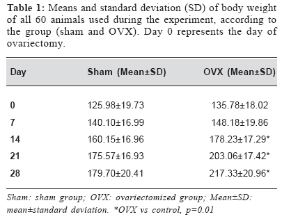
Brazilian Journal of Oral Sciences, Vol. 3, No. 8, Jan/Mar. 2004, pp. 425-427 Conventional X-ray densitometry detects osteopenia in ovariectomized young rats. Short communication Karina Gottardello Zecchin 1 Michele Conceição Pereira 1 Pablo Agustin Vargas 1 Edgard Graner 1 Jacks Jorge Júnior 1 1Department of Oral Pathology, Piracicaba Dental School - UNICAMP - São Paulo, Brazil Correspondence to: Jacks Jorge Júnior Departamento de Patologia Oral Faculdade de Odontologia de Piracicaba Av. Limeira, 901 13414-018 Piracicaba SP Phone 0055.19.34125317 Fax 0055.19.34125218 email -jjorge@fop.unicamp.br Received for publication: December 5, 2003 Code Number: os04009 Abstract The present study investigated the reduction on bone density 4 weeks after ovariectomy in rats, with conventional X-ray densitometry. Eighty female Wistar rats underwent bilateral ovariectomy (OVX group) or sham operation (sham group) under general anesthesia. Animals were killed by cervical dislocation 4 weeks after surgery. The left tibia of each animal was dissected and radiographed using oclusal films. Radiographs were scanned and virtual squares on the proximal tibial metaphysis were analyzed with proper software. Higher OD values represent darker areas in the X-ray. After that the tibia were decalcified with EDTA and serial transversal sections with 6 µm of the mesial root of the first mandibular molar were stained with hematoxylin-eosin. Digital images were captured and the densitometric volume of bone was evaluated using software. A significant increase of dark areas in the radiographies of OVX animals was observed when compared with control group (control=1.136±0.020 vs OVX=1.269±0.027, t test, p=0.01). Histomorphometric analysis showed a significant reduction on bone density of OVX animals (control=125.8±20.5 vs OVX=65.4±0.0154, t test, p=0.01). Conventional X-ray densitometry is useful for the characterization of osteopenia in rats after ovariectomy. Besides, 4 weeks are sufficient to cause significant decrease on bone content after ovariectomy. Key Words: osteoporosis, bone densitometry, animal models, rat.
Recently, during experiments with osteoporosis in young female rats, we found an intense and significant reduction of bone density four weeks after ovariectomy using conventional X-ray densitometry. Similar reduction has been reported after 3 to 4 months of oestrogen suppression in animals1,2 and a decade after the menopause in women3. A few papers have previously showed that osteoporosis can be detected 5 weeks after ovariectomy4, but none in young female rats and all used relatively sophisticated methods such as single photon absorptiometry5, and dual-energy X-ray absorptiometry (DEXA)6. This research was approved by the Ethics Committee of Experimentation in Animals of Campinas University - UNICAMP. Eighty female rats (Ratus norvegicus albinus, Wistar), 4 weeks old and weighing 100g in average underwent bilateral ovariectomy (OVX group) or sham operation (sham group) under general anesthesia with intramuscular injection of 2% tiazine (Rompun® - Bayer, Brazil, 5mg/kg, intramuscular) and 10% ketamine (Dopalen® - Agribands, Brazil, 10mg/kg, intramuscular). All animals were housed four per cage with 12 hours day-night light conditions at 21ºC, and were feed ad libitum and weighed weekly. Animals were killed by cervical dislocation 4 weeks after surgery. The left tibia of each animal was dissected carefully, fixed with 10% buffered formalin for 24h and radiographed using oclusal films (Insight I0-41®, Kodak, EUA) with 65KVA, 0.5s of exposition, and 21cm of distance focus-film. A 1mm-thickness aluminum step was used to control variability. Films were individually marked and developed in an automatic processing machine (Level 360® J Morita, Japan) at 28oC with fresh solutions. Radiographs were scanned (GS 700® Bio-Rad - Hercules, USA) and analyzed with proper software (Molecular Analyst, V 1.5® - Hercules, USA). Measurements used a virtual 1.806mm2 square on the proximal tibial metaphysis. The ratio of the tibia's optical density (OD) by the step's OD was used for the statistical analysis. Higher OD values represent darker areas in the X-ray. After that the tibia were decalcified with EDTA and serial transversal sections with six µm (lingual-buccal direction) were stained with hematoxylin-eosin. Digital images were captured and the densitometric volume of bone was evaluated using software (KS 400® Karl Zeiss, Germany). Three hundred points were counted on the proximal tibial metaphysis using a 10x ocular kpl with a 100 points reticule of integration II Zeiss, and a 10x objective. Efficiency of the ovariectomy procedure were confirmed by the increased body weight (Table 1), absence of proestrus and estrus phases on the estrous cycle on the 21o day after ooforectomy7 and verification of uterus atrophy at sacrifice day. X-ray densitometric analysis of bone trabeculae on the proximal tibial metaphysis showed a significant increase of dark areas in the radiographies of OVX animals when compared with control group (control=1.136±0.020 vs OVX=1.269±0.027, t test, p=0.01), while histomorphometric analysis showed a significant reduction on bone density of OVX animals when compared with control group (control=125.8±20.5 vs OVX=65.4±0.0154, t test, p=0.01). These results strongly indicate that four weeks after OVX the animals present a great degree of bone loss. Decreased bone density is only detectable in women one year after excision of ovaries. However, in rats the bone turnover is 3 to 5 times faster than in humans8 and osteoporosis can be seen earlier. Our data shows that young female rats have clear signs of osteopenia 4 weeks after ovariectomy, as observed by decreased bone density in X-rays and confirmed by histomorphometric analysis. Such precocious evidence may allow significant reduction of follow up time after surgery in research protocols for osteopenia in by estrogen deficiency. Additionally, such alterations can be detected by conventional X-ray densitometry. DEXA is the best resource to evaluate bone densitometry, but it is expensive and not available in all research centers. The use of a less expensive, still efficient, method of measuring osteopenia in experimental animals may help reducing costs in osteoporosis research. References
Copyright 2004 - Piracicaba Dental School - UNICAMP São Paulo - Brazil |
