 |
| About Bioline | All Journals | Testimonials | Membership | News |
|
||||||
|
||||||
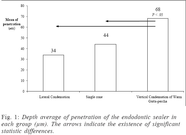
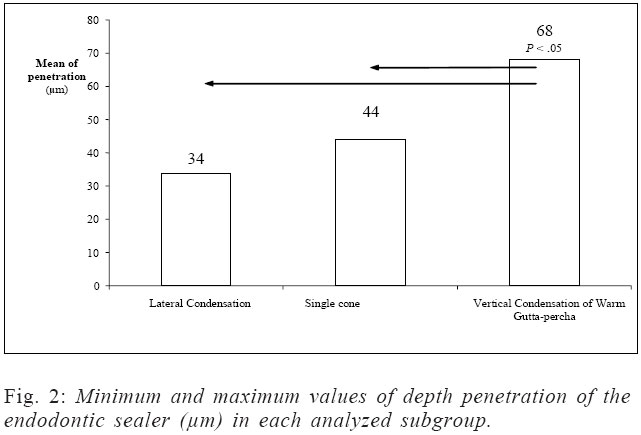
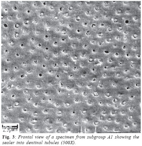
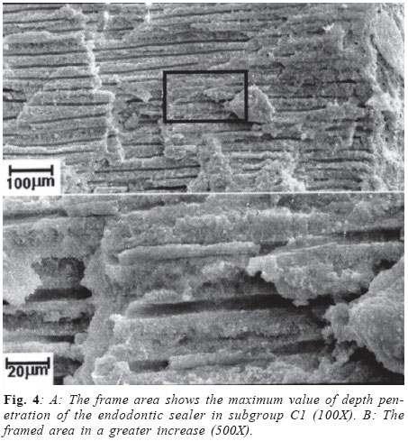
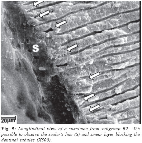
Brazilian Journal of Oral Sciences, Vol. 3, No. 9, April/June 2004, pp. 433-438 Influence of the filling technique on depth of tubular penetration of root canal sealer: a scanning eletron microscopy study Gustavo De-Deus1 Eduardo Diogo Gurgel-Filho2 Cláudio Maniglia-Ferreira2 Tauby Coutinho-Filho1 1 Department of Endodontics, Rio de Janeiro State University - UERJ, Brazil. 2 Department of Endodontics, University of Fortaleza, UNIFOR, Brazil. Correspondence to: Gustavo André De Deus Carneiro Vianna R. Afonso Pena, 33, ap.103 - Tijuca Rio de Janeiro - RJ ZIP CODE: 20270-240 e-mail: endogus@terra.com.br Received for publication: July 10, 2003 Code Number: os04011 Abstract The purpose of this study was to compare the depth of tubular dentinal penetration of sealer in three filling techniques. Seventy two teeth maxillary central incisors were instrumented and randomly divided in three groups A, B and C and obturated as following: A: lateral condensation; B: single cone technique and C: warm vertical compaction of guttapercha. Each sample was sectioned longitudinally and prepared for SEM analysis. The results revealed a depth of tubular penetration varying from 19 to 81mm, presenting an average post of 34 ± 16mm for G1; 26 to 101mm, presenting an average post of 44 ± 28mm for G2 and 22 to 188mm, presenting an average post of 67 ± 37mm for G3. A non-parametric ANOVA Kruskall-Wallis was used to determine whether there were significant differences among the groups, which identified significant differences between G3 and G1 (P = 0.021) and between G3 and G2 (P = 0.009). There were no significant differences between G1 and G2 (P> .05). The samples filled by warm vertical compaction of gutta-percha presented significantly deeper tubular sealer penetration than lateral condensation and single cone techniques. Key Words: filling technique, tubular penetration, root canal sealer. Introduction Most of the studies have been describing that microleakage usually happens from apical foramen to cervical root canal third and influences negatively on success of endodontic therapy1. It has been described that apical microleakage can take place in the interfaces between sealer and the dentin, sealer and gutta-percha, among the endodontic sealer itself or by its dissolution2. Although endodontic sealers continue to represent an important role for the control of microleakage, as they drain to the ramifications and improve adaptation of the filling to the irregularities of the interface between dentinal walls and gutta-percha3-4. Mc Comb and Smith5 predicted this increase of scientific interest in filling materials possessing affinity for dentin, or in other words, good adhesiveness. This characteristic could be able to increase filling efficiency. Penetration of root canal sealers into the dentinal tubules could improve sealing of the root canal system by increasing the surface area contact of filling materials to dentinal walls6. That theory justifies the scientific interest about the potential of dentinal tubule penetration of filling materials. Future trend idealizes these materials filling out dentinal tubules, binding intimately to organic and inorganic interfaces of dentin, destroying or neutralizing microorganisms and their by-products, inducing cement neoformation and strengthening root canal system. Inside this foresight, all currently sealers in use may be considered inadequate7. Many studies have analyzed adaptation of filling materials to dentinal walls2,8-9. Absence of the smeared layer enables consistently, root canal sealer penetration into dentinal tubules. The influence of smear layer on depth of penetration of three endodontic calcium hydroxide sealers into dentinal tubules was analyzed by Kouvas et al.10. The presence of smear layer in the dentinal walls impended penetration of the sealers in the control group. Such tubular penetration increases the interface between filling material and dentinal walls. This, in terms, may improve prevention of apical microleakage by filling techniques2,6. Furthermore, retention of filling material might be improved by this mechanical locking1. The purpose of this study was to compare the depth of tubular dentinal penetration of sealer using three filling techniques. Material and MethodsFor the present work seventy two teeth maxillary central incisors were selected from the tooth bank of Rio de Janeiro State University. The teeth were stored in 10% neutral formalin. Standard access cavities were made and the patency of each canal was confirmed by inserting a size #20 file through the apical foramen before and after completion of the root canal preparation. The working length was determined subtracted 1-mm from the apical foramen. The canals were shaped manually using a crown-down technique and stainless steel Flexofiles® (Dentsply-Maillefer, Ballaigues, Switzerland) and Gates Glidden burs (#6, #5, #4, #3). The coronal and middle third of each canal was preflared using Gates Glidden drills (Dentsply-Maillefer), sizes #6, #5, #4 and #3. The apical thirds were prepared with Flexofiles® (Dentsply-Maillefer), sizes #60, #55, #50 and #45 using a balanced force technique as described by Roane et al.11. The canals were irrigated between each file with 2mL of freshly prepared 5.25% solution of sodium hypochlorite using a syringe and 27-gauge needle. The teeth were randomly divided in three groups A, B and C and then subdivided in A1/A2, B1/B2 e C1/C2, with twelve specimens in each subgroup. In subgroups A1, B1 e C1 the teeth received a final flush of 10 mL of 17% EDTA (pH 7.7) for 3 minutes, followed by 10mL of 5.25% sodium hypochlorite to remove the smear layer12. In the subgroups A2, B2 e C2 the teeth did not receive a final flush with 17% EDTA. The canals were dried with paper points (Dentsply-Maillefer) and obturated as follows: A: Lateral Condensation Technique13; B: Lateral condensation with an accessory cone14 and C: Warm vertical compaction of gutta-percha15 associated with the Obtura II System (Obtura Corp., Fenton, MO, USA) in the backfilling phase. Pulp Canal Sealer EWT (Kerr, Sybron Dental Specialties, Romulus, MI, USA) was mixed manually according to the recommendations of the manufacturer and used for all groups. A size #40 file was used to pick up the measured spoon of sealer (1.25mL) one time from mixing pad and placed into the canal whilst rotating it counterclockwise16. In the lateral condensation group (A), a size #45 master guttapercha cone (Diadent Group International, Chongchong Buk Do, Korea) was coated with a measured spoon of sealer (1.25mL) and placed in the canal to the full working length. Lateral compaction was achieved in each canal by using accessory medium fine gutta-percha cones (Dentsply-Maillefer) and endodontic finger spreader size B (Dentsply-Maillefer). A heated instrument was used to remove the excess gutta-percha. In the single cone technique (B), the tip of a medium sized nonstandardized gutta-percha cone (Diadent Group International, Chongchong Buk Do, Korea) was trimmed back until tug-back was achieved (calibrated to a size #45) in the full working length. The trimmed gutta-percha cone was coated with a measured spoon of sealer (1.25mL). A heated instrument was used to remove the excess gutta-percha and then vertical force was applied with a plugger (1.0mm - Dentsply - Maillefer) to compact the gutta-percha in the coronal third of the canal. In the warm gutta-percha group (C), the tip of a medium sized non-standardized gutta-percha cone (Diadent Group International, Chongchong Buk Do, Korea) was trimmed back until tug-back was achieved in the full working length. The trimmed gutta-percha cone was coated with a measured spoon of sealer (1.25mL). At the level of cementum-enamel junction, the gutta-percha was scared off with the tip an activated heat carrier (Touch'n Heat - model 5004, Analytic Technology, Redmond, WA, USA). After deactivating the heat carrier, the cooling instrument was removed from the canal, bringing out an increment of gutta-percha. Vertical force was applied with a size 11 plugger (1.1mm diameter, Dentsply-Maillefer) to compact the gutta-percha in the coronal third of the canal. This procedure was repeated twice, first to a level 3-4mm deeper than cementum-enamel junction and vertically condensing the gutta-percha in the middle third of the canal using a size 7 plugger (0.7mm diameter, Dentsply-Maillefer), and secondly to the level 4mm short of the full working length and vertically condensing the guttapercha in the apical portion of the canal using a size 5 plugger (0.5mm diameter, Dentsply-Maillefer). Back-filling of the rest of the canal space was achieved by injecting warm guttapercha using Obtura II system (Obtura Corp., Fenton, MO, USA), each time injecting a 4-5-mm segment and condensing the gutta-percha with a prefitted plugger14,16. The teeth with apical root fillings were then stored in 100% humidity and at 37ºC for 2 weeks. After that, each sample was sectioned longitudinally using low-speed saw (Isomet, Buehler, Ltd. Lake Bluff, NY, USA) with a diamond disc (Æ 125mm x 0.35mm x 12.7mm - 330C) while constantly irrigated with water in order to prevent overheating. These sections were then placed in a small vial and dehydrated in series of 50-100% ethanol solutions for 10 minutes each. The specimens were then subjected to critical point drying (Critical point dryer, Seevac Inc, Florida, USA). After critical point drying the root sections were mounted on a labeled stub, and sputter coated with gold (Sputter Coater, Model s/ 50, Edwards, England). All sections were scanned by a Carl Zeiss 2500 scanning electron microscope (Carl Zeiss Vision Gmbh, Hallbergmoos, Germany). The penetration of filling material into dentinal tubules was examined and an assessment made as to whether the material in the dentinal tubules was sealer or smear plugs by comparing with the bulk of the sealer in the root canal. The measurements of the depth of the sealer penetration was made in the middle third of the root canal - at 6-mm from the apex - with increases range between X50 and X5000. For each sample, five measurements were made always in the deepest sealer penetration. The interface dentinal wall / filling material being always the observation focus. A statistic treatment was used to determine whether were significant differences among the groups. ResultsNo such tubular dentinal penetration was recorded in subgroups A2, B2 e C2. In the control samples were observed the smear layer occludes the dentinal tubules and thus blocked the penetration of the filling materials. Generally were observed with certain easiness sealer inside the dentinal tubules in experimental subgroups A1, B1 and C1. The measurements revealed a depth tubular penetration within a range of 19 to 81mm, showing an average post of 34 ± 16mm for G1 (lateral condensation). The measurements revealed a depth tubular penetration within a range of 26 to 101mm, showing an average post of 44 ± 28mm for G2 (single cone). The measurements revealed a depth tubular penetration within a range of 22 to 188mm, showing an average post of 67 ± 37mm for G3 (warm vertical compaction of gutta-percha). Average values of general measures of the depth tubular penetration are shown in Figure 1. The minimal and the maximum measures of the depth of tubular penetration in each group are showed in Figure 2. A multiple linear regression models (SPSS/PC + Statistics 4.0 software, SPDD International BV, Gorinchem, The Netherlands) were used and the data do not conform to a normal or Gaussian distribution. Since the data are not normally distributed a parametric statistical treatment of the raw data is inappropriate. A non-parametric ANOVA Kruskall-Wallis was used to determine whether were significant differences among the groups. A level of significance in all tests was set at P < .05. The statistical analysis identified significant differences between G3 and G1 (P = 0.021) and between G3 and G2 (P = 0.009). There were no significant differences between G1 and G2 (P >.05). Discussion It has been suggested that the quality of the apical seal may be improved by increasing the surface contact between the dentinal walls and sealer17. The findings obtained from both microleakage and SEM observation suggests that physical integrity of sealer matrix may also be important at providing resistance to leakage2. We can verify throughout the literature, an intensification of the interest for the study of some issues related to the interface between dentin and filling material, standing out among them, and dentinal tubule penetration of filling material1,8,10. Many researches evaluated the ability of endodontics sealers to penetrate into dentinal tubules by a scanning electron microscopic analysis2,8,10. The image created by the electron array from scanning electron microscope is able to show us a three-dimensional view, exhibiting not only the contour of the dentinal walls to be observed but also the endodontic sealer. Through this technique, all the surface of the dentinal wall can be scanned examined with rich details and the results can be characterized, measured and interpreted. Aiming better quality for the adaptation of filling material to dentinal walls, root canal sealers have been used as reinforcement for filling. In a previous research, Kerr Pulp Canal Sealer® demonstrated significantly less microleakage than the other sealers18. Since it is a sealer with decades of clinical and laboratory application, and also because it has been indicated by many as the ideal choice when techniques using some type of thermoplasticized procedure are used, Kerr Pulp Canal Sealer was the preferred sealer for this experiment7. A recently paper examined the depth penetration of Sealapex, Grossman's Sealer, AH Plus and Pulp Canal Sealer in the wave of condensation technique and conclude that Pulp Canal Sealer showed the maximum penetration depths into the dentinal tubules and Sealapex, the minimal depths penetration19. These finds reinforce the choice of Pulp Canal Sealer for this research. No such tubular dentinal penetration was recorded in subgroups A2, B2 e C2. This was expected as the smear layer occludes the dentinal tubules such as show in Figure 5. It was known that both gutta-percha and endodontic sealer possess the ability to penetrate dentinal tubules in the absence of smear layer4. This finding is closer to Kouvas et al.10, which studied the influence of smear layer on depth of penetration of three endodontic calcium hydroxide sealers (Sealapex, Roth 811 and CRCS) into dentinal tubules and concluded that the presence of smear layer in the dentinal walls impended penetration of the sealers in the control group. In this study, no such tubular dentinal penetration was recorded in subgroups A2, B2 e C2. This was expected as the smear layer occludes the dentinal tubules such as show in Figure 5. Another research found similar results with plastics filling materials1. They stated that the presence of smear layer prevent the entry of filling materials into dentinal tubules. The regime of irrigation with EDTA followed by NaOCl12 removed smear layer effectively. All experimental subgroups (A1, B1, and C1) showed tubular penetration to some extent. The effect of smear layer removal on dentinal-wall adaptation was done investigated in the literature9. Lateral condensation did not produce good adaptation to canal walls either with or without smear layer. Adaptation of both had surface stippling of the gutta-percha, which represented advancement of gutta-percha into dentinal tubules. Unlike lateral condensation, thermafil system produced a homogenous mass of gutta-percha in the canal15. This study demonstrated the deeper tubule penetration of sealer occur in samples filled by warm vertical condensation of guttapercha followed by lateral condensation with an accessory cone and finally by lateral condensation. With these data, we believe that filling technique may influence the ability of dentinal tubule penetration of filling material in accord with other studies20. This way, it was possible believe in a deeper tubule penetration of sealer by warm vertical condensation of gutta-percha techniques due to the thermoplasticized of gutta-percha. That process promotes better apical adaptation of filling material, decreasing this way, the apical and lateral microleakage indexes4. A non-parametric ANOVA test (Kruskall-Wallis) showed a significant differences between Vertical condensation of warm gutta-percha (C1) and cold lateral condensation techniques (A1 and B1). There are other factors that might be able to influence the capacity of dentinal tubule penetration of the endodontic sealer such as: the surface activity of the sealers, the contact angle between sealer and the dentinal walls, the diameter of the opened dentinal tubules and the employed obturation technique7. The humidity, temperature, content of dentinal tubules and also the existence of periapical and periodontal tissues may interfere the capacity of penetration of filling material into dentinal tissue21. The chemical and physical characteristics of root canal sealers might affect tubular penetration and adaptation of the material to dentinal walls following the removal smear layer20 The canal area evaluated was the middle third and it was easily observed that sealer penetration was deepest in this region6,21. This fact may be explained because the diameters of canal orifices at the middle third of the root are large enough in this area, so a clean surface results after treatment with chelating agent and because greater vertical forces may be applied in this area during root canal obturation. Normally, the literature related a numerical finding within a range of 35 to 80µm of depth of sealer penetration10,19-20. However, Sen et al.2 obtained measurements up to 800µm. That great difference of values can be due to some methodology deviation among those researches, mainly when consider the difficulty to get an appropriate cleaving, that might supplies us more homogeneous samplings as well as the instrumentation and filing techniques and the sealer used. The numeric results of our measurements were closer to those of Oksan et al.20 and Kouvas et al.10. The results of this work demonstrate that filling technique may influence the ability of dentinal tubule penetration of endodontic sealer. Vertical condensation of warm guttapercha showed a deeper tubule penetration than cold techniques. Future researches are necessary to evaluate, in a realist clinical approach the effect of tubular penetration of root canal sealer on the quality of the apical seal. AcknowledgementsThe authors thank the Department of Science and Materials Engineering (DCMM) of Pontifícia Catholic University (PUCRJ) for the essential technical assistance in this study, especially in memoriam to Engineer Maria de Fátima Lopes. References
Copyright 2004 - Piracicaba Dental School - UNICAMP São Paulo - Brazil |
