 |
| About Bioline | All Journals | Testimonials | Membership | News |
|
||||||
|
||||||
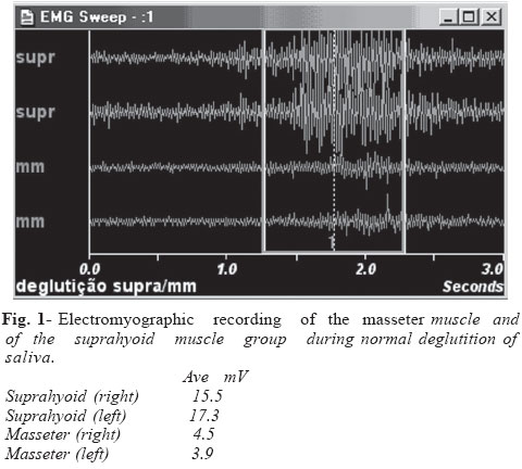
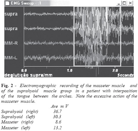
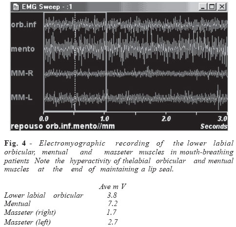
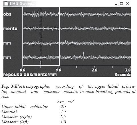
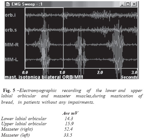
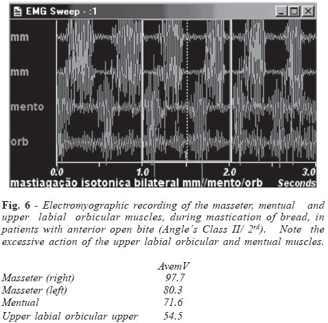
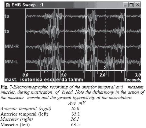
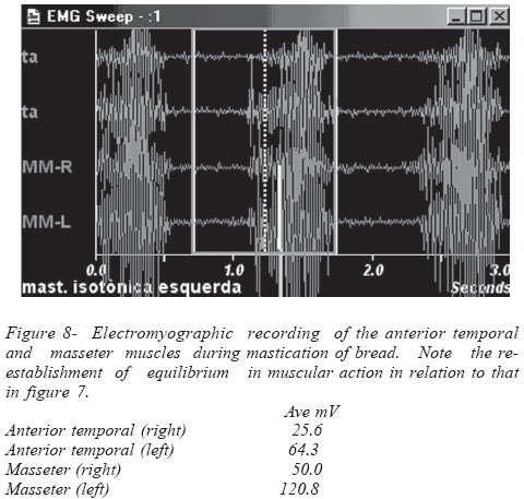
Brazilian Journal of Oral Sciences,Vol. 3, No. 10, July/September 2004, , pp. 506-509 Electromyography: applied in the phonoaudiology clinic Miriam Nagae1, Fausto Berzin2 1Language therapist, State University of Campinas, 2DDS, PhD, Professor of Piracicaba Dental School, State University of Campinas Correspondence to: Mirian Nagae, Universidade de Campinas- FOP/UNICAMP, Departamento de Morfologia, Av. Limeira, 901 Cep:13.414-903, Piracicaba- São Paulo- Brazil. Phone: +55 011 30321102, e-mail: mnagae@uol.com.br Received for publication: February 10, 2004 Code Number: os04025 Abstract The aim of this study was to discuss the utilization of surface electromyography in the practice of phonoaudiology to obtain a picture of the muscular compromise that occurs in patients with dental and skeletal alterations. The behavior of muscles in functions such as swallowing, chewing and respiration, and at rest, and possible deviations thereof, will be examined by electromyography based on the activated muscular chains. Compromises such as tension, flaccidity, palsy and rhythm could be detected quantitatively and qualitatively during the functions. Key Words: eletromyography, language therapist, TMD Introduction Phonoaudiology covers a vast area of clinical interests, among them oral motricity, which is basically at the center of impairments of muscle order. Knowledge of the condition of the musculature is of utmost importance in these cases. The tools that have been utilized to date for the handling of these cases are: palpation, visual inspection and the opinion of a professional. However, such an examination is rather subjective in character. With advances in technology, a series of instruments have permitted a clearer and more objective view of the actualcondition of the musculature. In the phonoaudiology clinic, electromyography, which records the electrical properties of muscles, has thereby been a valued tool1. To perform an electromyographic study, a professional needs a prior technical understanding in the handling of instruments as well as a profound knowledge of the anatomy and physiology of the musculature to be investigated. The choice of musculature would depend on the situation and the interest of the examiner. In phonoaudiology, the major focus is in the region of the head, chest and face (muscles: temporal , masseter, suprahyoid group, mentual, and labial orbicular). The examination could help in the diagnostics, treatment and even the prognostics of cases. Regarding its diagnostic value, it would be possible to determine for individual as well as groups of muscles both normal patterns and possible imbalances found in the musculature, such as in the following cases. Electromyography registration a) During deglutition (figure1), the signal of the suprahyoid muscle group is captured, which allows for a better visualization of the moment of deglutition, because of it very near to the musculature of the tongue. In cases of atypical deglutition, the anterior interposition of the tongue between the arches is common. This condition is found for example in subjects with an anterior open bite (Angle´s Class II/1rd division), where the masseter muscle exerts an intense pressure concomitant with the action of the suprahyoid muscle group (figure 2). b) In the mouth breather, we can observe that due to the flaccidity caused by the mouth open, the labial orbicular musculature needs to recruit many motor units to be able to maintain the lip seal (figure 4). It is interesting to see that after the patient is able to make nose breathing automatic and/or after myotherapeutic intervention, the muscles re-establish their normal pattern in the resting state (figure 3). c) During mastication, there is a significant action of the elevating muscles of the mandible, mainly the masseter muscle and little involvement of the perioral muscles (figure 5). When there is some alteration at the skeletal level, in Angle´s Class II/2rd cases for example, we can see that the musculature also undergoes a change in its pattern of activation, as can be observed in figure 6, where the mentual and labial orbicular muscles become significantly active. Regarding treatment, electromyography can be used to monitor development, but also as a therapeutic tool via biofeedback2, where patients learn to control their own neuromuscular responses. This resource is very interesting mainly in relapse cases after orthognathic surgery or orthodontic treatment, which could be caused by a muscular memory3 that does not permit a new pattern of muscle behavior to be established. In cases where there is pain or excessive tension in the mastication muscles produced by parafunction, the capability of monitoring the muscles is also very interesting. In relation to prognostics, electromyography permits us to determine the intensity of impairments (figure 7) and to obtain at the end of treatment quantitative evidence of the modifications that occurred (figure 8). Usually, an evaluation based on size and sensitivity of the musculature is not always possible. Phonoaudiology is capable of carrying out electromyography at least for the purpose of investigating the potential action of motor units by means of electrodes placed on the skin surface In the phonoaudiology clinic, the electromyographic interpretation is performed by means of a raw signal, which permits us to visualize qualitatively the size and shape of muscle action potentials. When there is an interest developed by more specific situations or for research, there is a need for prior handling of the signal, termed normalization4, for the purpose of obtaining a common language to be able to compare results. Electromyographic kinesiology is the procedure most indicated in such cases, since it allows us to study muscular activity for both individual and groups of muscles in various conditions, and to obtain a quantitative evaluation of the signal5. In phonoaudiology investigations, this has been achieved following this methodology6. In the phonoaudiology clinic, implementing the normalization of the signal in the future is inevitable. However, it is import to remember that this type of methodology is only intended for the quantitative investigation of isolated aspects (fatigue, stress, amplitude), and it is not sufficient for a more globalized evaluation necessary for the clinic. The raw signal in this aspect provides us with a more qualitative view, where nuances in shape, size and time of the recordings could furnish revealing information for a better understanding of cases. The ability to perform electromyographs in the clinic on a daily basis improved significantly the quality of all aspects, mainly with regard to the understanding of the causes of the imbalances encountered that will inevitably produce more rapid and accurate results. Currently, we are going through a very interesting phase, because due to better access to technology and interdisciplinary exchange of professionals such as physiotherapists, dentists and clinicians, we are getting closer to a sophistication that soon will permit us to see the various physiological modifications occurring in musculature, using such tests as electromyography. References
Copyright 2004 - Piracicaba Dental School - UNICAMP São Paulo - Brazil The following images related to this document are available:Photo images[os04025f5.jpg] [os04025f3.jpg] [os04025f6.jpg] [os04025f2.jpg] [os04025f7.jpg] [os04025f8.jpg] [os04025f1.jpg] [os04025f4.jpg] |
| |||||||||
