 |
| About Bioline | All Journals | Testimonials | Membership | News |
|
||||||
|
||||||
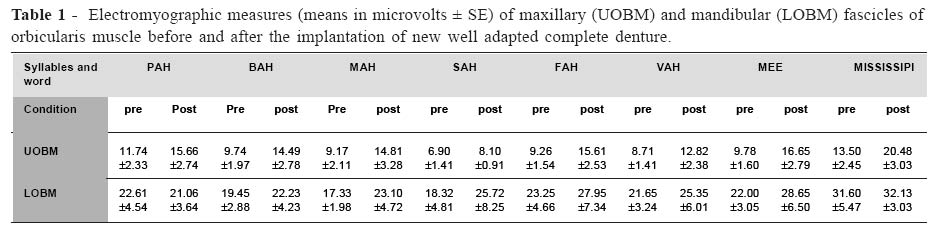
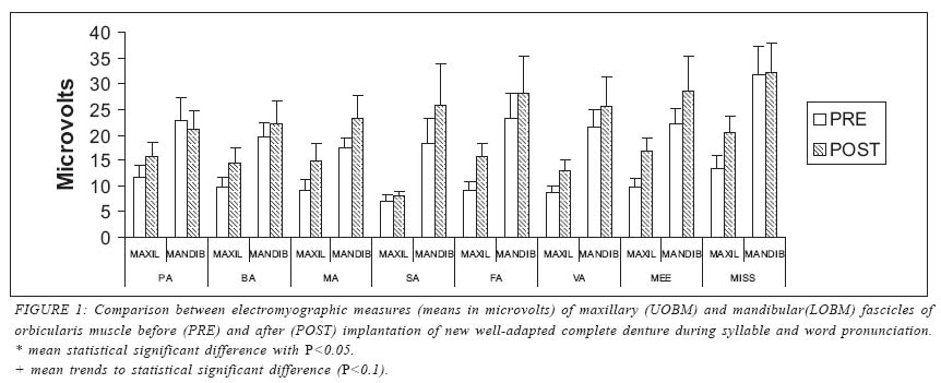
Brazilian Journal of Oral Sciences, Vol. 4, No. 14, July/Sept. 2005, pp. 801-805 Effect of denture quality on perioral muscle activity during speech Carla Moreto Santos1*Mathias Vitti2* Maria da Glória Chiarelo de Mattos3** Marisa Semprini4* Helena de Freitas Oliveira Paranhos5** Jaime Eduardo Cecilio Hallak6* Simone Cecilio Hallak Regalo7* *School of Dentistry, Department of Morphology, Stomatology and Physiology, USP, Ribeirão Preto, S.P., Brazil. **School of Dentistry, Department of Prosthesis and Dental Materials, USP, Ribeirão Preto, S.P., Brazil. 1PhD, Dental Surgeon 2PhD, Professor 3PhD, Professor 4PhD, Professor 5PhD, Professor 6MD, PhD Assistant Professor, Neuropsychiatry and Psychological Medicine Department, Faculty of Medicine, USP, Ribeirao Preto, S.P., Brazil and Clinical Research Fellow, Neuroscience and Psychiatry Unit, University of Manchester, Manchester, UK. 7PhD, Professor Correspondence to:Mathias Vitti Departamento de Morfologia Estomatologia e Fisiologia/FORP-USP Avenida do Café, s/n - Bairro - Monte Alegre 14040-904 –Ribeirão Preto SP –Brazil Fax: 55-02116-6330999 E-mail: mvitti@forp.usp.br Received for publication: March 03, 2005 Code Number: os05031 Abstract This study aimed to evaluate the electromyographic activity of the orbicularis oris muscle in patients using clinically inadequate conventional dentures before and after the insertion of clinically acceptable new convencional denture. Six patients, using inadequate dentures, were asked to pronounce the syllables PAH, BAH, MAH, SAH, FAH, VAH, MEE and the word MISSISSIPI. During this activity, we analyzed the Electromyographic activity of the orbicularis oris muscle. This was done before and after changing the inadequate denture for a acceptable new denture. A K6-I EMG Light Channel Surface Electromyograph was used (Myo-tronics Co). Results were analyzed by repeated measures ANOVA, with 3 sources of variation (Syllables, Muscles, and Clinical Conditions). ANOVA revealed higher electromyographic readings in the mandibular fascicle of the orbicular oris muscle, as compared to those of the maxillary fascicle (F = 79.02; P<0.01). The comparison regarding clinical conditions indicated higher electromyographic values after insertion of acceptable new denture (F= 32.64; P<0.01). Clinical condition after the implantation of a new well-adapted complete denture revealed higher electromyographic activity levels than those measured with the use of maladapted complete dentures. Key Words:denture, complete, electromyography, lip muscle Introduction Electromyography is conceptualized as the register and analysis of intrinsic electrical potentials produced by muscles and has been used as a research tool in Dentistry for over fifty years1- 2. Besides being used as a neuromuscular disorder diagnostic tool, one of the possible uses of EMG is to research mechanisms of speech. Since this technique has evolved, there has been a better recording and interpretation of signals, and a better understanding of speech has been achieved3-4. The syllables pronunciation is a reliable method used by dental surgeons for restoring the occlusal vertical dimension5. The labials, teeth labial and sibilants sounds make possible the intimae contact between teeth’s dentures and lips. According the dentures adaptation can be a different electromyography contraction. Lips and cheeks muscles act by symmetrically pressurizing the outer portion of dental arches, while the tongue applies pressure to the inner portion of dental arches (inside the mouth). Structure and function of dental arches are directly linked to the circumoral musculature4-9. Therefore, the circumoral musculature of conventional denture wearers is always subject to modifications. Circumoral musculature contributes to the definition of facial morphology, once its tonicity and the force of muscular contraction, particularly that of the orbicularis oris muscle, are influenced by frontal teeth and might affect teeth position10. The circumoral musculature of clinically inadequate prosthesis wearers does not show the same muscular tonus as that of dentulous patients with normal occlusion. The prosthesis clinically inadequate caused by reabsorption of the residual edge makes the prosthesis less compatible with lip function generating a wrong contraction of circumoral musculature11-12. Haraldson, Karlsson and Carlsson13 showed that inadequate conventional denture wearers have decreased strength in their occlusion, which could be a result of a global reduction of the circumoral musculature. Aging process produces characteristic structure deterioration of the stomatognathic system and also of muscles and nerves of the whole body14. In the situation of tooth loss, parts of the mandibular bone are reabsorbed and the oral mucosa loses its morphological characteristics, the muscular fibers become atrophic, motor neurons and their receptors are lost, and there is also a reduction of neurotransmitters15-17. The objective of this study is to observe the activity of maxillary and mandibular fascicles of the orbicularis oris muscle in conventional denture wearers, by means of electromyographic analysis, before and after the insertion of a clinically acceptable new conventional denture. Patients were evaluated during pronunciation of the syllables PAH, BAH, MAH, MEE (labials syllables), FAH, VAH (teeth labial syllables), and the sibilants sounds like mississipi and the syllable mee. Material and MethodsSix individuals, 1 man and 5 women, average ages of 58 years, using clinically inadequate conventional dentures were evaluated in this study. The maladaptation of the prosthesis was due to a failure in retention, absence of an adequate support, and absence of stability during use. This prosthesis had poor anterior projection, the anterior teeth was incorrectly positioned, causing a nasolabial sulcus increase. A decrease in the height of occlusion was noted, which caused a jaw anterior projection and circumoral muscle flabbiness. No situations of aches, temporomandibular dysfunction (TMD), para-functional habits, psychological problems, flabby tissue, or inadequate size ridge were registered among the patients. All individuals were fully informed of the nature of the investigation and agreed to take part in it. This research was approved by the Ethics Committee of the College of Odontology, USP- Ribeirão Preto-S.P., Brazil; process number: 00.1.215.58.0. Electromyographic tests were carried out at the Laboratory of Electromyography and Computerized Occlusal Diagnostic, of the Department of Morphology, Stomatology and Physiology of the College of Odontology, USP Ribeirão Preto, Brazil. An 8-channel K6-I EMG Light Channel Surface Electromyography device was used (Myo-tronics Co). Disposable double electrodes covered with silver chloride (Duotrodes; Myo-tronics Co) containing a conductor gel (Myogel- Myo-tronics Co) were used. The electrodes were placed on the maxillary and mandibular fascicles of the orbicularis oris muscle, parallel to the muscular fiber sheaf. Before electrode placement, the skin was cleaned with alcohol 70°, to eliminate dirt and fat residues. A reference electrode (ground) was placed close to the patient’s nape, to avoid any possible interference. During the measurements, individuals remained seated on a comfortable chair, in upright position, with their feet on the floor and their arms leaned on their legs. Their head was straightly positioned with the Frankfort plane parallel to the floor. A silent and partially illuminated environment was maintained, and patients were oriented to breathe deeply a couple of times before beginning the measurements and to remain relaxed in order to avoid any involuntary contractions that could affect muscular activity and rest positioning during syllable pronunciation. Electromyographic activity was recorded during the pronunciation of seven syllables (PAH, BAH, MAH, SAH, FAH, VAH, MEE) and one word (MISSISSIPI). For each record, the same syllable was slowly pronounced and repeated 6 times. Before pronouncing a new syllable, the voluntary was asked to lightly seal the lips, without pressing them. Two electromyographic tests were carried out; one before and one after the implantation of the well-adapted complete denture. The new dentures were made by a dentist at the Complete Denture Clinic according to the following principles: marginal seal and posterior palatal seal correction, adequate denture coverage, anterior teeth positioned further than incisive papilla with correct vertical angulations, and adequate height of occlusion. These details bring us retention, stability, support and ideal anterior teeth - orbicularis oris muscle relationship according each facial type of patients. Voluntaries were under observation along the process of prosthesis confection at the Conventional Denture Clinic. A 5-month period was defined as the time interval before performing new electromyographic analyses. The criteria and conditions for performing these tests were the same as described above. Statistical analysis was carried out using the Statistical Package for The Social Sciences (SPSS), version 10.0.General linear model of analysis of variance with repeated measures was used to analyze the electromyographic activity during the pronunciation of the 7 syllables before and after the insertion of clinically acceptable new conventional dentures, considering conditions (before and after insertion), syllables and word pronunciation, and the muscle fascicles (maxillaryand mandibular). The differences before and after the new prosthesis insertion,during the pronunciation for each syllable individually, wereanalyzed with a paired Student’s t-test for each fascicle ofthe oris orbicularis muscle (maxillary and mandibular). ResultsTable 1 shows the mean electromyographic activity during syllable pronunciation before and after the new conventional denture insertion. Significant statistical differences were found between conditions (prior to and following new prosthesis insertion–F= 32.64; P<0.01), between the maxillary and mandibular fascicles (F = 79.02; P<0.01), and the interaction between condition and muscular fascicles (F =14.71; P=0.04), but no differences were found between the syllables (F = 3.13; P=0.41) and in the interactions between condition and syllables (F = 0.34; P=6.58) and between syllables, condition and muscular fascicles (F = 0.28; P=3.82). The mandibular fascicle of the orbicularis oris muscle showed higher electromyographic means than the maxillary fascicle in both conditions (prior to and following new prosthesis) and the electromyographic measures were higher post insertion than pre insertion (Fig.1). The paired Student t-test comparing EMG activity during each of the different syllables and word pronunciation prior to and following the insertion of a new clinically acceptable conventional denture showed that, for the syllable MEE, the maxillary fascicle of the orbicularis muscle presented a significant increase in its electrical activity, and also showed trends toward significance (P<0.1) in the pronunciation of the syllable FAH and the word MISSISSIPI (Fig. 1). DiscussionIt has been seen an increase in life expectation worldwide14. Many studies report that elderly oral health is precarious and most elderly are edentulous12,17. The use of EMG as a research tool to reach a better comprehension regarding the stomatognathic system, particularly the circumoral musculature performance, is really important since the need for dental prosthesis is increasing6. This research found some limitations like a poor sample size considering the high variability in the EMG measures in this study. Some individuals that participated in this study didn’t use the new prosthesis, one of them dead, others didn’t want to continue the treatment. Then the researchers lost the great part of the initial sample. But this number of individuals brought us satisfactory results. The EMG assessment of the fascicles superior and inferior of the orbicularis muscle was made through syllables pronunciation because it has been proved to produce efficient muscular contraction enough to be recorded7-8. EMG analysis showed statistical significant differences before and after the implantation of a new clinically acceptable conventional denture when, during the use of a inadequate prosthesis, the orbicularis oris muscle presented a higher level of mandibular activity than after the change for a new one. The mandibular fascicle of the orbicularis oris muscle showed stronger electromyographic activity than the maxillary fascicle. This means that this fascicle is more active during speech. It is known that muscular performance during growth and orofacial development is limited by mandibular structure and growth pattern9. Both fascicles are independent and they interact in a complex way during the different lip positions during speech2. Although Ingervall and Redegard6 found mandibular activation of the mandibular fascicle compared to the maxillary fascicle previous to and after insertion new clinically acceptable prosthesis, their finding was during maximal intercuspidation position. During the syllables and word pronunciation, which emphasize the orbicularis oris muscle function, the presented study found a slight statistical difference in muscular activity, for the MISSISSIPI word and for the syllable MEE. During speech, the anterior position of the prosthetic teeth and the interaction between lips and the basis of the prosthesis might generate modifications in muscular contraction pattern. This increase in electromyographic activity with the new prosthesis probably happened because the teeth’s previous position was modified, once the bases of the new prosthesis are slightly thicker than the old ones. This is done in order to obtain better esthetics and functional results concerning lip support; decreasing lips’sulci and facial expression lines. From the analysis of the results obtained in this study, the following can be concluded: clinical condition after the implantation of a new well-adapted complete denture revealed higher electromyographic activity levels than those measured with the use of maladapted complete dentures; the inferior fascicle of the orbicularis oris muscle presented higher electrom yographic levels than those measured in the superior fascicle; syllable pronunciation is a reliable method for EMG analysis of the orbicularis oris muscle in patients with old and new complete dentures. AcknowledgmentsWe are grate by financial support to this research by FAPESP (process number: 00/05924-6). References
Copyright 2005 - Piracicaba Dental School - UNICAMP São Paulo - Brazil |
